Computed tomography and magnetic
resonance imaging in the osseous phase of
Nasu-Hakola disease
Tomografia computadorizada e ressonância magnética na fase óssea da doença de
Nasu-Hakola
Christiana Brenner
1, Carlos Eduardo Speck-Martins
1, Jaime Moritz Brum
1, Leandro Tavares Lucato
2,
Claudia da Costa Leite
2,3Nasu-Hakola disease (NHD-polycystic lipomembranous
osteodysplasia
with
sclerosing
leukoencephalopathy;
PLOSL) is a rare autosomal recessive disorder, caused by
mutations in two genes: TREM 2 and DAP 12. NHD is
characterized by a combination of diffuse bone cysts and
pre-senile dementia. Most of the NHD cases first present
in early adulthood with skeletal abnormalities (osseous
phase). Neurological symptoms manifest in the fourth
dec-ade of life as prefrontal syndrome
1,2,3. A 32 year-old male
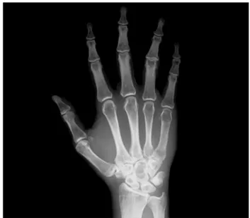
patient presented bone fractures, usually after minor
trau-mas and cystic lesions on X-Rays (Figure 1). He denied
any personality change or memory disturbances. The
neuro-logical examination was normal. The neuropsychoneuro-logical
tests displayed only easy distractibility. CT and MRI
dem-onstrate abnormalities in the basal ganglia and white matter
(Figure 2) showing that imaging findings precedes
neuropsy-chiatric symptoms.
1Rede Sarah de Hospitais, Brasília DF, Brazil;
2Departamento de Radiologia, Faculdade de Medicina, Universidade São Paulo, Sao Paulo SP, Brazil;
3University of North Carolina, Chapel Hill, United States.
Correspondence:Claudia Leite; Faculdade de Medicina da USP–Radiologia; Av. Dr. Ovídio Pires Campos, s/n Inrad Portaria 5 Ressonância Magnética; 05403-903 São Paulo SP, Brasil; E-mail: [email protected]
Conflict of interest:There is no conflict of interest to declare.
Received 26 January 2014; Received in final form 22 April 2014; Accepted 12 May 2014.
Figure 1.
R-xays of the right hand demonstrates multiple cystic
lesions in the carpal bones.
Figure 2.
Head CT discloses punctate basal ganglia calcification (arrows in A). Brain MRI, axial T2-weighted (B, C) and axial
FLAIR (D) images show diffuse cerebral atrophy, bilateral and symmetric hyperintensity in the posterior limbs of the internal
capsules (arrows in B), and in the parieto-occipital periventricular white matter and centrum semiovale (arrowheads in C and D).
DOI:10.1590/0004-282X20140081
IMAGES IN NEUROLOGY
References
1. Paloneva J, Autti T, Raininko R, et al. CNS manifestation of Nasu-Hakola disease: A frontal dementia with bone cysts. Neurology 2001;56:1552-1558.
2. Klünemann HH, Ridha BH, Magy L, et al. The genetic causes of basal ganglia calcification, dementia, and bone cysts: DAP12 and TREM2. Neurology 2005;64:1502-1507.
3. Kilic SA, Oner AY, Yuce C, Ozlu IC. Imaging findings of Nasu–Hakola disease: a case report. Clin Imag 2012;36:877-880.