Ambulatório de Neurologia do Desenvolvimento da Clínica Neurológica do Hospital das Clínicas da Faculdade de Medicina da Universidade de São Paulo, São Paulo SP, Brazil: 1MD; MSc; 2MD, PhD.
Received 25 May 2005, received in final form 5 August 2005. Accepted 30 September 2005.
Dr. Carlos O. Heise - Avenida Lacerda Franco 220 - 01536-000 São Paulo SP - Brasil. E-mail: [email protected]
PROGNOSTIC RELEVANCE OF RISK FACTORS
FOR OBSTETRICAL BRACHIAL PLEXOPATHY
Carlos O. Heise
1, José Luiz D. Gherpelli
2ABSTRACT - We did a case-control study to verify if the birthweight, forceps delivery or perinatal asphyx-ia have any significant effect on the prognosis of obstetrical brachasphyx-ial plexopathy. Group A was composed of 25 infants who completely re c o v e red at the age of 6 months. Group B was composed of 21 infants who w e re still not able to remove a blindfold from the face with the affected limb in the sitting position at the age of 12 months. There was no statistical diff e rence of the median birthweight or median first minute Apgar score between the groups. There was also no relation between birthweight higher than 4000g, first minute Apgar score lower than 6 or forceps delivery with a poor prognosis.
KEY WORDS: brachial plexus, obstetric paralysis, risk factors, birth weight, obstetrical forceps, Apgar score .
Relevância prognóstica dos fatores de risco para plexopatia braquial obstétrica
RESUMO - Realizamos um estudo caso-controle para verificar se o peso ao nascimento, parto forceps ou asfixia perinatal apresentam efeito significativo no prognóstico da plexopatia braquial obstétrica. O gru p o A foi composto por 25 lactentes que apresentavam recuperação completa aos 6 meses de idade. O grupo B foi composto por 21 lactentes incapazes de remover uma venda do rosto com o membro acometido na posição sentada aos 12 meses de idade. Não houve diferença significativa entre as medianas de peso ao nascimento ou do boletim Apgar do primeiro minuto entre os grupos. Também não foi observada re l a ç ã o e n t repeso ao nascimento maior que 4000g, boletim Apgar do primeiro minuto menor do que 6 ou part o forceps com um prognóstico desfavorável.
PA L AV R A S - C H AVE: plexo braquial, paralisia obstétrica, fatores de risco, peso ao nascimento, forceps obstétri-co, escore de Apgar.
Obstetrical brachial plexopathy (OBP) still is a com-mon consequence of birth trauma. Its incidence in developed countries is around 0.15%1,2and has not
been reduced despite pro g ress in obstetrics2 , 3. Fort
u-n a t e l y, most patieu-nts with OBP will fully recover after a few months, but 5% to 25% of them will re m a i n h a n d i c a p p e d2 , 4 - 8. The brachial plexus is formed by the
anterior branch of the spinal roots from C5 to T19.
Supraclavicular plexus lesions, such as OBP, can be cli-nically and anatomically divided into superior (C5-C6), middle (C7) and inferior (C8-T1) levels1 0. Pure
upper level plexopathy, or Erb palsy, accounts for 50% of the cases11. These patients have poor elbow
flexion, shoulder abduction, arm external ro t a t i o n , and fore a rm supination1 2. The wrist extension may
also be weak due to the involvement of the exten-sor carpi radialis muscles. The resulting posture is clas-sically described as “waiter’s tip”11. One third of the
patients with OBP have an upper and middle levels
p l e x o p a t h y. Middle level plexus involvement leads to poor elbow, wrist, and fingers extension1 2. Pure
lower level plexopathy, or Klumpke palsy, is extre m e-ly rare1 1. These patients have poor fingers flexion and
thumb opposition, and may also exhibit a Horner syn-d rome (miosis, ptosis, enophthalmos ansyn-d anhysyn-dro- anhydro-sis). One sixth of the patients with OBP have a com-plete brachial plexus lesion (from C5 to T1) and show a total limb paralysis (flail arm), with or without Hor-ner syndrome10.
Several risk factors for OBP have been identified, the most well know being high birthweight and assisted delivery1 , 2 , 4 , 1 3 , 1 4. Perinatal asphyxia may also
be a contributing factor, because the associated hypo-tonia would make the fetus more vulnerable to s t retch injuries1 0 , 1 5. Although the relation of these
The objective of this study was to verify if these risk factors have prognostic relevance or not.
METHOD
F rom July 2000 to December 2004, 79 infants with OBP (0-10 months old) were re f e rred to the child neurology out-patient unit of the Clinics Hospital of Sao Paulo. We did a c a s e - c o n t rol study based on two groups selected from these c h i l d ren. The re s e a rch ethical committee of the hospital approved this study.
G roup A (good prognosis) was composed of 25 infants who showed complete re c o v e ryand no strength asymme-t ry aasymme-t asymme-the age of 6 monasymme-ths. There were 21 paasymme-tienasymme-ts wiasymme-th C5-C6 palsy; 3 patients with C5-C5-C6-C7 palsy, and one patient with C8-T1 palsy. All patients were admitted before 60 days of age (median=16 days). The follow-up of these children was at least 4 months long (range: 4-12 months; median=6 months).
G roup B (poor prognosis) was composed of 21 infants who still were unable to remove a blindfold from the face with the affected limb in the sitting position at the age of 12 months. There were 10 patients with complete brachial plexus paralysis (C5-T1); 10 patients with C5-C6-C7 palsy (one of them bilateral), and one patient with C5-C6 palsy. Twelve patients were admitted before 60 days of age (medi-an=28 days), and nine patients were re f e rred later for sur-g e ry, at the asur-ge of 3 to 10 months (median=8 months). The follow-up of these children ranged from 2 to 52 months (median=32 months). Seventeen patients from this group w e re followed-up until at least two years of age and none of them developed good arm function. Five of these chil-d ren were submittechil-d to brachial plexus neurolysis before the age of 12 months, but there was no strength loss after the surg e ry. There f o re, the poor outcome could not be attri-buted to the surgical procedure.
The patients excluded from the study had an incom-plete follow-up (n=18), intermediate outcome (n=9), cere-bral palsy (n=3), or were submitted to brachial plexus g e ry with nerve grafts (n = 3). Patients submitted to g e ry with nerve grafts can lose muscle power after the sur-g e ry because the nerves are sectioned in order to place the grafts. Patients with intermediate outcom e were able to remove the blindfold from the face with the affected limb at 12 months of age, but still had clear strength
asymme-t ry (usually for supinaasymme-tion and arm exasymme-ternal roasymme-taasymme-tion) or scapular winging.
We compared the two groups in relation to birt h w e i g h t , first minute Apgar score (FMAS) and mode of delivery. FMAS was employed in order to evaluate the possible ro l e of fetal hypotonia secondary to birth asphyxia during deliv-e ry and not thdeliv-e deliv-evdeliv-entua l hypoxic- ischdeliv-emic deliv-encdeliv-ephalopa- encephalopa-t h y. Daencephalopa-ta on birencephalopa-thweighencephalopa-t and delivery mode were available in all patients. The FMAS was available in 24 patients of group A and 17 patients of group B. We used the ANOVA test to compare mean birthweight and Kru s k a l - Wallis test for median FMAS. Birthweight and FMAS were also trans-f o rmed in categorical variables to calculate the odds ratios. The patients exposed to risk factors should have birt h w e i g h t higher than 4000g, FMAS of less than 6 and forceps deliv-e ry. Thdeliv-e confiddeliv-encdeliv-e intdeliv-ervals for odds ratios wdeliv-erdeliv-e calculat-ed using 95% exact confidence limits. Statistic analysis was done using the Epi Info 2002 program (CDC, Atlanta, USA).
RESULTS
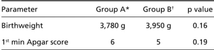
In group A, birthweight ranged from 2570 g to 4450 g (Fig 1). The median birthweight is shown in Table 1. Eight patients weighted more than 4000g ( Table 2). There were two pre - t e rm and 23 full-term infants. Eleven infants were larg e - f o r-gestational age, and 14 were adequate-for-gestational age. In gro u p B, birthweight ranged from 2000 g to 5515 g (Fig 1). The median weight is shown in Table 1. There were 9 patients who weighted above 4000g (Table 2). All infants were full-term. Nine infants were larg e - f o r-gestational age, one patient was small-for- g e s t a t i o n-al age, and 11 had adequate-for-gestationn-al age.
Table 1. Diff e rences of medians of birthweight and first minute Apgar scores between the two groups.
Parameter Group A* Group B† p value
Birthweight 3,780 g 3,950 g 0.16
1stmin Apgar score 6 5 0.19
*Good prognosis group;†Poor prognosis group.
Table 2. Pro p o rtion of patients exposed to the risk factors in the two groups, and odds ratio and confidence interval for a poor prognosis based on the presence of the risk factors.
Risk factor Group A* Group B† Odds Ratio C.I.‡
Birthweight > 4 Kg 32% 43% 1.59 0.41-6.30
1stApgar score < 6 46% 53% 1.33 0.32-5.52
Forceps delivery 36% 24% 0.56 0.12-2.38
T h e re was no statistically significant diff e rence of birthweight between the groups.
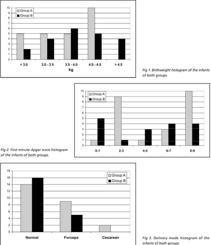
In the group A, the FMAS ranged from 1 to 9 (Fig 2). The median FMAS is shown in Table 1. There were 11 patients with a FMAS below 6 (Table 2). In the g roup B, the FMAS ranged from 0 to 8 (Fig 2). The me-dian FMAS is shown in Table 1. There were 9 patients
with a FMAS below 6 (Table 2). There was also no sta-tistically significant diff e rence between the gro u p s .
In the group A, there were 14 normal deliveries, 9 forceps deliveries, and 2 cesareans (Fig 3). In the group B, there were 16 normal deliveries and 5 for-ceps deliveries (Fig 3). There was no statistically sig-nificant difference between the groups (Table 2).
Fig 1. Birthweight histogram of the infants of both groups.
Fig 3. Delivery mode histogram of the infants of both groups
DISCUSSION
We could not demonstrate any relation between b i rthweight, perinatal asphyxia or forceps delivery and neurological prognosis of infants with OBP. Pa-tients with poor prognosis had slightly higher birt h-weight and slightly lower FMAS than patients with good prognosis, but this was not statistically signif-icant. Surprisingly, patients with good prognosis had a higher pro p o rtion of forceps deliveries, but this was also not statistically significant.
T h e reis a clear relation between the rate of re c o v-e ry and thv-e final prognosis of pativ-ents with OBP. Thv-e ideal age to define the final outcome would be thre e years, because the patients are not expected to im-p rove after this age1 6. However, Noetzel et al. could
c o rrectly predict the 18-24 months outcome of infants with OBP based on the neurological impairment at 6 month of age1 7and Basheer et al. found that 90%
of patients reach their final clinical status at 6 months of age1 8. Our criterion for poor prognosis relies on a
functional test at the age of 12 months. This test was based on the “cookie test” employed by Clarke and C u rtis at the age of nine months as a criterion for s u rgical interv e n t i o n1 9. It has the advantage of being
objective and easily perf o rmed. All children from the “good prognosis” group were able to perf o rm this test at 6-7 months of age.
Our study may not have enough power to detect the prognostic effect of birthweight and FMAS due to the small sample size. Larger studies or meta-analy-sis could possibly answer this question with more cer-titude. However, OBP is not a common condition and large series are difficult to provide. Meta-analysis is almost impossible since the follow-up period, the assessment protocol and the definitions of “good” and “poor” prognosis are so diff e rent among the publications dealing with OBP prognosis that the data cannot be compared16.
High birthweight is a very important risk factor for OBP1 , 2, but the effect of birthweight on the pro
g-nosis of affected newborns is controversial. Nehme et al. did a retrospective study with 30 patients and found that high birthweight was associated with poor prognosis in a multivariate analysis when asso-ciated with the neurological involvement2 0. Bager, in
a prospective cohort study with 41 patients, could not find any association between birthweight and the neurological outcome2.
Assisted deliveries, including forceps and ventouse, c a rry a higher risk for OBP1. Vacuum extraction
assist-ed deliveries are not usually perf o rmassist-ed in our
coun-t ry. Alcoun-though forceps delivery is clearly associacoun-ted with an increased risk of OBP1 4, Brown believes that
the forceps has no causal relation and that it is only another consequence of a difficult delivery21.
Evans-Jones et al. did a large study based on active s u rveillance for OBP in the United Kingdom and I re l a n d3. The outcome assessment was based on 322
q u e s t i o n n a i ressent to consultant pediatricians, fro m which 276 questionnaires returned. The assessment was done at the age of 23 weeks (range 18-27). There w e re 52% cases with full re c o v e ry, 46% with part i a l re c o v e ry, and 2% with no re c o v e ryat that age. They did not find a higher rate of incomplete re c o v e ry for m a c rosomic infants (relative risk: 1.37; confidence interval: 0.91-2.04) or patients with assisted deliver-ies (relative risk: 0.93; confidence interval: 0.72-1.22).
We are not aware of any study that specifically a d d resses the effect of FMAS on the prognosis of infants with OBP. Perinatal asphyxia is fre q u e n t l y associated with OBP4,7,14. The theory that hypotonia
induced by perinatal hypoxia predisposes the fetus to a brachial plexus stretch injury seems logical1 5, but
lacks experimental confirmation. Furt h e rm o re, the asphyxia should occur prior to the brachial plexus lesion, which is impossible to be sure in this study. Fetal blood pH is a better instrument than Apgar score to measure fetal hypoxia22.
In summary, the prognosis of OBP cannot rely on factors such as birthweight, delivery mode or peri-natal asphyxia. Nerve conduction studies may be help-ful for prognostic assessment2 3, but the best pro
g-nostic guides still are the neurological impairm e n t and the rate of recovery17,19,24.
REFERENCES
1. Gilbert WM, Nesbitt TS, Danielsen B. Associated factors in 1611 cases of brachial plexus injury. Obstet Gynecol 1999;93:536-540.
2. Bager B. Perinatally acquired brachial plexus palsy: a persisting chal-lenge. Acta Paediatr 1997;86:1214-1219.
3. Evans-Jones G, Kay SPJ, Weindling AM, et al. Congenital brachial pal-sy: incidence, causes, and outcome in the United Kingdom and Republic of Ireland. Arch Dis Child Neonatal Ed 2003;88:F185-F189.
4. G o rdon M, Rich H, Deutschberger J, Green M. The immediate and long-term outcome of obstetric birth trauma: I. Brachial plexus paralysis. Am J Obstet Gynecol 1973;117:51-56.
5. Greenwald AG, Schute PC, Shiveley JL. Brachial plexus birth palsy: a 10-year report on the incidence and prognosis. J Pediatr Orthop 1984;4: 689-692.
6. S j ö b e rg I, Erichs K, Bjerre I. Cause and effect of obstetric (neonatal) brachial plexus palsy. Acta Paediatr Scand 1988;77:357-364. 7. H a rdy AE. Birth injuries of the brachial plexus: incidence and pro g n
o-sis. J Bone Joint Surg Br 1981;63:98-101.
9. Ferrante MA, Wilbourn AJ. Electrodiagnostic approach to the patients with suspected brachial plexopathy. Neurol Clin N Am 2002;20:423-450.
10. Painter MJ, Bergman I. Obstetric trauma of the neonatal central and peripheral nervous system. Semin Perinatol 1982;6:89-104. 11. Van Dijk JG, Pondaag W, Malessy MJA. Obstetric lesions of the brachial
plexus. Muscle Nerve 2001;24:1451-1461.
12. Alfonso I, Alfonso DT, Papazian O. Focal upper extremity neuro p a t h y in neonates. Semin Pediatr Neurol 2000;7:4-14.
13. Donnelly V, Foran A, Murphy J, McParland P, Keane D, O'Herlihy C. Neonatal brachial plexus palsy: an unpredictable injury. Am J Obstet Gynecol 2002;187:1209-1212.
14. McFarland LV, Raskin M, Daling JR, Benedetti TJ. Erb/Duchenne's pal-sy: a consequence of fetal macrosomia and method of delivery. Obstet Gynecol 1986;68:784-788.
15. Server JW. Obstetric paralysis: report of eleven hundred cases. JAMA 1925;85:1862-1865.
16. Pondaag W, Malessy MJA, Van Dijk JG, Thomeer RTWM. Natural his-tory of obstetric brachial plexus palsy: a systematic re v i e w. Dev Med Child Neurol 2004;46:138-144.
17. Noetzel MJ, Park TS, Robinson S, Kaufman B. Prospective study of recovery following neonatal brachial plexus injury. J Child Neurol 2001; 16:488-492.
18. Basheer H, Zelic V, Rabia F. Functional scoring system for obstetric brachial plexus palsy. J Hand Surg Br 2000;25:41-45.
19. Clarke HM, Curtis CG. An approach to obstetrical brachial plexus injuries. Hand Clin 1995;4:563-581.
20. Nehme A, Kany J, Sales-De-Gauzy J, Dautel G, Cahuzac JP. Obstetrical brachial plexus palsy: prediction of outcome in upper root injuries. J Hand Surg Br 2002;27:9-12.
21. B rown KLB. Review of obstetrical palsies: nonoperative treatment. Clin Plast Surg 1984;11:181-187.
22. Marrin M, Paes BA. Birth asphyxia: does the Apgar score have diag-nostic value? Obstet Gynecol 1988;72:120-123.
23. Heise CO, Lorenzetti L, Marchese AJT, Gherpelli JLD. Motor conduc-tion studies for prognostic assessment of obstetrical plexopathy. Muscle Nerve 2004;30:451-455.