265
Arq Neuropsiquiatr 2013;71(4):264-268
Eagle’s syndrome
Síndrome de Eagle
Ana Beatriz Soldati1, Cláudia Miguelote1, Carla Quero2, Rita Pereira3, Roberto Santos4, Cristiane Soares5
1MD, resident in Neurology at Hospital Federal dos Servidores do Estado, Rio de Janeiro RJ, Brazil; 2MD, resident in Neuropediatry at Instituto Fernandes Figueira, Rio de Janeiro RJ, Brazil; 3MD, resident in Radiology at Hospital Federal dos Servidores do Estado, Rio de Janeiro RJ, Brazil; 4MD, Radiologist at Hospital Federal dos Servidores do Estado, Rio de Janeiro RJ, Brazil; 5MD, PhD, Neurologist at Hospital Federal dos Servidores do Estado, Rio de Janeiro RJ, Brazil.
Correspondence: Ana Beatriz Soldati; Rua Mário Viana 479 / apt. 802; 24241-000 Niterói RJ - Brasil; E-mail: biasoldati@gmail.com
Conflict of interest: There is no conlict of interest to declare.
Received 23 April 2012; Received in inal form 18 October 2012; Accepted 25 October 2012.
Eagle’s syndrome (ES) consists of craniofacial and neck pain due to elongation of the styloid process and/or calcii-cation of the stylohyoid ligament1. he normal length of the
styloid process ranges from 2 to 3 cm and it is hardly palpa-ble. ES is usually an asymptomatic disease that mostly afects women aged 30 to 50 years.
According to Eagle, two types of the syndrome have been described2: (A) the classic syndrome characterized by
pain in the tonsillar fossa, sometimes associated with dys-phagia and hypersalivation, and often followed by tonsillec-tomy and (B) the styloid-carotid syndrome: the styloid pro-cess compresses the carotid arteries and exerts particular
pressure on its sympathetic ibers. his subtype is not corre-lated with tonsillectomy.
We report the case of a young patient with ES and a sty-loid process of normal lenght, but with signiicant ligament calciication.
CASE
A 20-year-old female patient presented at the Neurology Service with a continuous, daily, pressure-type pain on the left side of the neck radiating to frontal and temporal regions.
DOI: 10.1590/0004-282X20130015
266 Arq Neuropsiquiatr 2013;71(4):264-268
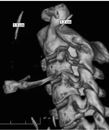
here was neither history of trauma or previous surgery, nor identiiable trigger points. Physical examination revealed pain on tonsillar fossa at palpation, as well as on the left side of the neck. he neurological examination including the cranial nerves was normal. hree-dimensional comput-ed tomography (CT) scan of the neck showcomput-ed bilateral stylo-hyoid ligament calciication of 1.9 cm on the right side and 1.2 cm on the left side (Fig 1). he length of the styloid pro-cess added to the length of the calciication was, respectively, 4.1 and 3.7 cm (Fig 2). he symptom of pain was solved after administration of ibuprofen for two months.
DISCUSSION
ES is a rare condition of unknown pathophysiology and etiology2. It can be idiopathic, congenital (due to persistence
Fig 1. 3D-CT scan image showing bilateral calciication of the stylohyoid ligament.: right stylohyoid ligament with 1.9 cm; left stylohyoid ligament with 1.2 cm.
Fig 2. CT scan image showing increased styloid processes: right with 4.1 cm; left with 3.7 cm.
of cartilage elements of the precursors of the styloid pro-cess) or acquired3.
Approximately 4% of the population has elongated styloid process, but only 4 to 10.3% of this group has its symptoms4.
Although many authors believe that this syndrome is more typical in adult life, the symptoms’ onset of the patient herein described began in her 20s.
Treatment for ES includes non-steroidal anti-inlamma-tory drugs, steroids and applications of anesthetic in the ton-sillar fossa4, in addition to surgical approach consisting of
re-moval of the styloid process intra or extraorally.
he patient reported full improvement with non-steroidal anti-inlammatory drugs. However, it is not known whether the symptoms may return in old age.
Dissection, aneurysm and pseudoaneurysm of the ca-rotid artery may occur in the course of ES. Further stud-ies are needed to investigate the relationship between the elongated styloid process and the development of such complications5.
1. Savranlar A, Uzun L, Uğur MB, Özer T. Three-dimensional CT of Eagle’s syndrome. Diagn Interv Radiol 2005;11:206-209.
2. Politi M, Toro C, Tenani G. A rare cause for cervical pain: Eagle’s syndrome. Int J Dent 2009;2009:781297.
3. Raina D, Gothi R, Rajan D. Eagle syndrome. Indian J Radiol Imaging 2009;19:107-108.
4. Murtagh RD, Caracciolo JT, Fernandez G. CT findings associated with Eagle syndrome. AJNR Am J Neuroradiol 2001;22: 1401-1402.
5. Dao A, Karnezis S, Lane JS, Fujitani RM, Saremi F. Eagle syndrome presenting with external carotid artery pseudoaneurysm. Emerg Radiol 2011;18:263-265.