INTRODUCTION
Nasolacrimal duct obstruction (NLDO) is a common ophthalmic presentation and has been reported to represent 3% of clinic visits(1). NLDO is typically treated using either external (EX-) or endoscopic (EN-) dacryocystorhinostomy (DCR) or using transcanalicular
multi-diode laser (TC-) to create a istula between the lacrimal sac and nasal cavity, allowing tear low. Each type of DCR is associated with speciic advantages and disadvantages(2,3). The traditional DCR procedure (EX-DCR) can cause cutaneous scarring, disruption of the medial can-thus, and excessive intra-operative bleeding. Advantages of EN-DCR
Bacteriological profile in conjunctival, lacrimal sac, and nasal specimens and
conjunctival normalization time following external, endoscopic, and
transcanalicular multidiode laser dacryocystorhinostomy
Peril bacteriológico e tempo de normalização conjuntival de espécimes de conjuntiva, saco lacrimal e
nasais após dacriocistorrinostomia externa, endoscópica e transcanalicular com laser de multi diodo
Melike Balikoglu-YilMaz1,2, aYse Banu esen3, Tolga YilMaz2, uMiT Taskin4, MuhiTTin Taskapili2, M. Faruk okTaY4, eMine sen5, TiMur kose6
Submitted for publication: August 10, 2015 Accepted for publication: February 10, 2016
1 Department of Ophthalmology, Faculty of Medicine, Izmir Katip Celebi University, Izmir, Turkey. 2 Department of Ophthalmology, Bagcilar Education and Research Hospital, Istanbul, Turkey. 3 Department of Microbiology and Clinical Microbiology Bagcilar Education and Research Hospital,
Istanbul, Turkey.
4 Department of Otorhinolaryngology, Bagcilar Education and Research Hospital, Istanbul, Turkey. 5 Department of Ophthalmology, Ulucanlar Eye Education and Research Hospital, Ankara, Turkey. 6 Department of Biostatistics and Medical Informatics, Faculty of Medicine, Ege University, Izmir, Turkey.
Funding: The present study was supported by the Institutional Review Board of Bagcilar Education and Research Hospital, Istanbul, Turkey (#1852).
Disclosure of potential conflicts of interest: None of the authors have any potential conflicts of interest to disclose.
Corresponding author: Melike Balikoglu-Yilmaz. Department of Ophthalmology, Izmir Katip Celebi University. Faculty of Medicine. Izmir - Turkey - E-mail: [email protected]
Approved by the following research ethics committee: Bagcilar Education and Research Hospital (#2011/13).
ABSTRACT
Purpose: To compare the conjunctival, lacrimal sac, and nasal flora cultures and conjunctival normalization time following external (EX-), endoscopic (EN-), and transcanalicular multidiode laser (TC-) dacryocystorhinostomy (DCR) and to evaluate the relationship between culture positivity and surgical success. We further performed antibiotic sensitivity analyses for lacrimal sac culture samples.
Methods: A total of 90 patients with primary acquired nasolacrimal duct obstruc-tion were recruited and divided into EX-DCR (n=32), EN-DCR (n=28), and TC-DCR (n=30) groups. Conjunctival, nasal, and lacrimal sac cultures and antibiograms were analyzed.
Results: In all three groups, coagulase-negative Staphylococcus (CNS) was predo-minantly isolated preoperatively from the conjunctiva, nose, and lacrimal sac and postoperatively from the conjunctiva. Preoperative and postoperative conjunctival culture positivity rates were similar between all the groups (p>0.05). A statistically signiicant diference in the growth rate of culture in the lacrimal sac was observed between the three groups (p=0.001). CNS and Staphylococcus aureus cultures were predominantly sensitive to linezolid, teicoplanin, tigecycline, vancomycin, and mupirocin. Conjunctival normalization times were similar between the three groups (p>0.05). Anatomical and functional success rates were not found to be significantly correlated with preoperative conjunctival and lacrimal sac culture positivity (p>0.05).
Conclusions: Similar rates of preoperative and 1-week postoperative conjunctival culture positivity were observed in all the groups; a significantly lower bacterial growth rate was observed in postoperative conjunctival cultures. CNS was the most commonly isolated organism. Bacterial growth rates in the lacrimal sac samples were significantly higher in the EN-DCR group. Bacterial growth rates obtained preoperatively from the conjunctival and lacrimal sac culture samples were not correlated with DCR success.
Keywords: Conjunctiva; Dacryocystorhinostomy; Nasolacrimal duct; Antibiotic sensitivity tests; Staphylococcus aureus,Transcanalicular multidiode laser
RESUMO
Objetivo: Comparar a flora conjuntival, do saco lacrimal e nasal com o tempo de normalização após dacriocistorrinostomia (DCR) externa (EX-), endoscópica (EN-) e transcanalicular a laser de multi diodo (TC-) para correlacionar a positividade da cultura com o sucesso cirúrgico, assim como identificar a sensibilidade aos antibió-ticos em amostras de saco lacrimal.
Métodos: Neste estudo prospectivo, 90 pacientes com obstrução do canal naso-lacrimal adquirida primária foram incluídos e divididos em grupos EX-DCR (n=32), EN-DCR (n=28) e TC-DCR (n=30). Culturas e antibiogramas conjuntivais, nasais e do saco lacrimal foram analisados.
Resultados:Staphylococcus coagulase-negativo (CNS) foi o organismo predo-minante isolado no pré-operatório (conjuntiva e nariz), no transoperatório (saco lacrimal) e pós-operatório (conjuntiva), nos 3 grupos. Taxas de positividade de cultura da conjuntiva pré- e pós-operatórias nos três grupos foram semelhantes (p>0,05). A diferença nas taxas de crescimento do saco lacrimal dos três grupos foi estatisticamente significativa (p=0,001). CNS e S. aureus foram mais sensíveis a linezolida, teicoplanina, a tigeciclina, vancomicina e mupirocina. O tempo de nor-malização conjuntival foi semelhante nos três grupos (p>0,05). Não houve relação estatisticamente significativa entre as taxas de sucesso anatômicas e funcionais e a positividade da cultura conjuntival e de saco lacrimal pré-operatória (p>0,05).
Conclusões: Pacientes submetidos a EX-DCR, EN-DCR, e TC-DCR apresentaram po sitividades de cultura conjuntival semelhantes no pré-operatório e na 1a semana pós-operatória. Houve uma redução significativa na taxa de crescimento das culturas da conjuntiva pós-operatórias. O organismo mais comumente isolado foi o CNS. A taxa de crescimento de bactérias a partir do saco lacrimal foi significativamente maior no grupo PT-DCR. O crescimento bacteriano da conjuntiva no pré-operatório e de amos-tras do saco lacrimal no transoperatório não se relacionaram com o sucesso da DCR.
and TC-DCR include the absence of a cutaneous scar and decreased operative duration. The new TC-DCR has demonstrated safe utility in elderly patients with systemic diseases(4-8).
A unique combination of stasis and moisture in NLDO may create an optimal environment for the growth of lacrimal sac lora. Nume-rous bacterial species have been implicated in chronic dacryocysti-tis(9). Furthermore, the types of isolated pathogens may change over time(10,11). Accurate identiication of the pathogens responsible for chronic dacryocystitis is critical for the selection of appropriate an-tibiotic agents(12). Therefore, to the best of our knowledge, we were the irst to compare the three types of DCR with respect to culture results with a view to select an appropriate antibiotic cover following each technique. Here we aimed to compare bacterial species preo-peratively isolated from the involved and contralateral conjunctival samples and from the involved and contralateral nasal and lacrimal sac samples together with those isolated from the involved side conjunctival samples after performing EX-DCR, EN-DCR and TC-DCR in patients. The present study also aimed to assess the relationship between culture results and success rates of each DCR type, identify the bacterial species most commonly responsible for dacryocystitis, and evaluate corresponding antibiograms of cultured isolates.
METHODS
This prospective, non-randomized, and comparative clinical study was conducted at the Departments of Ophthalmology and Otorhinolaryngology of Bagcilar Education and Research Hospital in Istanbul, Turkey, between February 2011 and December 2012. The present study was conducted in accordance with the ethical gui-delines of the Declaration of Helsinki after obtaining approval from the Institutional Ethics Committee. All patients provided informed consent.
We included patients with symptoms of epiphora who were diag-nosed with PANDO upon detection of obstruction on syringing and probing or on performing antero-posterior and lateral dacryocys-tography using lipiodol. Exclusion criteria were as follows: previous nasal or nasolacrimal system surgery; pre-saccal obstruction; ca-nalolithiasis; lacrimal system tumor; previous trauma to the ocular and nasal regions; bony deformity; abnormal intranasal anatomy, including an advanced deviated nasal septum, middle turbinate (MT) hypertrophy, or concha bullosa; nasal polyps; and previous treatment with topical or systemic antibiotics a month prior to undergoing DCR. A total of 90 patients scheduled to undergo primary DCR were included in the present study. Data regarding clinical outcomes of this study population were previously reported(13). Patients were non-randomly assigned into the EX-DCR (n=32), EN-DCR (n=28), and TC-DCR (n=30) groups according to the type of DCR requested. Patients were provided information regarding the EX-DCR, EN-DCR, and TC-DCR methods. Surgeries were performed in a standardized manner according to previously published methods(13). EX-DCR and TC-DCR were performed by three ophthalmic surgeons (M.B.Y., T.Y., and M.A.) who were highly experienced in EX-DCR and moderately experienced in TC-DCR. EN-DCR was performed by an experienced otolaryngologist (U.T.).
The techniques used in the present study were as follows:
EX-DCR: A 15-20-mm straight incision was created medial to the angular vein at the level of the anterior lacrimal crest. The lacrimal fossa was exposed by blunt dissection of the orbicularis muscle and opening of the periosteum prior to D-shaped osteotomy to create a bone window extending from the medial canthal tendon to the pro-ximal nasolacrimal duct, with the posterior lacrimal crest at the pos-terior aspect. An H-shaped incision was then created in the lacrimal sac and nasal mucosa using a surgical blade, and opposing mucosal laps were sutured with absorbable 6/0 vicryl. To prevent canalicular “cheese wiring,” a bicanalicular silicone tube was placed and tied gently and the skin was sutured with continuous 6/0 polypropylene to provide good cosmesis.
EN-DCR: The maxillary issure on the lateral nasal wall was initially identiied before the creation of a 2 × 1.5-cm mucoperiosteal lap. The lacrimal bone was then exposed and drilled into with a diamond burr to expose at least a 1 × 1-cm area of the sac while protecting the uncinate process. A lateral nasal wall opening was created through the MT axilla. The sac medial wall bulged from the new opening upon the application of pressure over the sac. A vertical incision was then created in the sac and the medial wall was excised, followed by the insertion of a bicanalicular silicone tube from both the upper and lower puncta and tying of the free ends in the nasal cavity.
TC-DCR: We irst dilated the superior and inferior puncta, follo-wed by the insertion of a semirigid 600-μm quartz multidiode laser iber (Multidiode S30 OFT, INTERmedic Arfran, Madrid, Spain) through the canaliculus. The iber was then rotated in an oblique orientation such that it rested against the medial lacrimal sac wall. Laser pa-rameters used were as follows: power, 10 W and pulse length, 400 milli seconds, adjusted as necessary with an interval between pulses of 400 milliseconds. We used a 0°, 4-mm rigid nasal endoscope as the nasal probe and a periosteal elevator to deviate the MT medially to provide good exposure for the last procedure. We were also able to provide protection from the laser probe using this technique. The red aiming beam of the laser probe was then directed at the lateral nasal wall and used to create an ostium of adequate size. The laser was then used to ablate the lacrimal bone and nasal mucosa. This os-teotomy site was immediately anterior and inferior to the root of the MT. Osteotomy was widened using the laser prior to the placement of bicanalicular silicone tubes once the laser probe was withdrawn.
Postoperatively, all patients were prescribed oral antibiotics, an anti-inlammatory drug, combination antibiotic-steroid eye drops four times a day for 1 week, and a nasal corticosteroid spray twice a day after nasal irrigation with 0.9% normal saline for 1 month to era dicate ibrin clots.
For each patient, conjunctival and nasal (inferior meatus) samples were obtained preoperatively from both the involved and contra-lateral sides. Lacrimal sac cultures were obtained directly from the lacrimal sac during DCR. Antibiograms for cultures from the lacrimal sac were also evaluated. Patients were re-examined weekly during the irst 2 postoperative months, and subsequently followed up on a monthly basis. We continued to postoperatively obtain and analyze conjunctival cultures from the involved side each week until negative culture results were obtained twice. Silicone tubes were removed at the 2-month postoperative visit. Anatomical success was deined as endoscopic endonasal ostium patency and successful nasolacrimal irrigation without relux. Functional success was deined as resolution of epiphora. Conjunctival normalization time was deined as the time from DCR until the second negative result of conjunctival culture from the involved side.
C
ONJUNCTIVAL,
LACRIMAL,
ANDNASALCULTURES
TATISTICALANALYSISIBM SPSS Statistics, version 20.0 software (SPSS Inc. Chicago, IL, USA) was used for all statistical analyses. Continuous variables were presented as mean ± standard deviation and categorical data were presented as numbers and percentages. The chi-square (χ2)
test or Fisher’s exact test was used to compare categorical variables between the groups. Numerical variables were compared between the three groups using analysis of variance (one-way ANOVA) or the Kruskal-Wallis test. The Mann-Whitney U test or Fisher’s exact test was used for pairwise comparisons of variables found to signiicantly difer between groups according to the Kruskal-Wallis test. The McNemar test was used to analyze culture positivity before and after DCR. All hypothesis controls were applied at a signiicance level of α=0.05
(p valuesof≤0.05 were considered to be statistically signiicant).
RESULTS
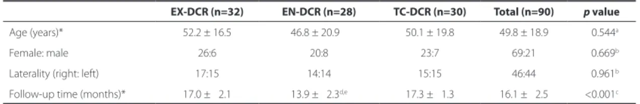
The present study included 69 (77%) female and 21 (23%) male patients, with a mean age of 49.8 ± 18.9 (range, 4-86) years. EX-DCR, EN-DCR, and TC-DCR were used in 32, 28, and 30 patients, respecti-vely. The mean follow-up time was 16.1 ± 2.5 (range, 10-20) months. Descriptive patient characteristics are presented in table 1.
Bacteria isolated from conjunctival, nasal, and lacrimal sac sam-ples are presented in table 2. Coagulase-negative Staphylococcus
(CNS) was the predominant organism isolated from pre- and post-ope ratively obtained irst-week conjunctival samples (14.4% for involved eyes and 12.2% for other eyes preoperatively, and 11.1% for involved eyes postoperatively) as well as from the preoperative nasal mucosa (67.8% for both sides) and lacrimal sac samples (22.2%).
Bacteroides fragilis was the only anaerobic bacterial strain isolated from lacrimal sac samples; that sample was from a single patient in the EX-DCR group.
EX-, EN- and TC-DCR groups had similar conjunctival culture po sitivity rates of 46.9%, 42.9%, and 30%, respectively, at the irst preoperative week and 18.8%, 3.6%, and 13.3%, respectively, at the irst postoperative week (preoperative, p=0.372; and postoperative, p=0.196; Table 3). Conjunctival culture positivity rates were signiican-tly lower after EX- and EN-DCR (p=0.022 and p=0.001, respectively). Although there was a trend toward a lower conjunctival culture positivity rate after TC-DCR, this trend was not statistically signiicant (p=0.267). The conjunctival growth rate was 40.0% (36 patients) at the involved site and 23.3% (21 patients) at the contralateral site, with a statistically signiicant diference observed (p=0.004). In contrast, the nasal growth rate was 98.9% (89 patients) at the involved site and 97.8% (88 patients) at the contralateral side, with no statistically signiicant diference observed (p=1.000).
The lacrimal sac culture growth rate was signiicantly higher in the EN-DCR (85.7%) group than in the EX-DCR (40.6%) and TC-DCR groups (46.7%, p=0.001). Comparison between conjunctival lora and lacrimal sac lora isolated from the involved side demonstrated a sig-niicantly higher lacrimal sac growth rate (56.7%, 51 patients) than the
conjunctival growth rate (40.0%, 36 patients; p=0.011). Conjunctival normalization times were similar between the three groups (1.5 ± 1.1, 1.0 ± 0.2, and 1.2 ± 0.5 weeks for EX-, EN-, and TC-DCR, respectively; p=0.166; Table 3).
Anatomical and functional success rates were not found to be associated with preoperative conjunctival and lacrimal sac culture positivity rates within the three groups and among all patients inclu-ded in the present study (p>0.05 for all; Table 4).
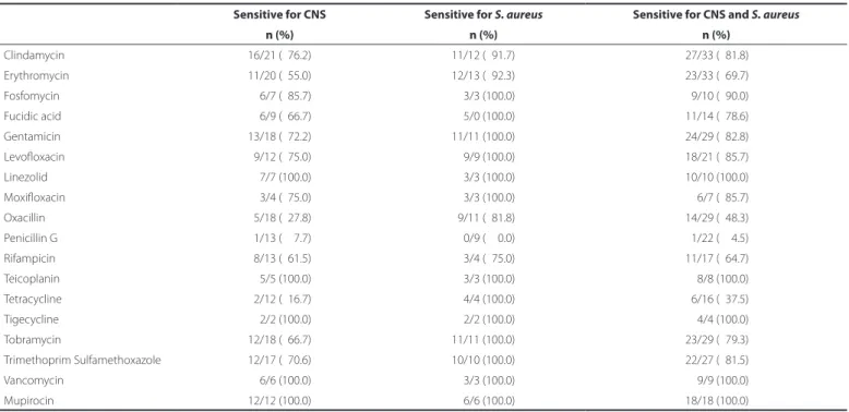
CNS and Staphylococcus aureus were most sensitive to linezolid, teicoplanin, tigecycline, vancomycin, and mupirocin (antibiotic sen-sitivity, 100% for all) (Table 5) and most resistant to penicillin G and tetracycline (antibiotic resistance, 95.5% and 62.5%, respectively).
DISCUSSION
The microbiological properties of the lacrimal sac in PANDO pa-tients has been a topic of interest in recent years as the spectrum of organisms constituting the lacrimal lora appears to have changed due to many factors(11,14,15). Despite a large number of comparative studies on DCR, none have previously compared three separate me-thods in terms of microbiological indings. Accordingly, we believe that the present study is the irst to compare clinico-bacteriological outcomes between conjunctival, nasal, and lacrimal sac samples isolated following three types of DCR (EX, EN, and TC).
The pathogens responsible for chronic dacryocystitis are typically gram-positive bacteria, including CNS, S. aureus, and Streptococci(16). Staphylococci, particularly S. aureus, have now replaced Streptococci as the most common cause of chronic dacryocystitis following the discovery of efective antibiotics, such as penicillin and cephalospo-rins, to which they have demonstrated greater resistance(10,11). Coden
et al.(17) evaluated culture samples from purulent lacrimal sac contents in 236 patients with dacryocystitis who were undergoing DCR and reported that the most common bacteria were S. epidermidis (27.3%) and S. aureus (22.1%), with a positive culture rate of 52%. Owji et al.(15) studied lacrimal sac cultures from the involved side and conjunctival cultures from the involved and normal sides of patients with NLDO and chronic dacryocystitis, and they found that the most frequently isolated organisms from the lacrimal sac and conjunctiva of the in-volved side were S. aureus (47.5% and 47.5%, respectively)and S. epi-dermidis (22.5% and 20%, respectively). On the other hand, the most frequently isolated organisms from the conjunctiva of the normal side and from that of the control healthy subjects were S. epidermidis
(60% and 60%, respectively)and S. aureus (47.5% and 30%, respective-ly)(15).Pradeep et al.(18) reported that the most common isolates from lacrimal sac specimens in chronic dacryocystitis cases were CNS and
S. aureus (71% and 14%, respectively). In the present study, the most commonly isolated organisms from the involved conjunctival side during the preoperative and irst postoperative week, involved nasal side preoperatively, lacrimal sac preoperatively, and contralateral conjunctival and nasal sites preoperatively were S. epidermidis (14.4%, 11.1%, 67.8%, 22.2%, 12.2%, and 67.8%, respectively) and S. aureus
Table 1. Patient characteristics
EX-DCR (n=32) EN-DCR (n=28) TC-DCR (n=30) Total (n=90) p value
Age (years)* 52.2 ± 16.5 46.8 ± 20.9d,e 50.1 ± 19.8 49.8 ± 18.9 <0.544a
Female: male 26:6 20:8 23:7 69:21 <0.669b
Laterality (right: left) 17:15 14:14 15:15 46:44 <0.961b
Follow-up time (months)* 17.0 ± 02.1 13.9 ± 02.3d,e 17.3 ± 01.3 16.1 ± 02.5 <0.001c EX-DCR= external dacryocystorhinostomy; EN-DCR= endoscopic dacryocystorhinostomy; TC-DCR= transcanalicular dacryocystorhinostomy with mul-tidiode laser.
Table 2. Results of preoperative conjunctival, nasal, and lacrimal sac cultures as well as of postoperative conjunctival cultures
EX-DCR (n=32) EN-DCR (n=28) TC-DCR (n=30) Total (n=90)
I C I C I C I C
Preoperative conjunctival culture
No bacteria isolated 17 (53.1) 21 (65.6) 16 (057.1) 20 (71.4) 21 (070.0) 28 (93.3) 54 (60.0) 69 (76.7) MSSA 01 (03.1) 02 (06.3) 01 (003.6) 03 (10.7) 03 (010.0) 01 (03.3) 05 (05.6) 06 (06.7) MRSA - - 01 (003.6) 01 (03.6) 01 (003.3) 01 (03.3) 02 (02.2) 02 (02.2)
Staph. epidermidis 06 (18.8) 07 (21.9) 06 (021.4) 04 (14.3) 01 (003.3) - 13 (14.4) 11 (12.2)
Staph. hominis - 01 (03.1) - - - 01 (01.1)
Staph. lugdunensis 01 (03.1) - - - 01 (01.1)
-Staph. haemolyticus - - - - 02 (006.7) - 02 (02.2)
-Staph. warneri - - 01 (003.6) - - - 01 (01.1)
-Strep. pluranimalium 01 (03.1) - - - 01 (01.1)
-Strep. sanguinis 01 (03.1) - - - 01 (01.1)
-Strep. pneumoniae - - 02 (007.1) - - - 02 (02.2)
-Haemophilus influenzae 01 (03.1) - - - 01 (01.1)
-Pseudomonas aeruginosa 01 (03.1) - - - 01 (01.1)
-Proteus mirabilis 01 (03.1) 01 (03.1) - - 01 (003.3) - 02 (02.2) 01 (01.1)
Citrobacter koseri 01 (03.1) - - - 01 (003.3) - 02 (02.2)
-Sphingomonas paucimobilis 01 (03.1) - - - 01 (01.1)
-Neisseria elongata - - 01 (003.6) - - - 01 (01.1)
-Preoperative nasal culture
No bacteria isolated - 01 (03.1) 01 (003.6) 01 (03.6) - - 01 (01.1) 02 (02.2) MSSA 05 (15.6) 07 (21.9) 08 (028.6) 07 (25.0) 04 (013.3) 03 (10.0) 17 (18.9) 17 (18.9) MRSA - - - - 03 (010.0) 03 (10.0) 03 (03.3) 03 (03.3)
Staph. epidermidis 25 (78.1) 23 (71.9) 16 (057.1) 19 (67.9) 20 (066.7) 19 (63.3) 61 (67.8) 61 (67.8)
Staph. hominis 01 (03.1) 01 (03.1) 01 (003.6) - - - 02 (02.2) 01 (01.1)
Staph. haemolyticus - - 01 (003.6) - 02 (006.7) 02 (06.7) 03 (03.3) 02 (02.2)
Strep. pneumoniae - - 01 (003.6) 01 (03.6) - 01 (03.3) 01 (01.1) 02 (02.2)
Pseudomonas aeruginosa - - - - 01 (003.3) 01 (03.3) 01 (01.1) 01 (01.1)
Serratia marcescens - - - 01 (03.3) - 01 (01.1)
Sphingomonas paucimobilis 01 (03.1) - - - 01 (01.1)
-Lacrimal sac culture
No bacteria isolated 19 (59.4) - 04 (014.3) - 16 (053.3) - 39 (43.3) -Aerobic bacteria
MSSA 01 (03.1) - 06 (021.4) - 04 (013.3) - 11 (12.2) -MRSA - - 01 (003.6) - 01 (003.1) - 02 (02.2)
-Staph. epidermidis 04 (12.5) - 11 (039.3) - 05 (016.7) - 20 (22.2)
-Staph. lugdunensis 01 (03.1) - - - 01 (01.1)
-Staph. haemolyticus - - 01 (003.6) - 01 (003.1) - 02 (02.2)
-Corynebacterium jeikeium - - 01 (003.6) - - - 01 (01.1)
-Strep. pluranimalium 01 (03.1) - - - 01 (01.1)
-Strep. mitis 01 (03.1) - 01 (003.6) - - - 02 (02.2)
-Strep. pneumonia - - 01 (003.6) - - - 01 (01.1)
-Strep. pyogenes - - 01 (003.6) - - - 01 (01.1)
-Haemophilus influenzae 01 (03.1) - - - 01 (01.1)
-Pseudomonas aeruginosa 01 (03.1) - - - 01 (003.1) - 02 (02.2)
-Serratia marcescens - - - - 01 (003.1) - 01 (01.1)
-Citrobacter koseri 01 (03.1) - - - 01 (003.1) - 02 (02.2)
-Sphingomonas paucimobilis 01 (03.1) - - - 01 (01.1)
-Kocuria kristinae - - 01 (003.6) - - - 01 (01.1)
Anaerobic bacteria
Bacteroides fragilis 01 (03.1) - - - 01 (01.1)
-First postoperative week conjunctival culture
No bacteria isolated 26 (81.2) - 27 (096.4) - 26 (086.7) - 79 (87.8)
-Staph. aureus - - - - 01 (003.3) - 01 (01.1)
-Staph. epidermidis 06 (18.8) - 01 (003.6) - 03 (010.0) - 10 (11.1)
-Second postoperative week conjunctival culture*
No bacteria isolated 27 (87.1) - 25 (100.0) - 29 (096.7) - 81 (94.1)
-Staph. aureus - - - - 01 (003.3) - 01 (01.2)
-Staph. epidermidis 03 (09.7) - - - 03 (03.5)
-Staph. hominis 01 (03.2) - - - 01 (01.2)
-Third postoperative week conjunctival culture*
No bacteria isolated 04 (66.7) - 02 (100.0) - 03 (100.0) - 09 (81.8)
-Staph. epidermidis 02 (33.3) - - - 02 (18.2)
-Fourth postoperative week conjunctival culture*
No bacteria isolated 02 (66.7) - - - 01 (100.0) - 03 (75.0)
-Staph. aureus 01 (33.3) - - - 01 (25.0)
(7.8%, 1.1%, 22.2%, 14.4%, 8.9%, and 22.2%, respectively). Anatomical and functional success rates did not correlate with preoperative con-junctival and lacrimal sac bacterial growth rates.
Methicillin-resistant S. aureus (MRSA) has been implicated in da-cryocystitis(19,20). In a study of dacryocystitis caused by community-on set MRSA, Kotlus et al.(20) stated that all their patients were at a risk of developing community-onset MRSA infections due to a hospital-ac quired strain of MRSA as they had been hospitalized for at least 3 months for chronic or comorbid conditions prior to presenting with dacryocystitis symptoms. They detected seven patients with acute or subacute MRSA dacryocystitis between 2001 and 2003(20). In con-trast, Pradeep et al.(18) found no MRSA in their series and stated that the lora of their patients was community acquired and not hospital acquired as they had been admitted to hospital on the day immedia-tely prior to surgery.In the present study, only 2 patients (2.2 %) had dacryocystitis caused by MRSA. Similar to Kotlus et al.(20), we believe obtaining cultures and performing sensitivity testing to determine whether antibiotic treatment is important for reducing the risk of exposure to MRSA in patients with dacryocystitis who do not respond to conservative treatment.
Gram-negative and anaerobic organisms have been reported to be present in 20%-27% and 7%-16% of patients with dacryocystitis, respectively(11,17,21,22), and the incidence of these infections appears to be rising(10). Gram-negative bacteria, including Pseudomonas
aeru-ginosa, Enterobacter, Citrobacter spp., Haemophilus influenzae, and
Escherichia coli, have also been reported as causative agents of
da-cryocystitis(10). Coden et al.(17) reported P. aeruginosa as the most com-mon gram-negative organism in dacryocystitis at an incidence rate of 8.7%. Brook et al. assessed the aerobic and anaerobic microbiology of 62 patients with dacryocystitis and reported a pure anaerobic growth rate of 32%, with Peptostreptococcus spp. and Propionibacterium spp. being the most frequently isolated species among anaerobes. These authors obtained specimens intraoperatively and used an anaero bic transport medium to transport them to the laboratory(23). Coden et
al.(17) found an anaerobic organism growth rate of 7.0%, with
Pro-pio nibacterium acnes as the most commonly isolated anaerobic organism. Proteus spp. and B. fragilis have been reported as other pathogens responsible for dacryocystitis(11,17,24,25). In our series, the only anaerobic bacteria isolated from the lacrimal sac was B. fragilis
(3.1%), and this patient was in the EX-DCR group. The lower anaerobic growth rate in the present study may be attributable to diiculty in ensuring the growth of anaerobes despite lacrimal sac samples being inoculated preoperatively.
Owji et al.(15) reported that microorganism growth rates in sam-ples from the lacrimal sac and involved and contralateral sides of the conjunctiva in a group of patients with NLDO and chronic dacryocystitis were 100%, 100%, and 97.5, respectively, while those in samples from the conjunctiva of healthy subjects was 82.5%. The same organism was isolated from the lacrimal sac and conjunctiva of the involved side in 90% of patients. They further reported that anaerobes, gram-negative bacteria, Corynebacterium, and Strepto-coccus spp. were isolated slightly more frequently from the involved Table 3. Bacterial growth rate and normalization time according to study group
EX-DCR (n=32) EN-DCR (n=28) TC-DCR (n=30) Total (n=90) p value
Preoperative conjunctival site bacterial growth, n (%)
Involved side 15 (046.9) 12 (42.9) 09 (030.0) 36 (40.0) 0.372a,j
Contralateral side 11 (034.4) 08 (28.6) 02 (006.7) 21 (23.3) 0.026a,j
p value 0.004b,e
Preoperative nasal site bacterial growth, n (%)
Involved side 32 (100.0) 27 (96.4) 30 (100.0) 89 (98.9) 0.326a,j
Contralateral side 31 (096.9) 27 (96.4) 30 (100.0) 88 (97.8) 0.596a,j
p value 1.000b.f
Intraoperative lacrimal sac bacterial growth, n (%)
Involved side 13 (040.6) 24 (85.7) 14 (046.7) 51 (56.7) 0.001a,j
p value 0.727b,g 0.002b,g 0.180b,g 0.011b,g
First postoperative week conjuntival site bacterial growth, n (%)
Involved side 06 (018.8) 01 (03.6) 04 (013.3) 11 (12.2) 0.196a,j
p value 0.022b,h 0.001b,h 0.267b,h <0.001b,h
Conjunctival normalization time (weeks, mean ± S.D.)
Involved side 1.47 ± 1.08 1.04 ± 0.19 1.17 ± 0.46 1.23 ± 0.72 0.166d,j
Conjunctival lora growth
No growth 1.29 ± 0.85 1.00 ± 0.00 1.24 ± 0.54 1.19 ± 0.59 Growth 1.67 ± 1.29 1.08 ± 0.29 1.00 ± 0.00 1.31 ± 0.89
p value 0.292c,k 0.248c,k 0.168c,k 0.659c,k
Lacrimal sac lora growth
No growth 1.26 ± 0.81 1.00 ± 0.00 1.06 ± 0.25 1.15 ± 0.59 Growth 1.77 ± 1.36 1.04 ± 0.20 1.29 ± 0.61 1.29 ± 0.81
p value 0.159c,k 0.683c,k 0.218c,k 0.264c,k EX-DCR= external dacryocystorhinostomy; EN-DCR= endoscopic dacryocystorhinostomy; TC-DCR= transcanalicular multidiode laser dacryocystorhinostomy.
Table 5. Sensitive susceptibility testing results for Staphylococcus spp. isolated from the lacrimal sac
Sensitive for CNS Sensitive for S. aureus Sensitive for CNS and S. aureus
n (%) n (%) n (%)
Clindamycin 16/21 (076.2) 11/12 (091.7) 27/33 (081.8) Erythromycin 11/20 (055.0) 12/13 (092.3) 23/33 (069.7) Fosfomycin 6/7 (085.7) 3/3 (100.0) 9/10 (090.0) Fucidic acid 6/9 (066.7) 5/0 (100.0) 11/14 (078.6) Gentamicin 13/18 (072.2) 11/11 (100.0) 24/29 (082.8) Levoloxacin 9/12 (075.0) 9/9 (100.0) 18/21 (085.7) Linezolid 7/7 (100.0) 3/3 (100.0) 10/10 (100.0) Moxiloxacin 3/4 (075.0) 3/3 (100.0) 6/7 (085.7) Oxacillin 5/18 (027.8) 9/11 (081.8) 14/29 (048.3) Penicillin G 1/13 (007.7) 0/9 (000.0) 1/22 (004.5) Rifampicin 8/13 (061.5) 3/4 (075.0) 11/17 (064.7) Teicoplanin 5/5 (100.0) 3/3 (100.0) 8/8 (100.0) Tetracycline 2/12 (016.7) 4/4 (100.0) 6/16 (037.5) Tigecycline 2/2 (100.0) 2/2 (100.0) 4/4 (100.0) Tobramycin 12/18 (066.7) 11/11 (100.0) 23/29 (079.3) Trimethoprim Sulfamethoxazole 12/17 (070.6) 10/10 (100.0) 22/27 (081.5) Vancomycin 6/6 (100.0) 3/3 (100.0) 9/9 (100.0) Mupirocin 12/12 (100.0) 6/6 (100.0) 18/18 (100.0)
CNS= coagulase-negative Staphylococcus.
Table 4. Preoperative conjunctival and lacrimal sac bacterial growth according to surgical success.
Preoperative conjunctival bacterial growth, n (%)
p value
Lacrimal sac bacterial growth, n (%)
p value
+ - +
-EX-DCR (n=32)
Anatomical success Successful 13 (86.7) 13 (76.5) 0.659b 11 (84.6) 15 (078.9) 1.000b
Unsuccessful 02 (13.3) 04 (23.5) 02 (15.4) 04 (021.1)
Functional success Successful 13 (86.7) 13 (76.5) 0.659b 11 (84.6) 15 (078.9) 1.000b
Unsuccessful 02 (13.3) 04 (23.5) 02 (15.4) 04 (021.1) EN-DCR (n=28)
Anatomical success Successful 08 (66.7) 13 (81.3) 0.418b 17 (70.8) 04 (100.0) 0.545b
Unsuccessful 04 (33.3) 03 (18.8) 07 (29.2)
-Functional success Successful 08 (66.7) 12 (75.0) 0.691b 16 (66.7) 04 (100.0) 0.295b
Unsuccessful 04 (33.3) 04 (25.0) 08 (33.3) -TC-DCR (n=30)
Anatomical success Successful 07 (77.8) 16 (76.2) 1.000b 10 (71.4) 13 (081.2) 0.675b
Unsuccessful 02 (22.2) 05 (23.8) 04 (28.6) 03 (018.8)
Functional success Successful 06 (66.7) 16 (76.2) 0.666b 09 (64.3) 13 (081.0) 0.417b
Unsuccessful 03 (33.3) 05 (23.8) 05 (35.7) 03 (020.0) Total (n=90)
Anatomical success Successful 28 (77.8) 42 (77.8) 1.000a 38 (74.5) 32 (082.1) 0.394a
Unsuccessful 08 (22.2) 12 (22.2) 13 (25.5) 07 (017.9)
Functional success Successful 27 (75.0) 41 (75.9) 0.920a 36 (70.6) 32 (082.1) 0.210a
Unsuccessful 09 (25.0) 13 (24.1) 15 (29.4) 07 (017.9)
EX-DCR= external dacryocystorhinostomy; EN-DCR= endoscopic dacryocystorhinostomy; TC-DCR= transcanalicular multidiode laser dacryocystorhinostomy. *= values are presented as number (percentage); a= Chi-square (χ2) test; b= Fisher’s exact test.
side than the contralateral normal side of the conjunctiva; however, this diference was not statistically signiicant, most likely due to the small sample size.
signiicant similarity between isolates from lacrimal and nasal/con-junctival samples and stated that the commensal lora of the nose and conjunctiva may have a direct role in the pathogenesis of chro-nic dacryocystitis. They obtained culture specimens directly from the lacrimal sac under an operating microscope and emphasized the reduced contamination during sample collection associated with this technique compared to the other methods of collection, such as applying pressure over the lacrimal sac or waiting for puru-lent material to exit the lacrimal sac via relux(9). The microorganism growth rates from lacrimal sac samples, involved and contralateral side conjunctival and nasal samples obtained preoperatively, and involved conjunctival samples obtained at irst and second postope-rative week were 56.7%, 40%, 23.3%, 98.9%, 97.8%, 12.2%, and 5.9%, respectively. All bacterial growth rates were similar between the three groups, except in culture samples obtained preoperatively from the lacrimal sac and the contralateral conjunctiva. The culture positivity rate from the lacrimal sac was signiicantly higher in the EN-DCR than in the EX-DCR and TC-DCR groups and was higher than the culture positivity rate of samples obtained preoperatively from the involved side conjunctiva for EN-DCR cases and all cases together. The similar positivity rates for lacrimal sac cultures during EN-DCR and preope-rative nasal cultures indicate that lacrimal sac samples may become contaminated with nasal lora while the lacrimal sac sample is being obtained intranasally. The positivity rate of lacrimal sac culture in endoscopic DCR may have been high for this reason. After DCR, conjunctival culture positivity rates were signiicantly reduced in all groups except in the TC-DCR group; this result is in close agreement with previous reported results(26). The most signiicant reduction in conjunctival culture positivity after DCR was observed in the EN-DCR group. Although not statistically signiicant, a reduction was also ob-served in the TC-DCR group. This reduction may be associated with the use of topical and oral antibiotics prescribed to patients during the irst postoperative week and with the elimination of the infection source (lacrimal sac) via lacrimal surgery.
In a study on 40 consecutive adult NLDO patients by Owji et al.(15), the mean conjunctival normalization time after EX-DCR was 4.5 (range, 3-8) weeks. While the authors reported signiicant as-sociations among normalization times, type of organismisolated from the lacrimal sac (particularly anaerobes and Streptococcus), a colony count of ≥103, and presence of a silicone tube, they found no relationship between normalization times and the presence or duration of epiphora or the presence of previous attacks of acute dacryocystitis(15). Furthermore, Owji et al.(15) stated that the delay period should be at least 4 weeks after DCR as the conjunctival lora normalized after 4 weeks in 67.5% of their patients. In another study by Eshraghi et al.(26) on the conjunctival lora and changes following EX-DCR, the mean normalization time was reported as 3.8 (range, 1-7) weeks in 38 patients with purulent regurgitation, 2.6 (range, 1-5) weeks in 33 patients without purulent regurgitation, and 3.3 weeks in all 71 patients. The authors reported signiicant associations among normalization times, pathogenic bacterial growth, higher colony counts, presence of a silicone tube, and purulent regurgitation(26). The most common organism to grow in the conjunctival cultures in patients with and without purulent regurgitation was S. epidermidis
(26.3% and 42.4%, respectively)(26). Eshraghi et al.(26) also suggested that cataract surgery can be performed 7 weeks after DCR as conjunc-tival cultures were negative by this time in their series. The authors in both studies deined conjunctival normalization time as the interval between undergoing DCR and obtaining a negative culture result or as the time to achieve a colony count below that of the normal side(15,26). The mean conjunctival normalization time was 1.47 (range, 1-5) weeks for EX-DCR, 1.04 weeks for EN-DCR (range, 1-2), and 1.17 (range, 1-3) weeks for TC-DCR in the present study. These results in-dicate that intraocular surgery may be scheduled after waiting for a conjunctival normalization time of approximately 5 weeks following DCR due to the risk of endophthalmitis. Furthermore, we observed
no signiicant association between mean conjunctival normalization times and type of surgery. However, we deined conjunctival norma-lization time, which difers from the deinitions for the same in the two above-mentioned studies(15,26).
Pinar-Sueiro et al. reviewed the clinical records of 697 patients who had undergone EX-DCR and found S. aureus to be most sensitive to gentamicin, co-trimoxazole, rifampicin, clindamycin, vancomycin, tobramycin, mupirocin, cefuroxime-axetil, chloramphenicol, te tra-cycline, fusidic acid, and cefalotin (100% sensitivity to all), while pe-nicillin demonstrated the worst activity against S. aureus with 83.3% resistance(27). In a study by Pradeep et al.(18), antibiogram results de-monstrated that Staphylococci represented the majority of cultured organisms (85%), and the most efective antibiotics against it were vancomycin, amikacin, third-generation cephalosporins, and amo-xyclav (100%, 89%, 83%, and 78% sensitivity, respectively). Penicillin (72% resistance) and erythromycin (75% resistance) were the least efective antibiotics. The authors(18) suggested that amoxyclav and third-generation cephalosporins should be used to treat chronic da-cryocystitis, while vancomycin and amikacin should be preferred in severe cases as they can be administered parenterally. Kotlus et al.(20) reported that tetracycline, trimethoprim/sulfamethoxazole, and van-comycin were the most efective antibiotics against MRSA dacryocys-titis.In the present study, CNS and S. aureus, the most commonly isolated (36.7%) microorganisms from the lacrimal sac, demonstrated the highest sensitivity to linezolid, teicoplanin, tigecycline, vancomy-cin, and mupirocin (antibiotic sensitivity rates of 100% for all).
The following are the strengths of the present study: prospective design; irst report to compare the culture results of EX-DCR, EN-DCR, and TC-DCR; long follow-up period; assessment of the postopera-tive microorganism growth rate; inclusion of anaerobic cultures of lacrimal samples; and evaluation of the antibiotic susceptibility of microorganisms isolated from the lacrimal sac. The present study also had a few limitations. We were unable to evaluate fungal patho-gens and patients could not be randomized, which may be seen as a limitation; however, patient preference was considered a priority in the present study.
In conclusion, PANDO patients had similar conjunctival lora preoperatively and during the irst week after undergoing EX-, EN-, and TC-DCR. A decrease in the growth rate of conjunctival cultures was observed after EX- and EN-DCR, but not after TC-DCR. CNS was the predominant organism isolated from pre- and post-operative conjunctival, pre-operative nasal, and per-operative lacrimal sac sam-ples in all groups. The most signiicant bacterial growth in the culture sample from the lacrimal sac was observed in the EN-DCR group. Bac-terial growth in pre-operative conjunctiva and pre-operative lacrimal sac samples was not associated with the success rate of DCR. Mean conjunctival normalization times were similar between the three groups at 1.47 weeks for EX-DCR, 1.04 weeks for EN-DCR, and 1.17 weeks for TC-DCR. It is necessary to wait for approximately 5 weeks for conjunctival normalization after DCR before planning intraocular surgery due to the risk of endophthalmitis. Greater understanding of the association between lacrimal lora and lacrimal surgery outcomes may facilitate the development of new strategies in treating PANDO. Further studies on the clinico-bacteriological outcomes of diferent surgical techniques in larger study samples are therefore required.
REFERENCES
1. Shun-Shin GA1, Thurairajan G. External dacryocystorhinostomy--an end of an era? Br J Ophthalmol. 1997;81(9):716-7.
2. Massaro BM, Gonnering RS, Harris GJ. Endonasal laser dacryocystorhinostomy. A new approach to nasolacrimal duct obstruction. Arch Ophthalmol. 1990;108(8):1172-6. 3. Woog JJ, Metson R, Puliaito CA. Holmium:YAG endonasal laser
dacryocystorhinos-tomy. Am J Ophthalmol. 1993;116(1):1-10.
5. Athanasiov PA, Prabhakaran VC, Mannor G, Woog JJ, Selva D. Transcanalicular ap-proach to adult lacrimal duct obstruction: a review of instrument and methods. Ophthalmic Surg Lasers Imaging. 2009;40(2):149-59.
6. Meister EF, Otto M, Rohrwacher F, Mozet C. [Current recommendations of dacryocys-torhinostomy]. Laryngorhinootologie. 2010;89(6):338-44. German.
7. Roithmann R, Burman T, Wormald PJ. Endoscopic dacryocystorhinostomy. Braz J Otorhinolaryngol. 2012;78(6):113-21.
8. Piédrola Maroto D, Franco Sánchez J, Reyes Eldblom R, Monje Vega E, Conde Jiménez M, Ortiz Rueda M. [Endonasal versus trans-canalicular endoscopic dacriocystorhinos-tomy using diode laser. Surgical techniques and outcomes]. Acta Otorrinolaringol Esp. 2008;59(6):283-7. Spanish.
9. Pinar-Sueiro S, Sota M, Lerchundi T, Gibelalde A, Berasategui B, Vilar B, Hernandez JL. Dacryocystitis: systematic approach to diagnosis and therapy. Curr Infect Dis Rep. 2012;14(2):137-46.
10. Briscoe D, Rubowitz A, Assia EI. Changing bacterial isolates and antibiotic sensitivities of purulent dacryocystitis. Orbit. 2005;24(2):95-8.
11. Hartikainen J, Lehtonen OP, Saari KM. Bacteriology of lacrimal duct obstruction in adults. Br J Ophthalmol. 1997;81(1):37-40.
12. Chaudhary M, Bhattarai A, Adhikari SK, Bhatta DR. Bacteriology and antimicrobial susceptibility of adult chronic dacryocystitis. Nepal J Ophthalmol. 2010;2(2):105-13. 13. Balikoglu-Yilmaz M1, Yilmaz T, Taskin U, Taskapili M, Akcay M, Oktay MF, Eren S.
Prospective comparison of 3 dacryocystorhinostomy surgeries: external versus en-doscopic versus transcanalicular multidiode laser. Ophthal Plast Reconstr Surg. 2014; 31(1):13-8.
14. Badhu BP, Karki BS, Khanal B, Dulal S, Das H. Microbiological patterns of chronic dacryocystitis. Ophthalmology. 2006;113(12):2377.e1-2.
15. Owji N, Khalili MR. Normalization of conjunctival lora after dacryocystorhinostomy. Ophthal Plast Reconstr Surg. 2009;25(2):136-8.
16. Bharathi MJ, Ramakrishnan R, Maneksha V, Shivakumar C, Nithya V, Mittal S. Compa-rative bacteriology of acute and chronic dacryocystitis. Eye (Lond). 2008;22(7):953-60. 17. Coden DJ, Hornblass A, Haas BD. Clinical bacteriology of dacryocystitis in adults.
Ophthal Plast Reconstr Surg. 1993;9(2):125-31.
18. Pradeep AV, Patil SS, Koti SV, Arunkumar JS, Garag SS, Hegde JS. Clinico-bacteriolo-gical study of chronic dacryocystitis cases in northern karnataka, India. J Clin Diagn Res. 2013;7(11):2502-4.
19. Kubo M, Sakuraba T, Arai Y, Nakazawa M. Dacryocystorhinostomy for dacryocystitis caused by methicillin- resistant Staphylococcus aureus: report of four cases. Jpn J Ophthalmol. 2002;46(2):177-82.
20. Kotlus BS, Rodgers IR, Udell IJ. Dacryocystitis caused by community-onset methicillin-re sistant Staphylococcus aumethicillin-reus. Ophthal Plast Reconstr Surg. 2005;21(5):371-5. 21. Huber-Spitzy V, Steinkogler FJ, Huber E, Arocker-Mettinger E, Schifbänker M.
Ac-quired dacryocystitis: microbiology and conservative therapy. Acta Ophthalmol (Copenh). 1992;70(6):745-9.
22. Seal DV, Barrett SP, McGill JI. Aetiology and treatment of acute bacterial infection of the external eye. Br J Ophthalmol. 1982;66(6):357-60.
23. Brook I, Frazier EH. Aerobic and anaerobic microbiology of dacryocystitis. Am J Oph-thalmol. 1998;125(4)552-4.
24. Evans AR, Strong JD, Buck AC. Combined anaerobic and coliform infection in acute dacryocystitis. J Pediatr Ophthalmol Strabismus. 1991;28(5):292.
25. McKeag D, Kamal Z, McNab AA, Sheorey H. Combined coliform and anaerobic infection of the lacrimal sac. Clin Experiment Ophthalmol. 2002;30(1):52-4.
26. Eshraghi B, Masoomian B, Izadi A, Abedinifar Z, Falavarjani KG. Conjunctival bacterial lora in nasolacrimal duct obstruction and its changes after successful dacryocys-torhinostomy surgery. Ophthal Plast Reconstr Surg. 2014;30(1):44-6.
27. Pinar-Sueiro S, Fernández-Hermida RV, Gibelalde A, Martínez-Indart L. Study on the efectiveness of antibiotic prophylaxis in external dacryocystorhinostomy: a review of 697 cases. Ophthal Plast Reconstr Surg. 2010;26(6):467-72.