Clinicoradiological Session
Case 2/2009- Eight-year-old child with pulmonary valve insufficiency
and left pulmonary artery stenosis, as residual defects after
Tetralogy of Fallot repair
Edmar Atik
Hospital Sírio-Libanês de São Paulo
Correspondencia: Edmar Atik •
InCor - Av. Dr. Enéas Carvalho de Aguiar, 44 - 05403-000 - São Paulo, SP - Brasil E-mail: [email protected]
Key Words
Heart defects, congenital; pulmonary valve insufficiency; pulmonary valve stenosis; tetralogy of Fallot / complications.
Clinical data
Tiredness at moderate exertion, unaltered for 2 years, despite the use of digoxin and spironolactone. Patient underwent Tetralogy of Fallot repair at 8 months of age, with augmentation of the right ventricular outflow tract with bovine pericardium graft and monocusp placement.
At physical examination, the patient was eupneic, had normal skin color and the peripheral pulses were normal. Weight: 32 kg; height: 130 cm; BP: 100/60 mm Hg; HR: 88 bpm. The aorta was not palpable at the sternal notch. There were slight precordial impulses at the left sternal border and the ictus cordis was palpable at the 4th and 5th
left intercostal spaces, in the hemiclavicular line and limited by two digital pulps. The heart sounds were normal and the second heart sound was constant, with its two components of equal intensity. A mild proto-meso-diastolic murmur was identified through auscultation along the left sternal border, not accompanied by thrill. There was no systolic murmur. The liver was not palpable.
The electrocardiogram showed signs of complete right bundle branch block with QRS complexes with a duration of 0.12 seconds. ÂP:+200, ÂQRS: undetermined, ÂT:+800.
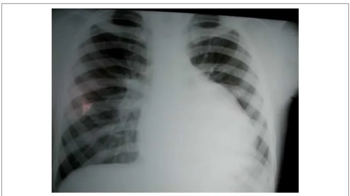
Radiographic image
shows slightly to moderately increased cardiac area, at the expense of the right cavities, corresponding mostly to a larger protusion of the right ventricular outflow tract between the mid arch and left lower arch, with slightly increased pulmonary vascular net on the right in contrast with the decreased aspect on the left (Figure 1).
Diagnostic impression
This image, in a patient previously submitted to Tetralogy
of Fallot repair, is compatible with right ventricular (RV) outflow tract dilation, augmented at the time of the surgery. The enlargement of the right ventricle and of the pulmonary vascular net on the right is caused by the pulmonary valve insufficiency. The decrease in the vascular net on the left indicates the possibility of left pulmonary artery stenosis.
Differential diagnosis
left atrial enlargement can also protrude in this region of the RV outflow tract, but the other elements rule out this possibility.
Diagnostic confirmation
the clinical elements lead to the diagnosis of pulmonary valve insufficiency and stenosis of the left pulmonary artery, after Tetralogy of Fallot repair. The echocardiogram (Figure 2) showed RV dilation (30 mm) caused by the marked pulmonary insufficiency. The left ventricle (38 mm), left atrium (28 mm), aorta (30 mm) and ejection fraction (71%) were normal.
Conduct
At the surgical intervention through the pulmonary trunk, the calcified monocusp was removed. The left pulmonary artery, obstructed at first, was enlarged with a bovine pericardium graft. Homograft number 21 was implanted between the RV and the pulmonary arteries in ECC of 100’ and period of anoxia of 60’. The immediate post-surgical evolution was good, with the disappearance of the heart murmur, despite the remaining cardiomegaly.
Comments
This case is interesting due to the occurrence of left pulmonary artery stenosis as a late complication of Tetralogy of Fallot repair. Additionally, it is worth mentioning the favorable evolution after the placement of a pulmonary homograft with no residual insufficiency, which is often found after this procedure.
Clinicoradiological Session
Edmar Atik
Arq Bras Cardiol 2009;92(2):157-158
Figure 1 - Chest x-ray, 8 years after Tetralogy of Fallot repair, showing slight to moderately increased cardiac area at the expense of the left lower arch and dilation of
the right ventricular (RV) outlow tract, in addition to pulmonary vascular net contrast, increased on the right and decreased on the left (pulmonary artery stenosis).
Figure 2 -Echocardiogram showing in the left parasternal projection in a cross-sectional view, the dilation of the right ventricular (RV) outlow tract and the pulmonary
arteries, with interposition of calciied monocusp in A and signs of marked pulmonary regurgitation by color Doppler, in B.