152
Saraiva et al
Primary mediastinal seminoma
Arq Bras Cardiol 2001; 76: 152-4.
Universidade Federal de Pernambuco and Serviço de Ecocardiografia do Recife (SECOR)
Mailing address: Lurildo R. Saraiva - Hospital das Clínicas da Universidade Federal de Pernambuco - Av. Moraes Rego, SN - 50670-420 - Recife, PE - Brazil English version by Stela Maris C. Gandour
Lurildo R. Saraiva, Djair Brindeiro Fº, Thiago B. Saraiva, Mauro B. Arruda, Vital Lira
Recife, PE - Brazil
Cardiac Extension of Primary Mediastinal Seminoma
Compressing the Right Ventricular Outflow Tract
Case Report
We report the case of a 33-year-old male with prima-ry seminoma of the anterior mediastinum with initial clini-cal manifestations suggestive of heart disease.
Tumors of the anterior mediastinum are currently re-presented by seminoma or primary germinoma 25 to 30% of the time, with a marked preponderance in the male sex1.
These neoplasms are invasive and poorly differentia-ted, characteristics accounting for their excellent regressive response to chemotherapy or radiotherapy. These neo-plasms are almost always related to the thymus, hence the name thymic seminoma has also been proposed for their de-signation 1.
Involvement of the pericardium by tumoral tissue is not rare 2, but myocardial and endocardial invasion up to
the point of obstructing the right ventricular outflow tract and compressing the pulmonary artery, mimicking a steno-tic lesion, is an uncommon event, rarely reported in the lite-rature. Describing the occurrence of such a tumor is the motive of this report.
Case Report
The patient is a 33-year-old male laborer from Jaboa-tão dos Guararapes, Pernambuco State, who sought the car-diology outpatient clinic complaining of fatigue on exertion, tachycardic palpitations, and precordialgia for 50 days. The patient reported that the precordial pain increased in inten-sity with inspiration, and was alleviated with ordinary anal-gesics. He also reported asthenia, anorexia, and recent wei-ght loss of approximately 2 kg in the last 15 days. He denied a rheumatic antecedent, alcohol abuse, smoking, venereal diseases, and diabetes mellitus.
On physical examination, the patient weighed 62.0kg
and was 1.73m tall (body mass index = 20.7kg/m2). He was in
regular condition, eupneic, anicteric, and with no peripheral cyanosis. His pulses were symmetrical and regular, his blood pressure was 140/90mmHg, and his heart rate was 88bpm. No jugular stasis occurred at 45°.
On precordial examination, clear systolic impulsions were observed on the pulmonary area, and no thrill existed. The cardiac rhythm was regular; the first cardiac sound had a normal intensity in the mitral area, and the second cardiac sound was split, variable, with a muffled pulmonary compo-nent. A moderate (++/4) rude ejection systolic murmur was auscultated in the pulmonary, aortic, and mesocardial areas. A forth cardiac sound could be heard in the mitral area.
The lungs were clear and no hepatosplenomegaly existed. Laboratory tests were as follows: hemoglobin, 14.3g/ dL; hematocrit, 43%; leukocytes, 5,700/mm3; normal levels
of urea and creatinine; antistreptolysin O, 200 U Todd. Chest X-ray in the posteroanterior view depicted a suggestive enlargement of the right ventricle, bulging of the middle arch (+/2), decreased pulmonary circulation, and mild enlargement of the mediastinum.
The electrocardiogram showed sinus rhythm and was compatible with right ventricular hypertrophy and clockwi-se rotation of the heart, and an elevation of the ST clockwi-segment with superior concavity in the inferior and lateral leads (QRS axis of +100°) was observed.
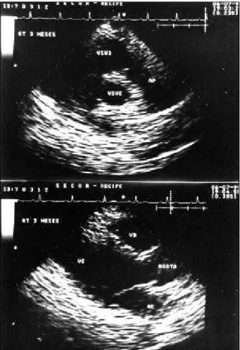
Doppler echocardiography (fig. 1) revealed enlarge-ment of the right chambers, hypertrophy of the interventri-cular septum, a small pericardial effusion, and a tumor inva-ding the right ventricular outflow tract, compressing the pul-monary artery trunk at the origin of its branches. Systolic pressure in the right ventricle was estimated in 95mmHg.
On thoracotomy, a large nonresectable tumor was ob-served involving the pericardial sac and the entire heart. A histopathological examination of a section of the tumor (fig. 2) revealed a probably primary neoplasia of the thymus comprising sheets of polygonal cells and septa in a delicate fibrous stroma with lymphocytic infiltrate. The neoplastic cells had large nuclei with fine chromatin and clear slightly acidophilic cytoplasm.
Arq Bras Cardiol 2001; 76: 152-4.
Saraiva et al Primary mediastinal seminoma
153 The patient was referred to the oncology unit, where
he underwent periodical cycles of chemotherapy consisting of ectoposide, bleomycin, and cisplatin. The outcome was successful with a normal echocardiogram 3 months after chemotherapy (fig. 3). One year and 3 months after his ad-mission to the hospital, the patient had no complaints and his cardiac assessment was normal.
Discussion
The curious finding in this case is that the attention of the physician was first drawn to a probable diagnosis of heart disease in a young male, who, in reality, had a malig-nant neoplasia of the mediastinum. For this initial false im-pression, findings resulting from the physical examination
Fig. 1 – Two-dimensional echocardiogram, paraesternal longitudinal view, at the level of the pulmonary valve: a large tumor mass is seen in the superior part of the anterior mediastinum invading the right ventricular outflow tract (RVOT, asterisks), reaching the pulmonary valve (arrows), and compressing the pulmonary artery (PA) at the origin of its 2 branches.
Fig. 2 – Polygonal cells in an alveolar pattern with pleomorphic nuclei and fibrous
154
Saraiva et al
Primary mediastinal seminoma
Arq Bras Cardiol 2001; 76: 152-4.
1. Kitami A, Suzuki T, Susuki S, Hori G. Primary seminoma in the middle mediastinum: case report in a 69-year-old male. Jpn J Clin Oncol 1998; 28: 142-4.
2. Hachiya T, Koizumi T, Hayasaka M, Kubo K, Sekiguchi M, Hamyuuda M, Honda T. Spontaneous regression of primary mediastinal germ cell tumor. Jpn J Clin Oncol 1998; 28: 281-3.
3. Pedra SRFF, Fontes VF. Estenose pulmonar. In. Porto CC (ed). Doenças do coração. Prevenção e tratamento. Editora Guanabara Koogan. Rio de Janeiro, 1998, cap. 75, pp. 393-8.
of the heart contributed, as did the electrocardiogram and the nature of the alterations in the cardiac silhouette on roentgen examination, which were very suggestive of narro-wing of the pulmonary valve. However, precordialgia with pleural features and recent onset does not constitute a usu-al symptom of such a disease 3, except for the retrosternal
pain in severe congenital stenotic forms 4. This pain along
with the mild enlargement of the mediastinum oriented the clinical investigation in a diverse direction.
The echocardiographic alterations (fig. 1) are very sig-nificant. In addition to providing a rapid diagnosis of neo-plasia, its localization in the right ventricular outflow tract and pulmonary artery trunk allows understanding of the se-miologic findings, such as the systolic impulsions, the rude systolic murmur, the decreased intensity of the second heart sound in the pulmonary area, and the precordialgia. These findings resulted from the right ventricular obstruc-tion and compression of the pulmonary artery in one side, and from pericardial involvement in the other.
According to Roberts 5, extension of the neoplasia to
the heart is most frequently seen in leukemias and breast and lung carcinomas; in the latter, pericardial constriction may occur. According to Cox 6, the primary malignant
ger-minal tumors, formerly included under the broad term “teratomas”, clinically manifest as chest pain with a pleural component and cough. Hemoptysis, pneumonia, and dysp-nea on exertion are rarer findings in these tumors, which are preferentially located in the anterior mediastinum, and more rarely in the middle mediastinum 1.
Even though this is not the case of a cardiac
metasta-sis – the term “extension” here is more appropriate 5 – in the
14 truly metastatic cases of diverse neoplasias (pancreatic and colon adenocarcinomas, and melanoma, among others) reported by Labid et al 7, the finding of systolic murmur with
characteristics similar to those of our patient’s cardiac mur-mur in the presence of dyspnea on exertion is the major clini-cal feature. Another unique observation in their patients was the usual electrocardiographic finding compatible with complete or incomplete right bundle-branch block and right ventricular hypertrophy, as seen in our patient, who also had morphological changes in the ST segment, recalling pericardial involvement, which might have contributed to the precordialgia.
Of unknown histogenesis, the scarce extragonadal germinomas, according to Kitami et al 1, are currently
ma-naged with initial surgical biopsy and subsequent chemo-therapy. Hachiya et al 2 recognized the rare possibility of
spontaneous regression in a 22-year-old Japanese male, in whom the severe necrosis related to tumor aspiration could explain regression due to restriction in blood flow.
Even though the neoplasia is radiosensitive, one third of the patients may relapse or even show metastases, hence the use of chemotherapy, which is in fact currently more appropriate 1.
On the basis of what has been reported, we think that exc-lusion of such a neoplasia is necessary whenever clinical fin-dings compatible with obstruction of the right ventricular out-flow tract are present in a young male complaining of asthenia, dyspnea, and thoracic pain with pleural characteristics, accompanied by radiological mediastinal enlargement.
References
4. Brickner ME, Hillis LD, Lange RA. Congenital heart disease in adults (first of two parts). N Engl J Med 2000; 342 (4): 256-69.
5. Roberts WC. Cardiac neoplasms. In: Topol EJ (ed) - Texbook of Cardiovascular Medicine. Philapelphia: Lippincott - Raven Publishers, 1999; 971-88. 6. Cox JD. Primary malignant germinal tumours of the mediastinum. Cancer 1975;
36: 1162-8.