381
LETTERS
Cervical spinal cord infarct associated with
patent foramen ovale
Infarto medular cervical associado a forame oval patente
Renan Barros Domingues1,2, Mayara Ferreira Vilas-Novas1
1 Escola Superior de Ciências da Saúde da Santa Casa de Misericórdia de Vitória (EMESCAM), Vitória ES, Brazil;
2 Programa de Pós-graduação em Neurociências, Universidade Federal de Minas Gerais (UFMG), Belo Horizonte MG, Brazil.
Correspondence: Renan Barros Domingues; Rua Prof. Almeida Cousin 125 / sala 1309; 29050-565 Vitória ES - Brasil; E-mail: [email protected]
Conflict of interest: There is no conflict of interest to declare. Received 15 January 2012; Accepted 23 January 2012
Spinal cord infarct is a rare entity representing only one to two percent of all strokes1,2. here are only two previous reports
of spinal cord infarct associated with patent foramen ovale (PFO), one in the anterior spinal artery (ASA) distribution and the other in the posterior spinal artery (PSA)3,4. Here, we report
the case of a woman with spinal cord infarct in which the only possible etiology found was a PFO.
CASE REPORT
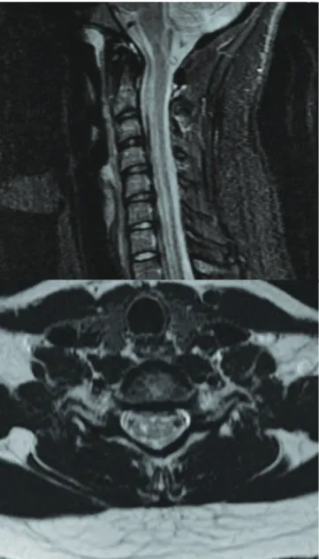
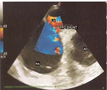
A 20-year-old woman with any previous known disease had a sudden interscapular pain and abdominal paresthesias. One day after that, she noted arms and legs weakness that persisted during the following weeks. Neurological examination revealed mild and symmetric legs, and arms paresis with exaggerat-ed tendon relexes and bilateral Babinski sign. here were no signs of lemniscal sensory deicits. here was a trunk and four limbs slight reduction of tactile and thermal sensory function. Magnetic resonance imaging (MRI) showed slight asymmetric T2-weighted hypersignals on the central H region of spinal cord extending from C5 to C7, compatible with spinal cord infarct (Fig 1). She was submitted to computed tomography angiog-raphy of the thoracic and cervical regions, which was normal. Holter was normal. he transesophageal ecocardiography re-vealed PFO with paradoxical interatrial shunt (Fig 2). All labo-ratorial data, including thrombophilias search, antinuclear an-tibodies, VDRL, sorologies for hepatitis, HIV, HTLV-1, anti-Ro and anti-La, were negative. he cerebrospinal luid (CSF) analy-sis was normal. Routine laboratorial tests, including blood cell count, glucose, cholesterol and triglycerides, renal and hepatic function, were normal. Cranial MRI was normal.
Based on these results, acetylsalicylic acid at a dose of 100 mg a day was introduced. Physical rehabilitation was ini-tiated. he patient improved, and a minimal strength deicit and hyperrelexia persisted.
Fig 1. Magnetic resonance imaging showing slight asymmetric
382
LETTERS
DISCUSSION
Several diseases that can lead to an acute spinal cord lesion were investigated. Demyelinating diseases were ex-cluded by normal cranial MRI and normal CSF examination. Infection myelitis was excluded by the normal CSF examina-tion and negative serologies. Spinal cord compression and tu-mors were excluded by the spinal MRI. Also, the spinal MRI was consistent with infarct diagnosis.
Several etiologies of spinal cord infarct were investigated. Aortic and vertebral dissections were excluded by computed tomography angiography. Vasculitis was excluded by normal CSF and negative autoantibodies. here was no risk factor for atherosclerosis. Cervical spine disease was excluded by cervi-cal MRI. here were not triggering movements. he only risk factor found was the PFO.
here are two reported cases of spinal cord infarct associ-ated with PFO, one in ASA territory and other in PSA terri-tory, both in the thoracic region. To our knowledge, this is the irst case of cervical spinal cord infarct in the ASA territory associated with PFO already described.
he best approach to prevent recurrences in patients with PFO is not yet established, but there are reports of treat-ment with acetylsalicylic acid, warfarin, and closure of PFO5.
RA: right atrium; LA: left atrium; Ao: Aorta; PFO Shunt: patent foramen ovale shunt.
Fig 2. Transesophageal ecocardiography showing patent
foramen ovale with paradoxical interatrial shunt.
1. Novy J, Caruzzo A, Maeder P, Bogousslavsky J. Spinal cord ischemia: clinical and imaging patterns, pathogenesis, and outcomes in 27 patients. Arch Neurol 2006;63:1113-1120.
2. Kumral E, Polat F, Güllüoglu H, Uzunköprü C, Tuncel R, Alpaydin S. Spinal ischaemic stroke: clinical and radiological findings and short-term outcome. Eur J Neurol 2011;18:232-239.
3. Petruzzellis M, Fraddosio A, Giorelli M, et al. Posterior spinal
artery infarct due to patent foramen ovale: a case report. Spine 2010;35:155-158.
4. Mori S, Sadoshima S, Tagawa K, Iino K, Fujishima M. Massive spinal cord infarction with multiple paradoxical embolism: a case report. Angiology 1993;44:251-256.
5. Mattle HP, Meier B, Nedeltchev K. Prevention of stroke in patients with patent foramen ovale. Int J Stroke 2010;5:92-102.
References
his last option was considered due to insuicient evidence. Acetylsalicylic acid was chosen because of the lower risk of bleeding comparing to warfarin.
PFO should be investigated in cases of spinal cord infarct in which the most common etiologies are ruled out.
Complex movement disorder in an elderly
patient and the chimera effect
Distúrbio do movimento complexo em uma paciente idosa e o efeito quimera
Marco A. T. Utiumi, Renato P. Munhoz, Caroline Cartaxo, Hélio A. G. Teive
Movement Disorders Unit, Neurology Service, Internal Medicine Department, Hospital de Clínicas, Federal University of Paraná (UFPR), Curitiba PR, Brazil.
Correspondence: Hélio A. G. Teive; Rua General Carneiro 1103/102; 80060-150 Curitiba PR - Brasil; E-mail: [email protected]
Conflict of interest: There is no conflict of interest to declare. Received 27 December 2011; Accepted 03 January 2012
Movement disorders (MD) are traditionally divided into hy-pokinetic syndromes or parkinsonism, and hyperkinetic syn-dromes, including several involuntary movements, such as chorea,