Inductio n o f Fo s pro te in
im m uno re activity by
spinal co rd co ntusio n
Departamentos de 1Cirurgia, O rtopedia e Traumatologia, Hospital das Clínicas, 2Farmacologia, Faculdade de Medicina de Ribeirão Preto, and
3Morfologia, Estomatologia e Fisiologia, Faculdade de O dontologia de Ribeirão Preto,
Universidade de São Paulo, Ribeirão Preto, SP, Brasil E.A. Del-Bel3,
C.A.G. Borges1,
H.L.A. Defino1 and
F.S. Guimarães2
Abstract
The objective of the present study was to identify neurons in the central nervous system that respond to spinal contusion injury in the rat by monitoring the expression of the nuclear protein encoded by the
c-fos gene, an activity-dependent gene, in spinal cord and brainstem regions. Rats were anesthetized with urethane and the injury was produced by dropping a 5-g weight from 20.0 cm onto the exposed dura at the T10-L1 vertebral level (contusion group). The spinal cord was exposed but not lesioned in anesthetized control animals (lami-nectomy group); intact animals were also subjected to anesthesia (intact control). Behavioral alterations were analyzed by Tarlov/ Bohlman scores, 2 h after the procedures and the animals were then perfused for immunocytochemistry. The patterns of Fos-like immu-noreactivity (FLI) which were site-specific, reproducible and corre-lated with spinal laminae that respond predominantly to noxious stimulation or injury: laminae I-II (outer substantia gelatinosa) and X and the nucleus of the intermediolateral cell column. At the brain stem level FLI was detected in the reticular formation, area postrema and solitary tract nucleus of lesioned animals. No Fos staining was de-tected by immunocytochemistry in the intact control group.However, detection of FLI in the group submitted to anesthesia and surgical procedures, although less intense than in the lesion group, indicated that microtraumas may occur which are not detected by the Tarlov/ Bohlman scores. There is both a local and remote effect of a distal contusion on the spinal cord of rats, implicating sensory neurons and centers related to autonomic control in the reaction to this kind of injury.
Co rre spo nde nce
E.A. Del-Bel
Departamento de Morfologia, Estomatologia e Fisiologia
FO RP, USP
14040-904 Ribeirão Preto, SP Brasil
Presented at the International Neurobiology Course, Faculdade de O dontologia de Ribeirão Preto,
Universidade de São Paulo, Ribeirão Preto, SP, Brasil, May 20-27, 1998.
Part of a Master’s thesis presented by C.A.G. Borges to the Departa-mento de Cirurgia, O rtopedia e Traumatologia, Ribeirão Preto, SP. Research supported by FAPESP,
CNPq and FAEPA.
Received December 9, 1998 Accepted February 8, 2000
Ke y wo rds
·Fos immunohistochemistry ·Intermediolateral column
neurons
·Reticular formation ·Area postrema ·Dorsal horn ·Solitary tract nuclei ·Laminectomy ·Spinal cord injury
The neurons of the mammalian central nervous system (CNS) respond to a lesion with an intense cell body reaction that may result in neuronal survival or death, with the acute post-injury phase being of critical im-portance (1,2). The changes in blood flow and tissue perfusion that produce vascular
damaged pathways (3,4). However, little is known about the molecular mechanism that initiates cellular reaction after injury.
Among the molecular and cellular mechanisms that may contribute to the medi-um/long-term neuronal response are changes in gene expression (4,5). Immediate early genes (IEG) are expressed transiently and rapidly in nerve cells by a broad array of stimuli. Among the best studied IEG are the Fos fos, fos B, fra-1, fra-2) and c-jun (c-jun, jun B, jun D) families. The expression of Fos protein in nerve cells is activity-depend-ent and related to the stimulus applied. In addition, the localization of the IEG may reflect a pattern of neuronal activation re-lated to the type of stimulus and to the long-term variation in neuronal physiology (4,6). Axotomy of the CNS was reported to induce c-Fos immunoreactivity in the red nucleus, the locus coeruleus and motor cor-tex (3,7). Recently, Ruggiero et al. (8) em-ployed the Fos gene to monitor peripheral and central responses to spinal cord transec-tion. Patterns of c-fos gene expression were site-specific and correlated with laminae that respond predominantly to noxious stimula-tion and contain sympathetic interneurons. Unfortunately, these studies do not mention the remote effects of a distal spinal cord lesion in brain stem nuclei correlated with autonomic control. Therefore, we investi-gated here the expression of Fos-like immu-noreactivity (FLI), used as a marker of neu-ral activity, at local and remote sites follow-ing spinal cord contusion injury.
Male Wistar rats weighing 250-300 g were used. Animals had free access to water and food and were kept on a 12-h light-dark cycle (lights on at 7:00 a.m.), under con-trolled temperature conditions. All experi-ments were performed between 9:00 and 12:00 a.m., and complied with the Guide
for the Use and Care of Animalsfrom the
Brazilian Society of Neurosciences and Be-havior. The site of lesion selected was the vertebral segment located between T10 and
L1. The animals were anesthetized with ure-thane (25%, 2 ml/kg), placed in a stereotaxic apparatus and submitted to the procedure. A total of 9 animals was used, divided into 3 groups. Group 1 (intact control, N = 3) con-sisted of similarly anesthetized unoperated rats; in group 2 (laminectomy group, N = 3), the spine was approached along the poste-rior surface and bilateral laminectomy of the T10-L1 segment was performed to expose the dura mater, which was left intact. In group 3 (contusion group, N = 3), the dura mater was exposed as described above and a lesion was produced by impact on the spine (9). To produce this lesion, a metal disk was placed on the meninges and a 5-g weight was dropped on it by free fall from a height of 20.0 cm inside a Teflon tube. After surgery, the muscular and subcutaneous layers and the skin were sutured and the animals were allowed to rest in individual boxes.
Sensorimotor function was evaluated in each animal 2 h after the experimental cedure for 5 min according to the scale pro-posed by Tarlov and modified by Bohlman (9). Grade zero indicates complete paraple-gia without movements, grade I small articu-late movements, grade II large movements, grade III indicates that the animal can stand on its feet, grade IV indicates that the animal can walk, and grade V indicates that the animal can walk on a plane with a 20-degree inclination. No obvious signs of pain or stress were seen in any group.
Fos immunohistochemistry as described by Del Bel et al. (10). Briefly, tissue sections were successively washed and incubated for 40 h with the primary Fos antibody (1:2000, Cambridge Research Biochemical, Cam-bridge, UK). Sections were then processed by the avidin-biotin immunoperoxidase method (Vectastain ABC kit, Vector Labo-ratories, Inc., Burlingame, CA, USA) and FLI was visualized by the addition of the chromogen 3,3-diaminobenzidine (DAB, Sigma Chemical Co., St. Louis, MO, USA) and hydrogen peroxide. The slices were mounted on slides and coverslipped for mi-croscopic observation. FLI could be visual-ized as a brown reaction product inside neu-ronal nuclei whose location was determined using the Paxinos and Watson atlas (11). Segments of the thoracic and lumbar spinal cord and regions of the brain stem were analyzed. Spinal laminae and brain stem nu-clei were distinguished on Nissl-stained ad-jacent sections.
Tissue from the laminectomy and contu-sion groups was processed at the same time for immunohistological staining; tissue from the intact control group was processed sepa-rately, but together with an FLI-positive con-trol (FLI was induced in rats with 2 mg/kg
haloperidol, ip). Stained neuronal bodies
were analyzed using a computerized image analysis system. Images were captured from slides using a Leika microscope and CCD camera, together with the National Institutes of Health Image software 9.0 (W. Rasband, National Institute of Mental Health). Fos-positive neuronal bodies were counted from a fixed-size area belonging to the spinal cord (laminae I-II and X) and midbrain (reticular formation, solitary tract nuclei and area pos-trema), measured bilaterally. For each treat-ment, three to five sections from each animal (N = 3 animals/group) were evaluated. A mean value for cell density in each region of the animal was then calculated. The statisti-cal analysis was performed using the t-test, and differences were considered significant
when P<0.05.
The evaluation of sensorimotor function showed no evidence of spinal cord injury due to the procedure in animals of the group submitted to anesthetic alone or anesthesia and laminectomy, with scores of 4 or 5 on the Tarlov/Bohlman scale in both cases. The group submitted to the spinal cord contusion showed partial or total paraplegia and all animals scored 1 or 2 on the Tarlov/Bohlman scale.
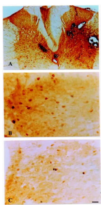
No Fos protein expression was detected in the intact control group; however, FLI was detected in the laminectomy and contu-sion groups, mainly in the spinal segments located between T10 and L1. In the contu-sion group, FLI was detected around the central spinal cord canal, in lamina X, in laminae I and II (Figure 1B and C) of the dorsal horn, and in laminae V, VI, VII VIII, and IX/accessory nucleus of the ventral horn of the rat spinal cord. In these animals, this FLI pattern was also observed in thoracic segments of the spinal cord above the level of injury, with the same intensity. However, in the laminectomy group we observed some FLI in laminae V-VIII, IX/accessory nucleus of the ventral horn of the rat spinal cord, but only around the surgery site. In contrast, there was no Fos protein expression in lami-nae I or II (Figure 1C), either locally or outside the surgical site (upper thoracic level). The detection of FLI in the laminectomy group indicated that some microtraumas may have occurred during the surgical procedure, as supported by macroscopic signs of tissue damage.
Although the presence of FLI was ob-served in both the laminectomy and contu-sion groups, its levels and molecular distri-bution in the spinal cord were higher in the contusion group (Figure 1A,B; Figure 2D,E and F).
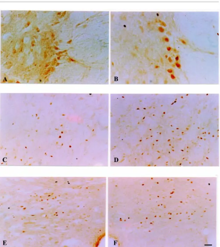
neuronal cell bodies and processes of the principal intermediolateral nucleus, a well-defined neuronal population located in the lateral horn along the borderline between white and gray matter, stained intensely for Fos-immunoreactivity in the contusion group (Figure 2B), but not in either control group (Figure 2A).
In addition, FLI occurs in some brain stem centers involved in central autonomic control such as the ventrolateral reticular area of the medulla oblongata (Figure 2E), the area postrema and the solitary tract nucleus (Figure 2F). Less intense FLI ex-pression was observed in tissues from the laminectomy group in the same areas (Fig-ure 2B and C, respectively). No FLI was observed in the intact control group.
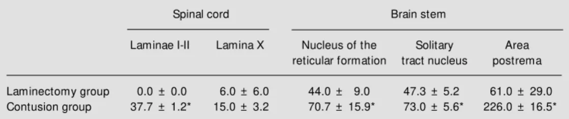
Differences in intensity of immunostain-ing between experimental and control ani-mals were striking and clear cut. However, quantitative analysis of the number of FLI cells in the laminae of the spinal cord and midbrain nucleus shows that spinal cord con-tusion produced a large and consistent pat-tern of FLI cells (Table 1), with significant differences between the contused group and the laminectomy group in laminae I-II and X of the spinal cord, solitary tract nucleus, area postrema and nuclei of the reticular forma-tion in the medulla oblongata (P<0.05).
The contusion injury produced by this procedure is heterogeneous (Figure 1A), a fact that should be considered in the analysis of the results (13). The impact zone con-tained bleeding and tissue damage; immuno-cytochemistry showed local expression of FLI 2 h after experimental spinal cord contu-sion; a less intense expression was seen in rats submitted to laminectomy. The topo-graphic distribution of neurons containing FLI was not observed in intact control ani-mals.
The precise mechanisms by which the effects of injury are distributed throughout the cord are unknown, but similar evidence was found by Yerzierski et al. (14),
son et al. (15) and Ruggiero et al. (8). Anes-thesia is not thought to affect Fos expres-sion; the results showed an effect of surgical stress, but the greater expression of Fos pro-tein following spinal cord contusion sug-gests that spinal cord injury causes an in-crease in Fos expression.
The detection of Fos protein in animals submitted only to laminectomy supports the evidence that microtraumas of the spinal cord may occur during surgery. These microtraumas were not reflected in behav-ioral tests using the Tarlov/Bohlman scale for sensorimotor analysis. Sensitive behav-ioral tests should be tried in order to evaluate the functional importance of these minor injuries.
FLI expression presented a different pat-tern of distribution at the spinal cord laminar level in Rexeds laminae I-II of the dorsal horn, only in the animals submitted to contu-sion injury, in agreement with previous re-sults (8). Yerzierski et al. (14) described an increased excitability in response to mechan-ical stimuli, elevated levels of background activity and long afterdischarge response af-ter excitotoxic spinal cord injury. The func-tional changes and location of these cells support the importance of neurons in the superficial laminae of the injured cord for long-term physiological changes after con-tusion injury.
In animals submitted to spinal cord
in-jury, Fos protein was also detected in the nuclei of the intermediolateral spinal cord that regulate the autonomic nervous system, and at higher levels (reticular formation of the brain stem, area postrema and solitary tract nuclei). These results suggest that the injury can affect (bilaterally) segments far away from the site of contusion. This is not surprising given the complexities of propri-ospinal connections and the likely involve-ment of propriospinal circuits in the distribu-tion of the descending influences on the bulbospinal pathway. The nuclei of the auto-nomic nervous system of the reticular forma-tion in which Fos protein was detected after impact injury are involved in blood pressure regulation, which is altered in tetraplegic and paraplegic patients (2). Cells of the in-termediolateral column represent pregangli-onic sympathetic fibers that emerge via the ventral roots and project to various sympa-thetic ganglia. Krens and Weaver (16) sug-gested that the increased response to sensory stimulation below the level of spinal cord injury may result in an increase in muscle tonus and aberrant movements, elevation of blood pressure accompanied by headache, convulsive seizures and even a cerebrovas-cular accident. According to these investiga-tors, these alterations may not result exclu-sively from the absence of descending inhi-bition of motor and autonomic neurons, but may also be under the influence of
supraspi-Table 1 - Effect of spinal cord contusion injury on the number of Fos protein-immunoreactive cells in the thoracic spinal cord and brain stem structures.
The quantitative analysis w as performed in laminae I-II and lamina X of the spinal cord and in the nucleus of the reticular formation, in the solitary tract nucleus of the brain stem, and in the area postrema. N = 3 animals/ group and 3 sections/region in each animal w ere analyzed to obtain each individual value. Results are reported as means ± SEM . * Indicates significant difference betw een groups (P<0.05, t-test).
Spinal cord Brain stem
Laminae I-II Lamina X Nucleus of the Solitary Area
reticular formation tract nucleus postrema
Laminectomy group 0.0 ± 0.0 6.0 ± 6.0 44.0 ± 9.0 47.3 ± 5.2 61.0 ± 29.0
nal nervous centers. Our observations of Fos protein expression in the reticular formation, area postrema and solitary tract nuclei agree with this hypothesis. Also, our observations support the hypothesis of Lenz et al. (17) that the pathophysiology of spinal cord injury involves changes not only in the spinal cord but also in supraspinal levels of the neuroaxis. Ruggiero et al. (8), commented that sympa-thetic nerve activity is maintained after high spinal injury through circuits that remain in question. We conclude that brain stem neu-rons with sympathetic nerve-related activity are selectively activated by spinal cord con-tusion.
Traumatic or ischemic injury of the CNS triggers reactive biochemical variations, some of them being destructive and others neuro-protective (2,13). Munglani and Hunt (4) and Ikeda and Nakagawa (18) have sug-gested that proteins may be synthesized in response to injury and may participate in the neuronal response secondary to injury. Con-cerning the functional relevance of the
in-duction of the c-Fos proto-oncogene-posi-tive neurons after injury, the increased activ-ity of the damaged neurons may be involved in neural changes after the lesion, although it is not possible to distinguish between regen-erative and/or degenregen-erative processes (6,19). The initial reports cited here only men-tioned the detection of changes in neuronal phenotype and gene expression in spinal cord injuries. We believe that a complete quantitative evaluation of the functional state of supraspinal neurons with more accurate behavioral analysis should be performed for a better investigation and understanding of this process and of its possible interrelations with already known phenomena of spinal cord injury.
Ackno wle dgm e nts
We are indebted to José Carlos de Aguiar, Eleni L.T. Gomes, Paulo Castania, and Célia Aparecida da Silva for excellent technical assistance.
Re fe re nce s
1. Nicholls J & Saunders N (1996). Regen-eration of immature mammalian spinal cord after injury. Trends in Neuroscien-ces, 19: 229-234.
2. Schw ab M E & Bartholdi D (1996). Degen-eration and regenDegen-eration of axons in the lesioned spinal cord. Physiological Re-view s, 76: 319-370.
3. Jenkins R, Tetzlaff W & Hunt SP (1993). Differential expression of immediate early genes in rubrospinal neurons follow ing axotomy in the rat. European Journal of Neuroscience, 5: 203-209.
4. M unglani R & Hunt SP (1995). Proto-on-cogenes: basic concepts and stimulation induced changes in spinal cord. In: Wiesenfeld-Hallin S, Sharma N & Nyberg F (Editors), Neuropeptides in the Spinal Cord. Fundamental and Clinical Aspects. Elsevier Science B.V., Amsterdam, 283-298.
5. Hunt SP, Pini A & Evan G (1987). Induc-tion of c-fos-like protein in spinal cord neu-rons. Nature, 328: 632-634.
6. Smeyne RJ, Vendrell M , Hayw ard M ,
Baker SJ, M iao GG, Schilling K, Robertson LM , Curran T & M organ JI (1993). Con-tinuous c-fos expression precedes pro-grammed cell death in vivo. Nature, 363: 166-169.
7. Weiser M , Baker H, Wessel TC & Joh TH (1993). Axotomy induced differential gene induction in neurons of the locus coerule-us and substantia nigra. M olecularBrain Research, 17: 319-327.
8. Ruggiero DA, Anw ar M , Kim J, Gootman N & Gootman PM (1997). Induction of c-fos gene expression by spinal cord tran-section in the rat. Brain Research, 763: 21-29.
9. Bohlman HH, Eugene B, Field G & George T (1981). Spinal cord monitoring of exper-imental incomplete spinal cord injury: A preliminary report. Spine, 6: 428-436. 10. Del Bel EA, Silveira M CL, Guimarães FS,
Graeff FG & Garcia-Cairasco N (1998). C-FOS mRNA and protein distribution pat-terns in rat brain after restraint stress and pentylenetetrazol-induced seizures. Cellu-lar and M olecuCellu-lar Neurobiology, 18:
339-346.
11. Paxinos G & Watson DV (1982). The Rat Brain in Stereotaxic Coordinates. Academ-ic Press, New York.
12. Nadelhaf I & Booth AM (1984). The loca-tion and morphology of preganglionic neu-rons and the distribution of visceral affer-ent from the rat pelvic nerve: a horserad-ish peroxidase study. Journal of Compara-tive Neurology, 226: 238-245.
13. Taoka Y & Okajima K (1998). Spinal cord injury in the rat. Progress in Neurobiology, 56: 341-358.
14. Yerzierski RP, Liu S, Ruenes KJ, Kajander KJ & Brew er KL (1998). Excitotoxic spinal cord injury: behavioral and morphological characteristics of a central pain model. Pain, 75: 141-155.
15. Thompson FJ, Reier PJ, Lucas CC & Parmer R (1992). Altered patterns of re-flex excitability subsequent to contusion injury of the rat spinal cord. Journal of Neurophysiology, 68: 1473-1486. 16. Krens NR & Weaver LC (1996). Sprouting
transection. Abstracts of the Society for Neuroscience, 26t h Annual M eet ing, Washington, DC, Part 2: 1488.
17. Lenz FA, Tasker RR, Dostrovsky JO, Kw an HC, Gorecki JM , Hirayama T & M urphy JT (1987). Abnormal single unit activity re-corded in the somatosensory thalamus of
a quadriplegic patient w ith central pain. Pain, 31: 225-236.
18. Ikeda S & Nakagaw a S (1998). Spinal cord transection induced c-Fos protein in rat motor cortex. Brain Research, 792: 164-167.
19. Rhagupathy LJ, Rosemberg TK, M cIntosh