Modulation of hormone secretion by
functional electrical stimulation of the
intact and incompletely dysfunctional
dog pancreas
1ITIS d.o.o. Ljubljana, Center for Implantable Technology and Sensors, 2School of Medicine, Institute of Pathophysiology, and
3Veterinary Faculty, University of Ljubljana, Ljubljana, Republic of Slovenia
J. Rozman1,
M. Bunc2 and
B. Zorko3
Abstract
The purpose of the present study was to modulate the secretion of insulin and glucagon in Beagle dogs by stimulation of nerves innervat-ing the intact and partly dysfunctional pancreas. Three 33-electrode spiral cuffs were implanted on the vagus, splanchnic and pancreatic nerves in each of two animals. Partial dysfunction of the pancreas was induced with alloxan. The nerves were stimulated using rectangular, charge-balanced, biphasic, and constant current pulses (200 µs, 1 mA, 20 Hz, with a 100-µs delay between biphasic phases). Blood samples from the femoral artery were drawn before the experiment, at the beginning of stimulation, after 5 min of stimulation, and 5 min after the end of stimulation. Radioimmunoassay data showed that in the intact pancreas stimulation of the vagal nerve increased insulin (+99.2 µU/ml) and glucagon (+18.7 pg/ml) secretion and decreased C-pep-tide secretion (-0.15 ng/ml). Splanchnic nerve stimulation increased insulin (+1.7 µU/ml), C-peptide (+0.01 ng/ml), and glucagon (+50 pg/ ml) secretion, whereas pancreatic nerve stimulation did not cause a marked change in any of the three hormones. In the partly dysfunc-tional pancreas, vagus nerve stimulation increased insulin (+15.5 µU/ ml), glucagon (+11 pg/ml), and C-peptide (+0.03 ng/ml) secretion. Splanchnic nerve stimulation reduced insulin secretion (-2.5 µU/ml) and increased glucagon (+58.7 pg/ml) and C-peptide (+0.39 ng/ml) secretion, and pancreatic nerve stimulation increased insulin (+0.2 µU/ml), glucagon (+5.2 pg/ml), and C-peptide (+0.08 ng/ml) secretion. It was concluded that vagal nerve stimulation can significantly increase insulin secretion for a prolonged period of time in intact and in partly dysfunctional pancreas.
Correspondence J. Rozman ITIS d.o.o. Ljubljana Lepi pot 11 1000 Ljubljana Republic of Slovenia Fax: +38-6-1470-1939
E-mail: [email protected]
Research supported by the Ministry of Education, Science and Sport, Ljubljana, Republic of Slovenia (No. J2-3415) and HPRN-CT-2000-00030 from the European Commission.
Received March 11, 2003 Accepted November 18, 2003
Key words
•Functional electrical
stimulation
•Implanted electrodes •Autonomic nerves •Pancreatic secretion •Insulin
•Glucagon
Introduction
Since the discovery of insulin in the early twenties (1,2), research has been concen-trated on the elucidation of the mechanisms that control hormone secretion by the
nervous system innervating the pancreas (5-7). A problem is how to elicit increased insulin secretion in patients with diabetes whose disease is not as severe as when islets of Langerhans are no longer functional. In the latter cases there is no way to cure diabe-tes other than to inject insulin. In severe diabetes treated by insulin injection, severe complications occur much earlier and more often than in normal individuals. Therefore, by using functional electrical stimulation as an alternative method we may slow down the progression of diabetes during a specific stage of the disease, and possibly eliminate the need for insulin. If functional electrical stimulation could be applied to nerves that innervate the pancreas, the lives of individu-als with diabetes could be more normal, with a consequent reduction in the very high di-rect and indidi-rect costs involved in the treat-ment of diabetes.
It is well known that stimulation of the autonomic nerves and treatment with neuro-transmitters affect islet hormone secretion (8-12). Insulin secretion is stimulated by parasympathetic nerves or their neurotrans-mitters and inhibited by sympathetic nerves or their neurotransmitters (13,14). Recently, several investigators have reported that stim-ulation of the vagus nerve increases insulin secretion in dogs and baboons. Moreover, stimulation of the splanchnic nerves causes morphological changes within the islets and the cells that are compatible with increased secretory activity. Accordingly, glucagon secretion is increased after stimulation of the splanchnic nerve in cats and calves (8) and after stimulation of the mixed pancreatic nerve in dogs (10). Ahrén et al. (15) and Ahrén and Taborsky Jr. (16) have studied the mechanisms of vagal nerve stimulation of glucagon and insulin secretion in halo-thane-anesthetized dogs. Both ventral and dorsal branches of the thoracic vagi were stimulated for 10 min using square wave impulses of 5-s duration and 13.5-mA cur-rent intensity at a frequency of 10 Hz below
the heart. The authors concluded that vagal nerve stimulation produces a moderate in-crease of glucagon secretion and a marked increase of insulin secretion in the dog. Fur-thermore, arterial glucose levels were in-creased. Woods and Porte Jr. (14) pointed out that most vertebrate species have a rich supply of autonomic neurons reaching the islets of Langerhans. These islets are richly innervated by parasympathetic, sympathetic, and sensory nerves. Both adrenergic and cholinergic fibers innervate all types of islet secretory cells, although there are species differences (17). Stimulation of the para-sympathetic nervous system leads to insulin secretion, whether at the level of the lateral hypothalamic nuclei, the motor nuclei of the vagus, the vagal trunks, or the mixed pancre-atic nerves. Stimulation of the sympathetic nervous system inhibits insulin secretion at the level of the ventromedial hypothalamic nuclei, the splanchnic nerves, or the mixed pancreatic nerves. The opposite responses occur for glucagon.
in-crease in insulin that follows vagal stimula-tion, they are unnecessary.
The working hypothesis of the present study was that with the stimulation of super-ficial regions of nerves innervating the mam-malian pancreas it is possible to elicit in-creased secretion of glucagon, and espe-cially insulin, at different ratios into the blood. The hypothesis was that, using chronically implanted multielectrode spiral cuffs, it would be possible to stimulate superficial regions of nerves innervating the pancreas of a dog and to elicit an increased secretion of hormones, especially insulin, in a dog with an incompletely dysfunctional pancreas. Thus, the purpose of the study was to modu-late the secretion of insulin and glucagon into the blood of a healthy dog and a diabetic dog by stimulation of nerves innervating the pancreas.
Material and Methods
Multielectrode spiral cuff
A cuff was made by bonding together two 0.1-mm thick silicone sheets (18). Once the sheet was stretched and fixed in that position, it was covered with a layer of adhe-sive (MED-1511, NuSil, Carpinteria, CA, USA). A second unstretched sheet was placed on the adhesive and the composite was com-pressed to a thickness of 0.3 mm. When released, the composite curled into a spiral tube as the stretched sheet contracted to its natural length. The diameter of the cuff was related to the amount of stretch: the greater the stretch, the smaller the diameter. Thirty-three 0.6-mm wide, 1.5-mm long and 50-µm thick rectangular electrodes made of plati-num ribbon (99.99% purity) connected to lead wires (AS 631, Cooner Wire, Chats-worth, CA, USA) were then mounted on a third 0.1-mm thick silicone sheet. The elec-trodes were arranged in three parallel groups, each containing 11 electrodes spaced at a distance of 0.5 mm. The distance between
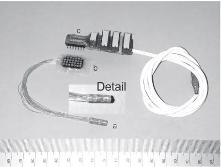
the spiral groups was 6 mm. Each electrode marked with the same number within each of the three parallel spiral groups was in the same position. Accordingly, 11 groups of three electrodes were placed along the same line in a longitudinal direction. All elec-trodes of the central and outer groups were then connected to the corresponding lead wires. The silicone sheet with the arranged electrodes was then bonded to the inner side of the mechanically opened cuff. The length of the cuff was optimized so that the surface of the nerve covered by the spiral cuff would be as small as possible to prevent damage associated with blood supply and excessive mechanical trauma of the nerve. Therefore, the cuff with an inner diameter of 2.5 mm was trimmed to the length of 18 mm as shown in Figure 1.
Finally, all lead wires were connected to a special common connector to be implanted within the lateral subcutaneous tissue for the time between experimental sessions. This common connector was designed to enable both the simple mechanical and electrical connections to the switch module to be also used during the stimulating sessions. The
Figure 1. Cuff used for stimulation: a) 33-electrode spiral cuff, b) subcutaneous common connector, and c) switch module.
c
b
a protocol approved by the Ethics Commit-tee of the Veterinary Faculty, Ljubljana, as described below. The animals were premedi-cated with 40 µg/kg medetomidine, im
(Domitor; Orion Corp., Espoo, Finland) and 0.2 mg/kg methadone, sc (Heptanon; Pliva,
Zagreb, Croatia). Induction was performed with 1.0 to 2.0 mg/kg propofol, iv (Diprivan;
Zeneca Pharmaceuticals Ltd., Macclesfield, Chesire, UK) (19). General anesthesia was maintained with 0.8 to 1.5 vol% isoflurane (Forane; Abbott Laboratories Inc., Abbott Park, IL, USA) in 100% O2. When neces-sary, analgesia was maintained during sur-gery with 0.5 to 2.0 mg/kg ketamine, iv
(Ketamine; Veyx-Pharma GmbH, Schwarzen-born, Germany). Antibiotics (20 mg/kg cefazolin, iv; Cefamezin; Krka, Novo Mesto,
Republic of Slovenia) were administered pe-rioperatively. Room temperature was kept between 23.4º and 24.4ºC, and the tempera-ture of the skin of the neck was also continu-ously monitored. According to our model, the cuff to be implanted on the vagus nerve should be installed on the nerve trunk just before bifurcation into the dorsal and ventral branches. However, to avoid potentially fa-tal trauma, the cuff was insfa-talled on the vagus nerve at the level of the neck as shown in panel A of Figure 2. The cuff on the splanchnic nerve was installed on the nerve trunk before the celiac ganglion as shown in panel B of Figure 2. Here the installation was complicated due to a very limited space.
The cuff on the pancreatic nerve was installed at the site just before the nerve enters the pancreas, as also shown in panel B of Figure 2. The leads of the implanted cuffs were routed between other organs and under the skin to the three corresponding common connectors fixed under the skin. Finally, the incisions were closed and the animals awak-ened. Analgesia during the early recovery period was provided with 0.3 to 0.5 mg/kg methadone, sc, three times a day. Tramadol,
8.0 mg/kg, sc (Tramal; Grünenthal GmbH,
Stolberg, Germany) was administered three Figure 2. A, X-ray of the 33-electrode spiral cuff implanted on the vagus nerve; B, X-ray of
the 33-electrode spiral cuffs implanted on the splanchnic (near the spine) and mixed pancreatic (below the spine) nerves of the dog.
A B
Vagus nerve Splanchnic nerve Pancreatic nerve
Connector
Switch
ECG electrodes
Stimulator Amplifier
Amplifier A/D
PC
Amplifier
Intravenous pressure transducer
Bladder pressure transducer
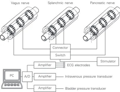
Figure 3. Schematic diagram illustrating an animal experiment in which the vagus, splanch-nic and pancreatic nerves were stimulated.
common connector was also designed to permit simple and reliable multiple use. This is very important because the common con-nector must be reimplanted several times between individual experiments without any damage.
Surgical implantation
times a day for two additional days. The first experiment was performed 30 days after im-plantation to allow the devices to stabilize.
Electrical stimulation of whole nerves
Stimulations of the intact pancreas were performed 2 (in the first dog) and 6 (in the second dog) months after implantation. The main purpose of these trials was to obtain data about normal levels of glucagon, insu-lin, and C-peptide in blood. However, stim-ulation of the pancreas, rendered partly dys-functional with alloxan, was conducted in both dogs 10 months after implantation and 17 days after disablement with alloxan. The period of the first 10 days was estimated to be necessary for the partial destruction of islets of Langerhans to be completed (20,21). When the subcutaneously implanted com-mon connector belonging to one of the im-planted cuffs was removed from the body, it was thoroughly cleaned and dried and then placed on the cleaned skin close to the wound. The common connector and the wound were then covered with self-adhesive surgical foil throughout the entire experiment. A special cable was developed to connect the common connector to the outlets of the stimulator. At one end of the cable to be connected to the common connector there was a switch mod-ule designed to fit its pins. The connection itself was made simply by perforating the self-adhesive surgical foil with the pins of the switch module and inserting them into the common connector. For the stimulations, it was of crucial importance that the connec-tion would permit reliable mechanical and galvanic connections. Moreover, the com-mon connector and the wound were insu-lated from the ambient atmosphere.
The 33-electrode spiral cuff also permits selective stimulation of the nerve fibers within the superficial regions close to the groups of the three electrodes. However, the aim of the present experiment was to stimulate all superficial regions close to the
uncovered groups of three electrodes of the aforementioned three nerves, as shown in Figure 3.
Accordingly, the switches of the switch-ing module belongswitch-ing to the cable were turned on so as to connect all electrodes in the central spiral group together and to form a single spiral electrode composed of 11 plat-inum electrodes. Similarly, all electrodes in both outer spiral groups were connected to-gether, forming two outer spiral electrodes, each consisting of 11 platinum electrodes. To obtain a “quasi-bipolar” stimulating con-figuration as described elsewhere (18), the two outer spiral electrodes were short-cir-cuited. The previously short-circuited outer electrodes of each spiral cuff were then con-nected through an explanted connector and through the switch module to one end of a custom-designed electrical stimulator, while the corresponding central electrodes were connected to the other end.
In the experiments, each nerve was stim-ulated with biphasic rectangular current pulses with the following parameters: width, 200 µs, amplitude adjustable between 10 and 20 mA, and frequency, 20 Hz. ECG, blood pressure, and pressure in the bladder were constantly monitored to assess the ef-fect of stimulation on the function of the cardiovascular system, lungs, and urinary tract.
Alloxan administration
To partly disable the pancreas, causing insulin-dependent diabetes mellitus or type I diabetes, the death of a certain portion of pancreatic islet ß-cells was induced (20-22). For this purpose, alloxan (Sigma, St. Louis, MO, USA) was dissolved in physiological saline (0.9% NaCl) at a concentration of 200 mg/ml. A quantity of freshly prepared solution was then injected iv at 50 mg/kg.
the protocols for treating naturally or experi-mentally induced type I diabetes are the same, insulin (Homofan 100, Pliva) therapy was applied (1 IE/kg animal weight). To stabilize blood glucose at a level approxi-mately four times higher than normal, a pe-riod of 7 days prior to the stimulation ses-sions was introduced. After the last experi-ment, both animals were sacrificed using the veterinary drug T61 (Hoechst, Frankfurt, Germany).
Blood sampling and analysis
Samples from the femoral artery were drawn before each experiment to obtain ini-tial data about blood glucagon, insulin, and C-peptide levels. Blood was obtained from the femoral artery at the beginning of stimu-lation, after 5 min of stimustimu-lation, and 5 min after the end of stimulation. The samples were placed on ice and centrifuged at 4oC immediately after the end of the experiments, and plasma was separated and frozen at -20ºC until radioimmunoassay. Plasma glucose was measured by the glucose oxi-dase method and hematocrit was determined at regular intervals throughout the experi-ments.
Kits from Linco Research, Inc. (St. Charles, MO, USA) were used for radioim-munoassay of canine C-peptide, human in-sulin, and glucagon.
Results
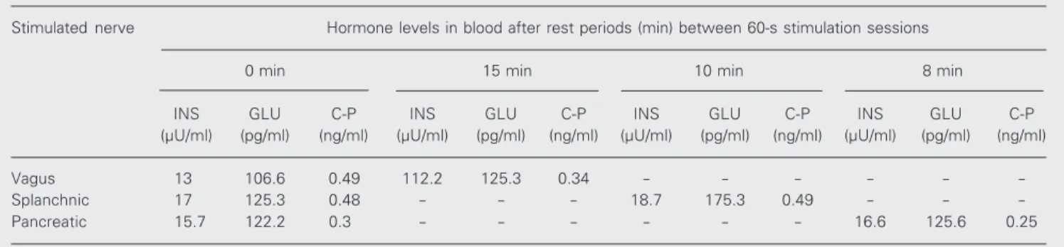
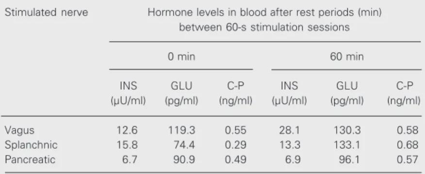
Since the results obtained for the first and the second dog were quite similar, we pres-ent them as an average of the data obtained for both dogs. Stimulation of the vagus nerve caused a statistically significant increase in insulin secretion from 13 to 112.2 µU/ml (Table 1). However, vagal nerve stimulation did not cause significant changes in gluca-gon secretion (from 106.6 to 125.3 pg/ml). Vagal nerve stimulation also decreased C-peptide secretion from 0.49 to 0.34 ng/ml. Splanchnic nerve stimulation of the intact pancreas did not cause marked changes in insulin secretion (from 17 to 18.7 µU/ml, on average) but caused a considerable increase in glucagon secretion from 125.3 to 175.3 pg/ml. Splanchnic nerve stimulation did not cause marked changes in C-peptide secre-tion (from 0.48 to 0.49 ng/ml, on average). Pancreatic nerve stimulation in the intact pancreas did not cause significant changes in the secretion of any of the three hormones. Vagal nerve stimulation of the dogs with partly dysfunctional pancreas caused an in-crease in insulin secretion from 12.6 to 28.1 µU/ml, as well as an increase in glucagon secretion from 119.3 to 130.3 pg/ml and a minor increase in C-peptide secretion from 0.55 to 0.58 ng/l (Table 2). Splanchnic nerve stimulation in the severed pancreas caused a minor decrease in insulin secretion from
Table 1. Effect of nerve stimulation on pancreatic hormones in the blood of the second dog with intact pancreas.
Stimulated nerve Hormone levels in blood after rest periods (min) between 60-s stimulation sessions
0 min 15 min 10 min 8 min
INS GLU C-P INS GLU C-P INS GLU C-P INS GLU C-P
(µU/ml) (pg/ml) (ng/ml) (µU/ml) (pg/ml) (ng/ml) (µU/ml) (pg/ml) (ng/ml) (µU/ml) (pg/ml) (ng/ml)
Vagus 13 106.6 0.49 112.2 125.3 0.34 - - -
-Splanchnic 17 125.3 0.48 - - - 18.7 175.3 0.49 - -
-Pancreatic 15.7 122.2 0.3 - - - 16.6 125.6 0.25
15.8 to 13.3 µU/ml and a considerable in-crease in glucagon secretion from 74.4 to 133.1 pg/m, as well as a small increase in C-peptide secretion from 0.29 to 0.68 ng/ml. Pancreatic nerve stimulation in the severed pancreas did not cause marked changes in insulin secretion (from 6.7 to 6.9 µU/ml) but caused a minor increase in glucagon and C-peptide secretion (from 90.9 to 96.1 pg/ml for glucagon and from 0.49 to 0.57 ng/ml for C-peptide).
In series of stimulations in which the current pulses ranged from 10 to 20 mA, measurable changes were observed in the splanchnic and vagus nerves. Splanchnic nerve stimulation with a current of 12 mA or more increased bladder pressure from 8 to 12.5 mmHg and precipitated generalized stomach contractions. Dramatic changes were observed during stimulation of the vagus nerve with a current of more than 18 mA. There was a rapid decrease of arterial blood pressure from 92 to 61 mmHg and an irregu-lar heart beat. A further increase of the vagal stimuli caused brachycardia, followed by asystolia and hypotension.
Discussion
The present study has demonstrated for the first time that multielectrode spiral cuffs chronically implanted on the nerves inner-vating the pancreas of a dog can elicit in-creased glucagon and insulin secretion from an intact or an incompletely dysfunctional pancreas.
The cuffs act on the nerve with a defined and constant pressure and permit monopolar and bipolar stimulation of the whole nerve as well as selective monopolar and bipolar stim-ulation of different superficial regions of the nerve with many combinations possible.The results of this study could be used in various types of animal and basic human studies of the neurophysiology of endocrine glands. Ultimately, the spiral cuffs could be used for both stimulation and recording. The present
study has provided new information about the electrophysiology of the pancreas and the regulation of both insulin and glucagon secretion. Recent studies have dealt with chemical and electrostimulating factors known to affect these hormones (8,10,15,16), but none has dealt with factors resulting in stimulation of the vagus, splanchnic, and mixed pancreatic nerves with multielectrode cuffs implanted for a prolonged period of time. The present study also contributed to the development of new implantable sys-tems for the stimulation of peripheral nerves in the autonomic nervous system, glands, and other organs innervated by the auto-nomic nervous system, as well as electrode systems and sensors for recording different biological signals.
It is well known that cardiovascular dis-ease is the leading cause of death in diabetes mellitus. Atherosclerosis, arteriosclerosis, and severe heart disease as a consequence of damaged coronary vessels appear much ear-lier in diabetics than in healthy individuals. By using a method of electrical stimulation of the pancreatic nerves it should be possible to slow down the progress of these diseases. In addition, with electrical stimulation, the doses of insulin could be reduced, and in cases in which islets of Langerhans still func-tion but do not secrete enough insulin, insu-lin treatment could even be abolished. A
Table 2. Effect of nerve stimulation on pancreatic hormones in blood of the second dog with partly dysfunctional pancreas.
Stimulated nerve Hormone levels in blood after rest periods (min) between 60-s stimulation sessions
0 min 60 min
INS GLU C-P INS GLU C-P
(µU/ml) (pg/ml) (ng/ml) (µU/ml) (pg/ml) (ng/ml)
Vagus 12.6 119.3 0.55 28.1 130.3 0.58
Splanchnic 15.8 74.4 0.29 13.3 133.1 0.68
Pancreatic 6.7 90.9 0.49 6.9 96.1 0.57
methodology as well as accompanying tech-nological solutions could be developed and used to transfer this animal model to an alternative human model for the treatment of diabetes mellitus. However, a requirement for the potentially successful transfer of this model to man is a good functional result demonstrated by increased and reliable insu-lin secretion over a prolonged period of time. Moreover, a methodology involving spiral cuffs could be successfully used in the trans-fer to humans of this animal model of vagal nerve stimulation. Since the vagus nerve connects the brain to the internal organs, including the heart and digestive tract, a wide-range of vagal nerve stimulation appli-cations should be possible.
However, the technique used to stimu-late the autonomic nerves has extensive sys-temic effects, such as changes in endocrine function of many internal organs and glands, and even a nonspecific stress condition. Therefore, all possible systemic effects must be considered before this technique can be transferred to humans. One possible solu-tion to reduce or even abolish these effects could be the technique of selective stimula-tion of autonomic nerves. One applicastimula-tion under study is the use of vagal nerve stimula-tion in slowing the progress of coronary artery disease. Another potential application is the control of Parkinsonian and essential tremor.
References
1. Banting FG & Gairns S (1924). Factors influencing the production of insulin. American Journal of Physiology, 68: 24-39.
2. Zunz E & La Barre J (1928). Influence de l’hyperglycémie et de l’hypoglycémie des centres nerveux supérieus sur la sécrétion interne du pancreas. Annals of Physiology, 4: 688-693.
3. Ahrén B (2000). Autonomic regulation of islet hormone secretion -implications for health and disease. Diabetologia, 43: 393-410. 4. Ahrén B (1999). Regulation of insulin secretion by nerves and
neu-ropeptides. Annals of the Academy of Medicine, Singapore, 28: 99-104.
5. Berthoud H-R & Powley TL (1991). Morphology and distribution of efferent vagal innervation of rat pancreas as revealed with antero-grade transport of Dil. Brain Research, 553: 336-341.
6. Love JA & Szebeni K (1999). Morphology and histochemistry of the rabbit pancreatic innervation. Pancreas, 18: 53-64.
7. Love JA & Szebeni K (1999). Histochemistry and electrophysiology of adult rabbit pancreatic neurons in primary culture. Pancreas, 18: 65-74.
8. Ahrén B, Veith RC & Taborsky Jr GJ (1987). Sympathetic nerve stimulation versus pancreatic norepinephrine infusion in the dog: 1. Effects on basal release of insulin and glucagon. Endocrinology, 121: 323-331.
9. Dunning BE, Ahrén B, Veith RC & Taborsky Jr GJ (1988). Nonadre-nergic sympathetic neural influences on basal pancreatic hormone secretion. American Journal of Physiology, 255: E785-E792. 10. Dunning BE & Taborsky Jr GJ (1989). Galanin release during
pancre-atic nerve stimulation is sufficient to influence islet function. Ameri-can Journal of Physiology, 256 (Part 1): E191-E198.
11. Love JA, Richards NR, Owyang C & Dawson DC (1998). Muscarinic modulation of voltage-dependent calcium channels in insulin-se-creting HIT-T15 cells. American Journal of Physiology, 274: G397-G405.
12. Roy MW, Le KC, Jones MS & Miller RE (1984). Neural control of pancreatic insulin and somatostatin secretion. Endocrinology, 115: 770-775.
13. Sha L, Love JA, Ma R & Szurszewski JH (1997). Cholinergic trans-mission in pancreatic ganglia of the cat. Pancreas, 14: 83-93. 14. Woods SC & Porte Jr D (1987). Neural control of the endocrine
pancreas. Physiological Reviews, 54: 596-619.
15. Ahrén B, Paquette TL & Taborsky Jr GJ (1986). Effect and mechan-ism of vagal nerve stimulation on somatostatin secretion in dogs. American Journal of Physiology, 250: E212-E217.
16. Ahrén B & Taborsky Jr GJ (1986). The mechanism of vagal nerve stimulation of glucagon and insulin secretion in the dog. Endocrinol-ogy, 118: 1551-1557.
17. King BF, Love JA & Szurszewski JH (1989). Intracellular recordings from pancreatic ganglia of the cat. Journal of Physiology, 419: 379-403.
18. Rozman J, Zorko B & Bunc M (1999). Stimulation of pancreatic nerves of a dog. In: Poster Abstracts. National Institute of Neuro-logical Disorders and Stroke, Bethesda, MD, USA, 3.
19. Havel PJ, Paquette TL & Taborsky Jr GJ (1986). Halothane is less suppressive than pentobarbital on reflex and neural activation of pancreatic F-cells. American Journal of Physiology, 251 (Part 1): E111-E116.
20. Ahrén B & Sundkvist G (1995). Long-term effects of alloxan in mice. International Journal of Pancreatology, 17: 197-201.
21. Herson PS & Ashford MLJ (1997). Activation of a novel non-selec-tive cation channel by alloxan and H2O2 in the rat insulin-secreting
cell line CRI-G1. Journal of Physiology, 501: 59-66.