Case Report
Left Atrial Myxoma Associated with Obstructive Coronary Artery Disease
Ronaldo Altenburg Odebrecht Curi Gismondi, Renato Kaufman, Gabriel Ângelo de Cata Preta Correa, César
Nascimento, Luiz Henrique Weitzel, José Oscar Brito Reis, Antônio Sérgio Cordeiro da Rocha, Ademir Batista da Cunha
Instituto Nacional de Cardiologia Laranjeiras - Niterói, RJ, Brazil
Mailng Address: Ronaldo Altenburg Odebrecht Curi Gismondi •
Rua Ministro Otávio Kelly, 185/701 - 24220-300 – Niterói, RJ, Brazil E-mail: [email protected]
Manuscript received November 28, 2005; revised manuscript received December 8, 2005; accepted December 8, 2005.
Key words
Myxoma; heart atria left; coronary disease.
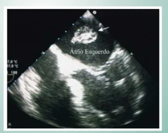
block, left anterior hemiblock, and secondary alterations of ventricular repolarization. Chest radiography showed lung hyperinflation and a normal cardiac silhouette. Transesophageal echocardiography showed a 4.7 x 1.7-cm mobile mass in the left atrium that was non-obstructive at Doppler. Its origin could not be visualized, but there seemed to be a pedicle originated in the right upper pulmonary vein (Fig. 1).
Myxomas are the most common type of cardiac tumors, accounting for 50% to 60% of the total in some case series, with an estimated incidence between 0.5 and one case per one million inhabitants-year1. The most common symptoms include dyspnea, atypical chest pain, and obstructive and embolic phenomena. Cases of sudden death have already been described, probably related to embolization to the coronary circulation2. Concomitant presence of coronary artery disease has been rarely described. The objective of this study is to report the case of a patient with left atrial myxoma and obstructive coronary lesions indicating the need of a coronary artery bypass grafting.
Case Report
Sixty-seven-year-old male patient with hypertension, dyslipidemia, chronic obstructive pulmonary disease and past history of smoking and acute myocardial infarction in 2000, treated with angioplasty without stenting of the anterior descending and right coronary arteries at the time. During preoperative assessment for an inguinal hernia repair, he underwent a transthoracic echocardiography that evidenced a tumor mass in the left atrium compatible with myxoma, and was referred to our institution for therapeutic management.
In the initial assessment, the patient was asymptomatic, with a normal physical examination. Electrocardiogram showed a sinus rhythm with complete left bundle branch We describe the case of a 67-year-old male patient with obstructive coronary artery disease who, in the preoperative assessment for an inguinal hernia repair, had undergone an echocardiography that showed a large, mobile, non-obstructive tumor in the left atrium, with a pedicle originated in the right superior pulmonary vein. The patient underwent a coronary angiography with left ventriculography that showed severe stenosis in the mid-third of the left anterior descending artery, moderate stenosis in the proximal third of the circumflex artery at the origin of the first marginal branch, and a non-obstructive lesion in the distal third of the right coronary artery. Moderate left ventricular dysfunction was also observed. The patient then underwent resection of the tumor and coronary artery bypass grafting. The histopathological examination revealed a myxoma.
Fig. 1 -Myxoma in the left atrium in a two-dimensional image of transesophageal echocardiography.
Considering the past history of infarction and the patient’s age, we decided to perform a preoperative coronary angiography that showed no lesions in the trunk, a severe lesion at the mid-third of the anterior descending artery, a moderate lesion in the circumflex artery at the origin of the first marginal branch, albeit with a thin irregular distal bed, and a non-obstructive lesion at the distal third of the right coronary artery (Figs. 2 and 3). Moderate left ventricular dysfunction was also observed.
The patient underwent resection of a myxoma in the left atrium and coronary artery bypass grafting with an anastomosis between the left internal thoracic artery and the anterior descending branch of the left coronary artery. His postoperative course was uneventful. The specimen was sent to histopathological analysis that confirmed a myxoma.
Case Report
Gismondi et al LEFT ATRIAL MYXOMA ASSOCIATED WITH OBSTRUCTIVE CORONARY ARTERY DISEASE
Arq Bras Cardiol 2007; 88(1) : e1-e2
Discussion
Myxoma is an usually asymptomatic disease with the triad of emboligenic, unspecific constitutional or obstructive symptoms.
The presence of symptoms of coronary artery disease in these patients can be explained, in many cases, by the embolization of tumor fragments. Reports of acute myocardial infarction and sudden death are not infrequent2. However, the high mean age and occurrence of other risk factors for atherosclerosis make the presence of a coronary artery disease an important diagnostic possibility. Some authors also support a hypercoagulability state in patients with myxoma, and in one report increased levels of interleukin-6 and 8 were demonstrated3.
The mean age of patients with myxoma is 56 years, and 70% are females1. Some previous publications showed a prevalence of coronary artery disease between 0 and 11% in patients with myxoma2. However, two recent studies showed values between 20.3 and 36.6%, more compatible with the age range of these patients for whom a higher percentage of risk factors for atherosclerosis such as systemic hypertension and dyslipidemia is observed1,2.
Echocardiography remains an excellent test for diagnosis and topographic definition, providing information such as size and location. Approximately 90% of the myxomas are isolated lesions and 86% are located in the left atrium1. The transesophageal is more accurate than the transthoracic echocardiography, and is routinely performed in the preoperative assessment in our service.
Some authors support the idea that all patients should undergo cardiac surgery as soon as the diagnosis is made, without previous angiographic examination, basing this assumption on the risk of sudden death2,4,5. Others consider coronary angiography a mandatory test only for patients with anginal symptoms or over forty years of age1,2,4. Venticulography is another controversial aspect because of the potential risk of embolization of tumor fragments from the atrium into the left ventricle2,4,5.
The patient reported had various risk factors for atherosclerosis, such as systemic hypertension, previous smoking habit, and past history of myocardial infarction, so that the hemodynamic study was imperative. In addition, since the echocardiography did not show the tumor prolapsed into the ventricle, a left ventriculography was performed uneventfully.
In the experience of our service the performance of a preoperative coronary angiography is key for all patients over 40 years of age with a past history of coronary artery disease or anginal symptoms, when undergoing cardiac surgery. The surgical procedure used is the same for conventional coronary artery bypass grafting.
Potential Conflict of Interest
No potential conflict of interest relevant to this article was reported.
1. Erdil N, Ates S, Cetin L, Demirkilic U, Sener E, Tatar H. Frequency of left atrial myxoma with concomitant coronary artery disease. Surg Today. 2003; 33: 328-31.
2. Li AH, Liau CS, Wu CC, Chien KL, Ho YL, Huang CH, et al. Role of coronary angiography in myxoma patients: a 14-year experience in one medical center. Cardiology. 1999; 92: 232-5.
3. Isobe N, Kanda T, Sakamoto H, Morishita Y, Suzuki T, Kobayashi I. Myocardial
infarction in myxoma patients with normal coronary arteries. Angiology. 1996; 47: 819-23.
4. Rice PL, Pifarre R. Left atrial myxoma and coronary artery disease: combined surgical treatment. Arch Surg. 1981; 116: 353-5.
5. Van Cleemput J, Daenen W, De Geest H. Coronary angiography in cardiac myxomas: findings in 19 consecutive cases and review of the literature. Cathet Cardiovasc Diagn. 1993; 29: 217-20.
References
Fig. 2 -Coronary angiography in right anterior oblique view, in which a severe lesion in the mid-third of the anterior descending artery is observed.
Fig. 3 -Coronary angiography in left anterior oblique caudal (spider) view showing severe lesion in the mid-third of the anterior descending artery.