Mailing Address: Ricardo Wang •
Praça Rui Barbosa 245 – Centro - 80010-030 - Curitiba, Paraná, Brasil E-mail: [email protected], [email protected]
Manuscript received August 23, 2008; revised manuscript received Septem-ber 13, 2008; accepted OctoSeptem-ber 10, 2008
Occlusion of the Left Coronary Trunk Secondary to Tertiary Syphilis
Ricardo Wang
1,2,3, Gustavo Blume
1, Newton Fernando Stadler Souza Filho
1,2, Lidia Zytynski Moura
1,3Hospital Santa Casa de Curitiba1; Instituito de Neurologia e Cardiologia de Curitiba2; Pontificia Universidade Católica do Paraná3, Curitiba, PR, Brasil
Summary
A 27-year-old patient with tertiary syphilis, manifested as myocardial ischemia, presenting unstable angina, secondary to left coronary trunk occlusion. The diagnosis was confirmed by the serological findings and the pathological assessment of the aorta fragment.
Introduction
Despite the increase in the incidence of coronary artery disease (CAD) in young patients, the acute myocardial infarction is still rare in this age range1. Even in this group,
the atherosclerotic etiology is by far the main cause of ischemic heart disease and we tend to treat all patients in a standardized way and forget that, in this group of patients, the non-atherosclerotic etiology must be considered. This group of patients presents distinctive characteristics in management, as well as in treatment and prognosis (in general, a better one).
The objective of this report is to present, discuss the propedeutics and therapeutic management of a young patient with Acute Coronary Syndrome, presenting an acquired, non-atherosclerotic etiology.
Case Description
A 27-year-old male patient, an adopted child, married, civil construction worker, reported presenting an episode of typical precordial pain five months before, during intense physical exertion and associated with dyspnea, which lasted for 60 minutes followed by spontaneous relief; at the time, he did not pay attention to the episode, considering it an episode of muscular pain. From this moment on, he started to present precordial pain after physical exertion (CCS Functional Class I); the pain had worsened in the two weeks before medical examination and started to present as precordial pain after moderate physical exertion (functional class II-III), now accompanied by dyspnea.
The patient denied other diseases or using medications. As he had been adopted, he reported a vague history of coronary disease by his biological father, but he did not know how old his father was at the time of the diagnosis. He reported presenting high-risk sexual behavior during the adolescence,
which included unprotected sexual intercourse and use of cocaine from 16 to 18 years old; he denied using drugs after this period.
At physical examination, he presented good general status, with no macroscopic alterations; BP was 110/60 mmHg, heart rate was 70 bpm, and the heart auscultation did not disclose significant alterations, as well as the vascular examination.
The alterations identified at the ECG and ergometric test for the detection of ischemia are shown in Figures 1 and 2. The echocardiogram showed a 29-mm aortic root, a 32-mm left atrium, a 16-mm right ventricle and 11-mm septum and posterior wall. Ejection fraction (EF) was 63% with mild diffuse hypokinesia.
Due to the presence of ischemia at the functional assessment, we decided to perform a coronary angiography (Figure 03), which showed a coronary circulation with a pattern of right coronary dominance, without significant lesions and the presence of intercoronary collateral circulation, originating from the conus arteriosus branch to the anterior descending artery (Vieussens’ collateral circulation) Rentrop grade III, and collateral circulation of the posterior ventricular branches to the circumflex artery. The left coronary artery was occluded at the origin (Figure 04). The ventriculography shows preserved ventricular function with an EF of 74%, by Simpson’s method.
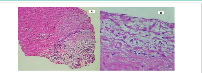
The myocardial revascularization surgery was indicated, with a right mammary artery graft to the anterior descending artery and left mammary artery graft to the first marginal branch of the circumflex artery with the help of extracorporeal circulation (ECC). The aortic cannulation showed an abnormal thickening of the its wall and a fragment was removed and sent to pathological assessment, which disclosed medial tunic degeneration and focal perivascular lymphoplasmacytic infiltrate in the adventitial tunic, compatible with syphilitic aortitis. Based on the anatomopathological diagnosis, the etiological treatment was initiated with crystalline penicillin. The CSF assessment did not disclose alterations compatible with neurosyphilis.
There were no complications in the immediate postoperative period and the patient was discharged on the 5th postoperative
day. At the four-month follow-up, the patient is now asymptomatic and has resumed his daily activities.
Discussion
Thoracic pain is responsible for approximately 4 million patients being examined annually. The pain of ischemic origin is particular in its morbidity and mortality and, therefore, must always be ruled out2. The atherosclerotic etiology represents
the main cause of myocardial ischemia; however, in patients younger than 35 years, the non-atherosclerotic etiology is
Key Words
Truncus Arteriosus / Abnormalities; Syphilis, Cardiovascular.
289
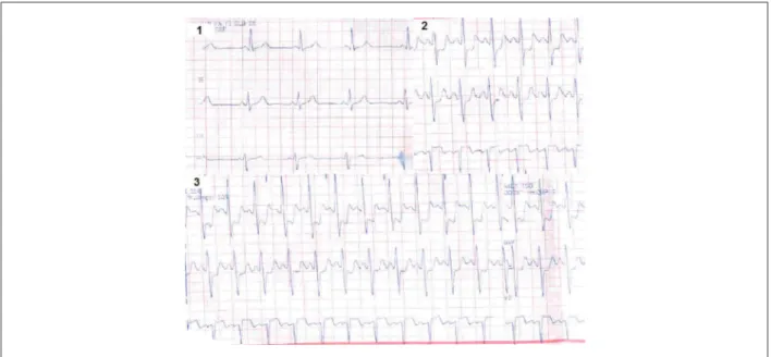
Figure 01 - Resting Electrocardiogram at hospital admission.
Figure 02 - Ergometric testing. Box 01 shows resting pre-stress recording. Box 02: recording on the third minute of the irst stage of Ellestad with pain and BP: 120/60mmHg.
Box 03: at 18 seconds of the second stage, BP: 130/60 mmHg.
4-fold higher, especially due to congenital coronary anomalies or extrinsic causes3.
The role of functional assessment in the approach of thoracic pain is not only to rule out the diagnosis of myocardial ischemia, but also to stratify the cardiovascular risk. The case in question presented clinical data and alterations at the ergometric test that met severity criteria, and thus, the coronary angiography was chosen.
The descriptions of chronic occlusions of the left coronary artery trunk are rare in the literature, and even more when associated with normal ventricular function, with an estimated incidence of 0.04 to 0.4% of the patients with coronariopathies4. To attain this clinical condition, the
patient must necessarily present a well-developed collateral circulation, as its absence is associated with severe ventricular dysfunction, which invariably leads to death, due to the large ischemic area. In this case, the patient presented the right coronary free from obstructive lesion, which did not limit the coronary flow, associated with a well-developed collateral circulation that was responsible for the preservation of the ventricular function4.
The surgical finding of aortic thickening associated with the absence of other coronary lesions at the previous coronary angiography prompted the search for a pathology that would primarily affect the aorta. Among the differential diagnoses, it is necessary to pay special attention to vasculitis, such as
Wang et al
Left Coronary Trunk Lesion Secondary to Syphilis
Arq Bras Cardiol 2009; 93(3) : 289-292
290
Figure 03 - Angiography of the right coronary artery in left anterior oblique projection, with selective injection in the conus arteriosus, demonstrating collateral circulation to the anterior descending artery (Vieussens’ anastomotic annulus).
Figure 04 - Angiography of the right coronary artery (RCA) in right anterior oblique projection, demonstrating collateral circulation of the distal branches
of the RCA to the circumlex artery (Cx).
Figure 05 - Contrast injection in the left coronary sinus, in left anterior oblique
projection, showing absence of opaciication of the left coronary artery.
Figure 06 - Left ventriculography in right oblique projection, showing normal
ventricular function and ejection fraction of 74%.
Takayasu’s arteritis, giant-cell arteritis, and Reiter’s syndrome5.
Another diagnosis that must be recalled is the syphilitic aortitis, which is currently increasingly rarer, mainly after the advent of antibiotic therapy. The diagnosis can be attained through the clinical and laboratory picture and confirmed by the pathological findings.
The cardiovascular involvement of syphilis can be divided in four main categories: 1. Syphilitic aortitis; 2. Aortitis with
aneurysm; 3. Aortitis with valvulitis and aortic regurgitation and 4. Stenosis of coronary ostia, which is the second most common cardiovascular complication3. The study by
Heggtveit6, which analyzed 100 cases of patients with tertiary
syphilis, showed that 40% presented aneurysm, 29% with valvular involvement and 26% with coronary involvement, with 11% of them being the isolated form. It presents an insidious characteristic, generally occurring 15 to 30 years
Wang et al Left Coronary Trunk Lesion Secondary to Syphilis
Arq Bras Cardiol 2009; 93(3) : 289-292
291
References
1. Hong MK, Cho SY, Hong BK, Chang KJ, Chung IM, Lee MH, et al. Acute myocardial infarction in young adults. Yonsei Med. 1994; 35 (2): 184-9.
2. Baracioli LM, Borges FA. Dor torácica. In: Cavalcanti EFA, Martins HS. (eds.). Clínica médica dos sinais e sintomas ao diagnóstico e tratamento. São Paulo: Editora Manole; 2007.
3. Waller BF. Nonatherosclerotic coronary heart disease. In: Fuster V, Alexander RW, O’Rourke RA. Hurst’s the heart 10th ed. Philadelphia: McGraw Hill;
2001.
4. Sugihita K, Shimizu T, Kinugawa K, Harada K, Ikenouchi H, Matsui H, et al. Chronic total occlusion of the left main coronary artery. Inter Med. 1997; 36: 471-8.
5. Scully RE, Mark EJ, McNeely WF, Ebeling SH, Phillips LD. Case 10-1998, Case record of the Masschusetts General Hospital. N Engl J Med. 1998; 338: 897-903.
6. Heggtveit HA. Syphilis aortitis: a clinical pathologic autopsy: study of 100 cases, 1950 to 1960. Circulation. 1964; 29: 346-55.
7. Virmani R, Burke AP. Nonatherosclerotic diseases of the aorta and miscellaneous diseases of the main pulmonary arteries and large veins. In: Silver MD, Gotlieb AI, Schoen FJ. Cardiovascular pathology. 3rd. ed. Saint
Louis: Churchill Livingstone; 2001.
8. Topaz O, Warner M, Lanter P, Soffer A, Burns C, DiSciascio G, et al. Isolated significant left main coronary artery stenosis: angiographic, hemodynamic, and clinical findings in 16 patients. Am Heart J. 1991; 122 (5): 1308-14. after the infectious onset of the disease7.In this case, in
addition to the early presentation of the cardiovascular manifestations of syphilis, the latter manifest as left coronary trunk occlusion, which is also rare.
The involvement of the coronary ostia is the typical lesion in syphilis, affecting the initial 3 to 4 mm segment, with distal segment involvement being rare3. The presentation
can range from stable angina, acute coronary syndrome or sudden death8. The pathological assessment discloses chronic
inflammation with lymphoplasmacytic infiltrate, together with medial destruction, accompanied by fibrosis. The preferred surgical approach is the bypass surgery, preferably with arterial conduct and the ostium approach, despite its initial good results, carries a risk of fibrosis recurrence and re-occlusion5.
The etiological treatment of syphilis presents a controversy about the ideal moment for treatment approach. Some authors defend that the treatment with beta-lactamics be started after the myocardial revascularization, as the antibiotic course can trigger a Jarisch-Herxheimer reaction, with aortic edema and even occlusion of the diseased coronary ostium5. There is also
the possibility of intracoronary thrombosis, which justifies the use of associated corticoid therapy.
Conclusion
The present report illustrates the need to perform an etiological investigation in young patients with coronariopathies, mainly those occurring in the LCT.
The syndromic treatment decreases the chances of therapeutic effectiveness and, as demonstrated in this case, can lead to more severe future complications, as it does not prevent the natural progression of the disease. The occurrence of syphilitic aortitis is rare, but its recognition and effective treatment decrease not only the progression of the disease, but also its transmission.
Potential Conflict of Interest
No potential conflict of interest relevant to this article was reported.
Sources of Funding
There were no external funding sources for this study.
Study Association
This study is not associated with any post-graduation program.
Figure 07 - Histological section of the aorta. A: section showing the endothelial surface with intimal thickening. B: Plasmocytic iniltrate in the adventitial layer of the
aorta (HE staining, kindly provided by Dr. Luiz Kortze).
Wang et al
Left Coronary Trunk Lesion Secondary to Syphilis
Arq Bras Cardiol 2009; 93(3) : 289-292