© 2013 Sociedade Brasileira de Hemodinâmica e Cardiologia Intervencionista. Published by Elsevier Editora Ltda. All rights reserved.
Emergency Unprotected Left Main
Percutaneous Coronary Intervention in a
Heart Transplant Patient
Frederico Thomaz Ultramari
1, Adrian Paulo Morales Kormann
2, Rafael Maestri
3, Anne Louise Marchi
4,
Marcos Vinícius Claussen Moura
4, Frederico Di Giovanni
6RESUMO
Intervenção Coronária Percutânea Emergencial de Tronco de Coronária Esquerda Não-Protegido em
Paciente com Transplante do Coração
A doença vascular do enxerto cardíaco é a principal causa de falência do enxerto e morte depois do primeiro ano após o transplante. O melhor tratamento para lesões de tronco de coronária esquerda não-protegido em corações transplantados ainda não está estabelecido. Descrevemos o caso de uma intervenção coronária percutânea emergencial de tronco de coronária esquerda não-protegido em coração transplantado após morte súbita revertida com sucesso em paciente que aguardava cirurgia de revascularização do miocárdio.
Descritores: Transplante de coração. Doença da artéria coro-nariana. Angioplastia. Stents farmacológicos.
ABSTRACT
Cardiac allograft vasculopathy is the leading cause of graft failure and death after the irst year of heart transplantation. The optimal therapy for unprotected left main coronary artery disease in transplanted hearts has not been established. We report a case of emergency unprotected left main percutaneous coronary intervention in a transplanted heart after an aborted sudden death in a patient who was waiting for coronary artery bypass graft surgery.
Descriptors: Heart transplantation. Coronary artery disease. Angioplasty. Drug-eluting stents.
Original Article
1 Interventionist Cardiologist Physician at the Hemodynamics Service of the Hospital Santa Isabel de Blumenau. Blumenau, SC, Brazil. 2 Interventionist Cardiologist Physician at the Hemodynamics Service of the Hospital Santa Isabel de Blumenau. Blumenau, SC, Brazil. 3 Interventionist Cardiologist Physician at the Hemodynamics Service of the Hospital Santa Isabel de Blumenau. Blumenau, SC, Brazil. 4 Pharmaceutical, coordinator of the Research Division of the Hemo-dynamic Service of the Hospital Santa Isabel de Blumenau. Blumenau, SC, Brazil.
5 Cardiologist Physician at the Cardiology Service of the Hospital Santa Isabel de Blumenau. Blumenau, SC, Brazil.
6 Cardiovascular Surgeon Physician at the Cardiovascular Surgery Ser-vice of the Hospital Santa Isabel de Blumenau. Blumenau, SC, Brazil. Correspondence to: Frederico Thomaz Ultramari. Rua Coronel Vidal Ramos, 400 – ap. 71 – Jardim Blumenau – Blumenau, SC, Brazil – CEP 89010-330
E-mail: [email protected]
Received on: 3/18/2013 • Accepted on: 5/26/2013
C
ardiac allograft vasculopathy is the leading cause of graft failure and death after the irst year of transplantation. There is gradual increase in the prevalence of vasculopathy in these patients: 8% in one year, 32% in ive years, and 43% in eight years. After detection of diffuse or multivessel disease, the risk of death or retransplantation is extremely high.1Drug treatment has shown signiicant improvement in the prevention of cardiac allograft vasculopathy, and consists in the aggressive control of classic risk factors for cardiovascular disease and treatment of acute re-jection and infection by cytomegalovirus. Therapeutic options include percutaneous coronary intervention
(PCI), coronary artery bypass graft (CABG) surgery, or a new heart transplant.
CABG may not be the best option, as diffuse intimal involvement frequently occurs, affecting distal vessels. Furthermore, perioperative mortality rates range between 40% and 80%, and graft patency rates in the long-term are unknown.2,3 A new heart transplant is hindered by
the scarcity of donors and high perioperative mortality. The survival rate for patients who undergo repeat cardiac transplantation (75% in one year) is lower when com-pared to the irst procedure, and half of these patients develop recurrent cardiac allograft vasculopathy.2,3 For
reports a case of emergency unprotected left main percutaneous coronary intervention in a transplanted heart after a successfully reversed sudden death in a patient awaiting CABG surgery.
CASE REPORT
Female patient, 50 years-old, former smoker, dys-lipidemic, with peripheral arterial failure, had a past history of CABG in 1993 and heart transplant in 2001, due to ischemic heart disease with heart failure classiied as New York Heart Association (NYHA) functional class IV, and Canadian Cardiovascular Society (CCS) class IV angina. She used atenolol, losartan, amiodarone, acetylsalicylic acid (ASA), atorvastatin, delazacort, cy-closporine, mycophenolate, cilostazol, and omeprazole.
From September 2012 onwards, she began to no-tice oppressive precordial discomfort chest pain and dyspnea on exertion, with progressive symptom wors-ening. An echocardiogram performed in October 2012 evidenced left ventricle with preserved dimensions and systolic function; ejection fraction of 59%; mild mitral regurgitation; dilated right ventricle, with moderate to severe dysfunction; moderate tricuspid regurgitation; and estimated pulmonary artery systolic pressure of 40 mmHg. The changes found were the same observed in the echocardiography performed in 2006.
A subsequent cardiac catheterization showed 50% lesion in the distal portion of the left main coronary artery, 40% lesions in the proximal third, and 70% and 95% lesions in the distal third of the left anterior descending artery. The left circumlex artery showed only mild and diffuse parietal irregularities, and the right coronary artery showed a 30% lesion in its distal third. The left ventricle showed normal end-diastolic volume and contractile function. The mitral valve did not allow for regurgitation. The recorded pressures were 85/12 mmHg in the left ventricle and 85/59 mmHg in the aorta. During the same procedure, three endomyocar-dial fragments were removed, whose histopathological analysis showed subendocardial ibrosis and absence of signs of rejection. In the previous catheterization, performed in 2008, the coronary arteries and the left ventricle showed no changes, and the biopsy found no rejection.
Her case was discussed at a meeting with the cardiology, hemodynamics, and cardiovascular sur-gery teams in November 2012. There were discordant opinions: the interventionists indicated PCI for the left
through an endotracheal tube and received fentanyl, midazolam, and dopamine through infusion pumps. Vital signs showed blood pressure of 106/60 mmHg, heart rate of 73 bpm, and 93% oxygen saturation. Segmental assessment showed isochoric and photoreactive pupils, rhythmic and mufled heart sounds with no murmurs, discrete crackling rales at the bases, abdomen without visceromegaly, lower limbs without edema and with good perfusion. The electrocardiogram showed no acute ischemic changes. Initial laboratory assessment showed the following signiicant changes: creatine phosphokinase (CPK) = 526 U/L (24 U/L to 170 U/L), creatine kinase – MB fraction (CK-MB) = 30.7 U/L (< 25 U/L), troponin = 0.18 µg/L (< 0.01 g/L), and D-dimer = 5,681.3 ng/mL (68 ng/mL to 494 ng/mL).
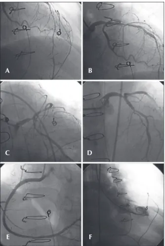
Due to the evolution with persistent hypotension, requiring start of noradrenaline infusion, the hemo-dynamic team was called to perform an emergency catheterization. The procedure, performed through the femoral artery, was uneventful and showed a 60% lesion in the distal third of the left main coronary artery, 60% lesions in the proximal third, 70% and 95% lesions in the distal third of the left anterior descending artery, 30% lesion in the proximal third of the left circum-lex artery, and 40% lesions in the middle and distal thirds of the right coronary artery. There were no new segmental contractility alterations in the left ventricle (Figure 1). The recorded pressures were 172/14 mmHg in the left ventricle and 172/83 mmHg in the aorta, and the norepinephrine infusion velocity was reduced. An arteriography was performed to rule out the suspicion of pulmonary thromboembolism; no thrombus or areas of peripheral hypoperfusion were found. The pressure measured in the left main coronary artery was 48/21 mmHg, with a mean of 31 mmHg.
the stent with a 4.0/20 mm compliant balloon at 16 atm. A 3.0/10 mm compliant balloon was positioned at the left circumlex artery ostium and the procedure was inished with a kissing balloon, both inlated to 10 atm, yielding satisfactory angiographic result (Figure 2). PCI was not performed in the other left anterior descending artery lesions, as they were located in the distal third, in a ine-calibre segment.
At the end of the procedure, the patient was hypertensive, allowing for a gradual reduction in the norepinephrine dose, which was suspended on the next day. Markers of myocardial necrosis showed a slight increase, reaching maximum values on December 25th,
2012 at the collection performed 3.5 hours after the PCI (CPK = 671 U/L and CK-MB = 48.1 U/L). The following samples showed a progressive decrease in these values.
Pneumonia was diagnosed on December 27th,
and treatment with piperacillin and tazobactam was initiated. On the following day, the patient developed supraventricular tachycardia reversed with electrical car-dioversion. On the same day, the patient once more had hypotension, requiring reintroduction of noradrenaline. There was progressive clinical and laboratory worsening, and the patient went into cardiac arrest with pulseless electrical activity on December 31st, 2012.
DISCUSSION
Cardiac allograft vasculopathy is not a single disease entity, but rather a set of combined changes characterized by intimal fibromuscular hyperplasia, atherosclerosis (accelerated, affecting both adults and children), and vasculitis (inflammation of one or more layers of the vessel wall). Moreover, the veins are Figure 1 – In A, B, C, and D, left coronary artery in left-anterior oblique,
caudal right-anterior oblique, caudal left-anterior oblique, and cranial left-anterior oblique projections, respectively. In E, right coronary artery in cranial left-anterior oblique projection. In F, left ventricular systole in the right-anterior oblique projection.
Figure 2 – In A, stent implantation in left main coronary artery lesions and proximal left anterior descending artery. In B, stent post-dilation. In C, control angiography in caudal right anterior oblique projection. In D, kissing balloon. In E, the inal result in caudal right anterior oblique projection. In F, the end result in cranial projection.
A
C
E
B
D
F
A
C
E
B
D
to be eccentric, to affect proximal epicardial vessels and spare the intramyocardial arteries. Intimal ibromuscular hyperplasia tends to be circumferential and may involve diffusely large and small epicardial coronary arteries, as well as intramyocardial vessels. This pattern of diffuse involvement of small epicardial arteries appears to cause most of the arterial occlusions. The intramyocardial arter-ies are affected in their medial or subepicardial portions, making it unlikely that the endomyocardial biopsy will obtain samples with the involved vessels. It is important to note that calciication, unlike atherosclerosis, is not a prominent inding in intimal ibromuscular hyperplasia, even in severely affected vessels.4
Many risk factors for the development of cardiac allograft vasculopathy have been identiied: systemic arterial hypertension, diabetes mellitus, dyslipidemia, obesity, insulin resistance, infection by cytomegalovirus, speciic serology for leukocyte antigens, donor’s age, donor’s brain death, and occurrence of cell or humoral rejection.5 Symptom onset may be insidious and, since the
heart is denervated, patients often do not report angina. Its existence suggests myocardial reinnervation, which occurs in 10% to 30% of recipients in the long-term. Manifestations may include silent ischaemia, conges-tive heart failure, ventricular arrhythmias, myocardial infarction, and sudden death.6,7
The diagnosis of cardiac allograft vasculopathy is hindered by silent ischemia and low sensitivity of non-invasive tests. Intravascular ultrasound (IVUS) is more sensitive than coronary angiography to detect early disease and allows for the evaluation of both the arterial lumen and vessel wall. However, only the proximal portions can be evaluated, which limits its role in cases of diffuse vasculopathy.8
The best treatment for cardiac allograft vasculopathy has not been established, but there is evidence that myocardium revascularization procedures are effective in transplanted hearts with evidence of ischaemia and/or high-risk lesions. Patients free of these conditions have a good prognosis; however, those with diffuse disease with no possibility of revascularisation have unfavoura unfavorable ble evolution.3
In transplanted patients, PCI presents excellent angiographic success rates, ranging from 90% to 100%, rare complications, and restenosis rates with drug-eluting stents between 9% and 19%, lower than those of bare-metal stents (which range between 18% and 31%).7-12 It is also a good therapeutic option for patients
with left main coronary artery lesions. An international
all patients, of whom 14 (67%) received drug-eluting stents. In a mean follow-up time of 4.9 ± 3.2 years, three patients (14 %) died, one patient (5%) had myocardial infarction, and four (19%) had restenosis requiring target-lesion revascularization. There were no cases of stent thrombosis. One individual (5%) was submitted to CABG and ive (24%), to a new transplant.11
In a review totaling ten publications and 34 pa-tients undergoing left main coronary artery PCI in the transplanted heart, including the publication mentioned in the previous paragraph, four subjects (11.8%) died within a mean follow-up of 43.1 ± 32.4 months. Con-sidering that 12 participants (35.3 %) had distal lesions in the left main coronary artery and 18 (52.9%) received bare-metal stents, repeat revascularization with PCI or CABG was required in nine subjects (26.4%). At 60 months, death- and revascularisation-free survival was 66%.12 To the present date, most articles published on
unprotected left main coronary artery PCI in patients undergoing cardiac transplantation consist in a limited number of case reports and small series of patients.11,12
In conclusion, PCI with drug-eluting stents is a therapeutic option for transplanted patients with un-protected left main coronary artery lesions, especially in emergency cases, and can delay the need for a new heart transplant. The procedure is safe and has high rates of immediate success; however, the long-term evolution requires further studies.
CONFLICTS OF INTEREST
The authors declare no conlicts of interest.
REFERENCES
1. Schmauss D, Weis M. Cardiac allograft vasculopathy: recent developments. Circulation. 2008;117(16):2131-41.
2. Musci M, Loebe M, Wellnhofer E, Meyer R, Pasic M, Hummel M, et al. Coronary angioplasty, bypass surgery, and retransplanta-tion in cardiac transplant patients with graft coronary disease. Thorac Cardiovasc Surg. 1998;46(5):268-74.
3. Prada-Delgado O, Estévez-Loureiro R, López-Sainz A, Gargallo-Fernández P, Paniagua-Martín MJ, Marzoa-Rivas R, et al. Percutaneous coronary interventions and bypass surgery in patients with cardiac allograft vasculopathy: a single-center experience. Transplant Proc. 2012;44(9):2657-9.
4. Lu W, Palatnik K, Fishbein GA, Lai C, Levi DS, Perens G, et al. Diverse morphologic manifestations of cardiac allograft vas-culopathy: a pathologic study of 64 allograft hearts. J Heart Lung Transplant. 2011;30(9):1044-50.
6. Di Cori A, Petronio A, Gemignani C, Zucchelli G, Di Bello V, Mariani M. Symptomatic acute myocardial infarction in a cardiac transplant recipient successfully treated with primary coronary angioplasty: evidence of prognostic importance of chest pain after cardiac transplantation. J Heart Lung Trans-plant. 2005;24(8):1146-9.
7. Bader FM, Kfoury AG, Gilbert EM, Barry WH, Humayun N, Hagan ME, et al. Percutaneous coronary interventions with stents in cardiac transplant recipients. J Heart Lung Transplant. 2006;25(3):298-301.
8. Cale R, Almeida M, Rebocho MJ, Aguiar C, Sousa P, Brito J, et al. The value of routine intracoronary ultrasound to assess coronary artery disease in cardiac allograft recipients. Ver Port Cardiol. 2010;29(2):231-41.
9. Lee MS, Tarantini G, Xhaxho J, Yang T, Ehdaie A, Bhatia R, et al. Sirolimus- versus paclitaxel-eluting stents for the treatment
of cardiac allograft vasculopathy. JACC Cardiovasc Interv. 2010;3(4):378-82.
10. Tanaka K, Li H, Curran PJ, Takano Y, Arbit B, Currier JW, et al. Usefulness and safety of percutaneous coronary interventions for cardiac transplant vasculopathy. Am J Cardiol. 2006; 97(8):1192-7.
11. Lee MS, Yang T, Fearon WF, Ho M, Tarantini G, Xhaxho J, et al. Long-term outcomes after percutaneous coronary inter-vention of left main coronary artery for treatment of cardiac allograft vasculopathy after orthotopic heart transplantation. Am J Cardiol. 2010;106(8):1086-9.