Article
0103 - 5053 $6.00+0.00*e-mail: [email protected]
Pharmacokinetic Proile of Liposome-Encapsulated Ropivacaine after Maxillary
Iniltration Anaesthesia
Michelle Franz-Montan,*,a Cristiane de Cássia Bergamaschi,a Eneida de Paula,b
Francisco C. Groppo,a José Ranali,a Leonardo Fernandes Fracetoc and Maria C. Volpatoa
aDepartment of Physiological Sciences, Piracicaba Dental School,
University of Campinas, Campinas-SP, Brazil
bDepartment of Biochemistry, Institute of Biology, University of Campinas,
Campinas-SP, Brazil
cDepartment of Environmental Engineering,Julio de Mesquita Filho State University,
Sorocaba-SP, Brazil
O objetivo do presente estudo foi determinar os parâmetros farmacocinéticos da ropivacaína encapsulada em lipossomas após anestesia local em 14 voluntários sadios. Neste estudo randomizado, cruzado e duplo-cego, os voluntários receberam anestesia iniltrativa na maxila de ropivacaína a 0,5% encapsulada em lipossomas e ropivacaína 0,5% com epinefrina a 1:200.000 em duas sessões distintas. Amostras de sangue foram coletadas antes e após (de 15 a 1440 min) a administração das formulações de ropivacaína. A quantiicação da concentração plasmática de ropivacaína foi feita por meio de HPLC com detecção por UV. Os parâmetros farmacocinéticos AUC0–24 (área sob a curva de concentração × tempo do tempo 0 até 24 horas) , AUC0–∞(área sob a curva de concentração x tempo do tempo 0 até o ininito), Cmax (concentração máxima da droga),
CL (clearance renal), Tmax (tempo em que ocorre a concentração máxima); t1/2 (meia vida de
eliminação) e Vd (volume de distribuição) foram analisados pelo teste de Wilcoxon. Nenhuma diferença (p > 0,05) foi observada entre as duas formulações em cada parâmetro farmacocinético avaliado e as concentrações plasmáticas de ropivacaína, considerando cada período de tempo. Ambas as formulações apresentaram peril farmacocinético semelhante, indicando que a formulação lipossomal poderia ser uma opção mais segura para o uso deste anestésico local, devido à ausência de vasoconstritor.
The aim of this study was to determine the pharmacokinetic parameters of liposomal ropivacaine after dental anesthesia in 14 healthy volunteers. In this randomized, double-blind and crossover study, the volunteers received maxillary iniltration of liposome-encapsulated 0.5% ropivacaine and, 0.5% ropivacaine with 1:200,000 epinephrine in two different sessions. Blood samples were collected before and after (from 15 to 1440 min) the administration of either ropivacaine formulation. HPLC with UV detection was used to quantify plasma ropivacaine concentrations. The pharmacokinetic parameters AUC0–24 (area under the plasma concentration × time curve from
baseline to 24 h), AUC0–∞(area under the plasma concentration–time curve from baseline to ininity), Cmax (maximum drug concentration), CL (renal clearance), Tmax (maximum drug concentration time), t1/2 (elimination half-life) and Vd (volume of distribution) were analyzed using the Wilcoxon signed-rank test. No differences (p > 0.05) were observed between both formulations for any of the pharmacokinetic parameters evaluated and plasma ropivacaine concentrations, considering each period of time. Both formulations showed similar pharmacokinetic proiles, indicating that the liposomal formulation could be a safer option for use of this local anesthetic, due to the absence of a vasoconstrictor.
Introduction
Prolonged-action local anesthetic is required when postoperative pain and discomfort are expected, especially after major surgical procedures.1 In many countries,
bupivacaine, the racemic mixture of S- and D-bupivacaine, is the only long-acting local anesthetic available in dental practice.
Ropivacaine (RVC), another long-acting local anesthetic of the cyclic aminoamide family, is synthesized in the
S-enantiomer form and presents lower toxicity to the cardiovascular and central nervous systems, compared to bupivacaine.2
Traditionally, most of local anesthetic formulations are administered together with a vasoconstrictor in order to increase anesthesia duration and to reduce systemic absorption rate.3 It was recently demonstrated that the use
of these formulations increased, especially those containing epinephrine 1:100.000.4 In spite of the known safety of
these drugs, there is evidence concerning adverse reactions involving the autonomic system, been the most common those associated to the medical status of the patient.5
Alternative drug delivery systems, such as liposomes, have been used to prolong the duration of action of many drugs, including local anesthetics.6 Liposomes are
phospholipid vesicles that have been demonstrated to be effective drug carriers, improving the effectiveness and reducing the toxicity of anesthetics in both cardiovascular and central nervous systems.6-10 These vesicles are
nontoxic and nonimmunogenic because their components (phosphatidyl choline and cholesterol) are also found in biological membranes.11 These characteristics have been
demonstrated in vivo (animal models) for liposome-encapsulated bupivacaine using multilamellar vesicles,12-16
and large unilamellar vesicles.17
Previous authors have shown that liposomal encapsulation of bupivacaine altered its pharmacokinetic proile after extradural injection in rabbits, resulting in lower concentrations of the drug in plasma, liver and myocardium.18 Grant and co-workers16 observed that when
encapsulated in liposomes, bupivacaine remained at the injection site for a signiicantly longer period of time after subcutaneous injection in mice.
Attempting to simulate an accidental intravascular injection of a local anesthetic, Boogaerts and co-workers8 assessed the acute CNS (central nervous
system) and cardiac toxicities induced by intravenous infusion in rabbits of 0.25% bupivacaine, with and without epinephrine (1:200,000), compared to liposome-encapsulated bupivacaine. They demonstrated a reduction of CNS and cardiac toxicities using liposome-encapsulated
bupivacaine. The addition of epinephrine to the plain solution did not decrease the CNS and cardiac toxicities induced by bupivacaine.
It was recently demonstrated in animal studies, which used sciatic and infraorbital nerve blockades, that encapsulation of ropivacaine into unilamellar vesicles increased the duration and the intensity of analgesic effects.6
Although long-acting local anesthetics are normally used in low doses in dentistry, high doses may be required for removal of four impacted third molars in a single session.19 According to Zink and Graf,20 ropivacaine seems
to have the greatest margin of safety of all the long-acting local anesthetics, and could be useful in lengthy dental procedures.
The present study is the irst attempt to measure the pharmacokinetic parameters of ropivacaine after maxillary infiltration anesthesia using a liposome-encapsulated formulation in healthy volunteers. The pharmacokinetic parameters of an RVC formulation containing epinephrine (vasoconstrictor) were also assessed for comparison.
Experimental
Materials
RVC hydrochloride was donated by Cristalia Prod. Quim. Farm. Ltda (Itapira, SP, Brazil). Egg phosphatidylcholine (EPC), cholesterol (Ch) and α-tocopherol (α-T) were purchased from Sigma Chemical Company (St Louis, MO). All other reagents used were of analytical grade.
RVC-liposome formulation
The liposomal RVC formulations were prepared as described by Araújo and co-workers.6 Briely, EPC-Ch-α-T
(4:3:0.07, molar ratio) ilms were obtained by evaporating stock chloroform solutions under a stream of wet nitrogen, followed by vacuum for 2 h. Films were suspended in HEPES buffer (20 mmol L-1, 154 mmol L-1 NaCl, pH 7.4),
and multilamellar vesicles were obtained after vortexing at ambient temperature (5 min, 25 °C). Large unilamellar vesicles were prepared by extrusion (15 cycles) of the multilamellar vesicles within 400 nm membrane ilters (25 °C), using a Lipex Biomembranes Inc. (Vancouver, Canada) extruder. RVC was added directly to the liposomes after extrusion, to reach a inal concentration of 0.5%. This formulation was sterilized by autoclaving (121 ºC, 1 atm during 15 min), as described previously by Cereda and co-workers. 10
The following liposomal characterization was previously determined by Araújo and co-workers.6 The
by photon correlation spectroscopy (Malvern Mastersizer- Malvern Instruments, France), showed an average size of 371 ± 7 nm (85% of the population), which did not change after RVC encapsulation. All the measurements presented a homogeneous distribution of the liposomal population obtained (polydispersity index of 0.12-0.17). The encapsulation eficiency of RVC into the liposomes, of samples containing 2 mmol L-1 RVC and 4 mmol L-1
liposomal suspensions determined by ultracentrifugation, was around 24%.
Subjects
This research was approved by the Ethical Committee of Piracicaba Dental School, University of Campinas (approval #164/2006). Fourteen healthy volunteers (seven males and seven females) aged 24 (± 3.1) years old were selected, and signed a written informed consent. The volunteers presented no systemic or oral disorders, had no history of allergy to any of the local anesthetics used, and were not taking any medication, as determined by oral questioning and by their documented health histories.
Prior to the beginning and right after the end of the study, all the subjects were submitted to laboratory tests to conirm that they were in good health and that the females were not pregnant. The same tests were performed at the end of the study to ensure that all results previously obtained were not altered by the anesthetic formulations tested. These tests included cross-reactive protein, blood-hemoglobin, lymphocyte count, platelet count, erythrocyte sedimentation rate, serum (S)-sodium,
S-potassium, S-chloride, S-albumin, S-alkaline phosphate,
S-gamma-glutamyl-transferase, S-aspartate transaminase,
S-alanine transaminase, S-creatine, plasma glucose, urea, cholinesterase, total protein, bilirubin, uric acid, urine glucose, urine leukocyte count, urine protein, and urine hemoglobin. Serology tests for human immunodeiciency virus and hepatitis B and C were also performed. Female subjects underwent a urine βHCG pregnancy test.
Ambulatory procedures
Anesthetic procedures
In this double-blind and crossover study, the volunteers randomly received 1.8 mL of 0.5% ropivacaine with 1:200,000 epinephrine, and liposome-encapsulated 0.5% ropivacaine, for iniltration anesthesia at the apex of the right maxillary canine, in two different sessions spaced one week apart.
Ropivacaine with 1:200,000 epinephrine was obtained by simple dilution of the commercially available solution
of ropivacaine (Naropin® 10 mg mL−1,AstraZeneca, São
Paulo, Brazil) immediately before application. Under sterile conditions, 5 mL of 1% ropivacaine was diluted with 5 mL of 1:100,000 (v/v) epinephrine (Drenalin®, Ariston Ind.
Quim. Farm. Ltda, São Paulo, SP, Brazil).
The local anesthetics (1.8 mL) were placed into coded sterile 3 mL Luer-Lok syringes (Becton Dickinson, Curitiba, Brazil) fitted with disposable needles (30G, one-inch, Becton-Dickinson Company, Franklin Lakes, NJ, USA). After topical anesthesia on the injection site with 20% benzocaine, the formulations were injected at the maxillary buccal fold of the right-canine region at an injection rate of 1 mL min−1. The same operator performed
the maxillary iniltration anesthesia in all the subjects.
Blood sampling and drug analysis
Blood samples (4.5 mL) from a forearm vein were collected with a heparinized cannula before and 15, 30, 45, 60, 75, 90, 120, 240, 420, 600 and 1440 min after administration of either of the ropivacaine formulations. 0.4 mL of a heparinized saline solution (0.9% NaCl and heparin, 9.8:0.2 v/v) was injected into the cannula after each blood sampling, to prevent clotting. The last sample was obtained using a sterile syringe and needle. Immediately after each blood collection, the samples were centrifuged at 3000 × g for 15 min, and plasma was removed and stored at −70 oC.
Detection of ropivacaine concentrations in the plasma samples was performed by high-performance liquid chromatography (HPLC), following a method adapted from Kawata and co-workers.21 Briely, chromatographic
separations were carried out using an ODS column (TSK-GEL, 150 mm, 4.6 mm i.d., TOSOH) at room temperature. The detection wavelength was set at 215 nm. The analytical calibration curve was obtained by diluting ropivacaine in drug-free human plasma (concentration range: 0.03-10 µg mL−1; concentration used 10.0,
5.0, 2.5, 1.25, 0.62, 0.31, 0.15, 0.075, 0.030 µg mL−1,
peak area = 20.21[RVC] + 0.50, r = 0.9998, n = 3). The speciicity was tested in the presence of plasma components, and it was demonstrated that these factors did not affect RVC quantiication. The limit of detection was determined according to the ICH guidelines (2005).22
The mobile phase consisted of acetonitrile, methanol and 0.05 mol L-1 phosphate buffer (10:30:60, v/v), adjusted
to pH 4.0 and pumped at a low rate of 1.0 mL min−1. The
Plasma samples (250 µL) were extracted by adding 125 µL of 0.1 mol L-1 sodium hydroxide in a 2.0 mL tube.
The mixture was submitted to agitation and addition of 1 mL ethylacetate in order to extract ropivacaine. The 2.0 mL tube was vortexed for 1.5 min and centrifuged at 1500×g for 6 min. The upper organic phase was transferred to another 2.0 mL tube, and 1 mL of ethylacetate was added. The upper organic phase was removed to a new 2.0 mL tube. After evaporation to dryness at room temperature, the residue was dissolved in 30 µL of the mobile phase, injected into the HPLC system and quantiied using the analytical curve.
Pharmacokinetic and statistical analyses
The following pharmacokinetic parameters were evaluated by computer software (PK Solutions, non-compartmental pharmacokinetics data analysis, 2001; Summit Research Services, Montrose, CO, USA): Cmax (maximum drug concentration); Tmax (maximum drug concentration time); AUC0–24, (area under the plasma concentration- time curve from baseline to 24 h); AUC0–∞ (area under the plasma concentration-time curve from baseline to ininity); CL (renal clearance); t1/2 (elimination half-life) and Vd (volume of distribution).
Statistical analysis was performed using the Student’s
t-test in order to compare the ropivacaine concentrations between the groups at each time interval. Pharmacokinetic parameters were compared using the Wilcoxon signed-rank test. The signiicance level was set at 5%, and the tests were performed with BioEstat 5.0 (Fundação Mamirauá, Belém, PA, Brazil)
Results and Discussion
In an earlier study, Araújo and co-workers6 demonstrated
that the size distribution of liposomal formulations containing RVC presented two modes, one with a maximum at 371 nm (85%), and another with a peak at 128 nm (15%). The eficiency of encapsulation was around 24%, which was suficient to modify the release proile of the pharmaceutical, with a reduction of the release rate over a one-hour period from 76 to 58%. In the same study it was also shown that, compared to RVC alone, the liposomal RVC formulation increased the duration and intensity of analgesic effects in sciatic and infraorbital nerve blocking experiments.
Extending the earlier work of Araújo and co-workers6
here we report on the first attempt to assess the pharmacokinetic parameters of ropivacaine after maxillary infiltration anesthesia using a liposome-encapsulated ropivacaine formulation in healthy volunteers, comparing
the results with a commercial RVC formulation containing epinephrine vasoconstrictor.
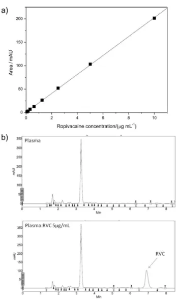
The calibration curve for determination of plasma ropivacaine (Figure 1a) was linear in the concentration range 0.030-10 µg mL−1 (R² = 0.9998), showing that the HPLC
procedure was suficiently sensitive to quantify ropivacaine in plasma. The concentration of RVC was determined using the equation: peak area = 20.21[RVC] + 0.50 (n = 3). The limit of detection of ropivacaine in plasma, determined as described by ICH guidelines (2005),22
was 0.030 µg mL−1. Its retention time was 7 min, and no
interference from other plasma components was observed (Figure 1b). Selectivity and sensitivity were similar to those previously reported by Kawata and co-workers.22
The detection limit for ropivacaine observed in our study (30 ng mL−1) was close to the limit observed by those
authors (25 ng mL−1).
Figure 1. a) Calibration curve of plasma concentration of ropivacaine and peak area as measured by HPLC (see Methods section); b) HPLC chromatogram of plasma and plasma with 5 µg mL−1 of RVC (HPLC
Mean plasma concentrations of RVC are plotted as a function of time in Figure 2 for both the liposomal RVC formulation, and for RVC with epinephrine. The pharmacokinetic parameters (Cmax, Tmax, AUC0–24, AUC0–∞, CL, t1/2 and Vd) were subsequently calculated (Table 1). No statistically signiicant differences (p > 0.05) were observed between the formulations for all of the pharmacokinetic parameters evaluated.
Kawata and co-workers22 studied the topical application
of 5 mL of 0.5% viscous ropivacaine, held in the mouths of only two volunteers for 10 min. They observed a Cmax of 107 (± 25.5) ng mL−1 and a T
max of 50 (± 14.1) min. Despite
methodological differences, these results were similar to those observed in the present study (Table 1).
Many substances are added to local anesthetics to improve their eficacy by modifying their pharmacodynamic
and pharmacokinetic properties, with epinephrine being the most commonly used. Lee and co-workers23 demonstrated
that the addition of epinephrine signiicantly reduced the concentration of ropivacaine after epidural anesthesia in humans during the irst hour, in both arterial and venous blood. Here, we have found no difference between the pharmacokinetic proiles of both formulations, showing that liposome encapsulation of ropivacaine was as effective as epinephrine in reducing ropivacaine absorption.
Several animal studies have also demonstrated that liposomal encapsulation of long-acting local anesthetics is able to alter their pharmacokinetic behavior, with lower plasma concentrations and toxicity compared to the plain solution.13,15,16,24
The use of a liposomal formulation instead of epinephrine containing local anesthetic could be an advantageous since, it was demonstrated that local anesthetic solutions containing sympathomimetic vasoconstrictors could promote changes in heart rate and blood pressure,25,26
and dysrrhythmias,27,28 increasing the risk of morbidity,
especially in cardiovascular patients when higher doses or more stressful procedures are being carried out.29
Despite differences in liposolubility, partition coeficient, and some other physico-chemical/pharmacokinetic parameters, ropivacaine and bupivacaine have some similarities, such as pka, protein binding and molecular weight. In addition, they have similar onset times and blocking durations when used in epidural blockade.2
No differences in anesthetic eficacy parameters were found between these two local anesthetics after maxillary iniltration.30
Table 1. Median pharmacokinetic parameters following maxillary iniltration of liposome-encapsulated 0.5% ropivacaine and 0.5% ropivacaine with 1:200,000 epinephrine.
Pharmacokinetic parameters Groups Median Quartiles
First Third p value
Cmax (ng mL−1)
liposome-encapsulated 0.5% ropivacaine 0.5% ropivacaine with 1:200,000 epinephrine
92.9 93.4 82.7 63.2 97.7 114.7 0.6378 Tmax (min)
liposome-encapsulated 0.5% ropivacaine 0.5% ropivacaine with 1:200,000 epinephrine
30.0 37.5 15.0 30.0 56.3 45.0 0.9645 AUC0-t (ng-min mL−1)
liposome-encapsulated 0.5% ropivacaine 0.5% ropivacaine with 1:200,000 epinephrine
40.4 32.4 26.3 20.1 55.2 44.0 0.6378
AUC0-∞ (ng-min mL−1)
liposome-encapsulated 0.5% ropivacaine 0.5% ropivacaine with 1:200,000 epinephrine
71.9 78.5 28.1 4.9 138.6 102.6 0.7794 Vd (mL kg−1)
liposome-encapsulated 0.5% ropivacaine 0.5% ropivacaine with 1:200,000 epinephrine
2.6 2.8 1.5 1.5 4.4 13.8 0.5754 T1/2 (min)
liposome-encapsulated 0.5% ropivacaine 0.5% ropivacaine with 1:200,000 epinephrine
869 868 349 142 1512 1498 0.9738 CL
(mL min−1 kg−1)
liposome-encapsulated 0.5% ropivacaine 0.5% ropivacaine with 1:200,000 epinephrine
0.0013 0.0017 0.001 0.0009 0.0029 0.0041 0.8182
Grant and co-workers13 compared 0.5% plain
bupivacaine with 2% liposomal bupivacaine, and even with a 4-fold higher concentration of bupivacaine in the liposomal formulation the plasmatic levels of bupivacaine decreased when the liposomal formulation was used for wound analgesia in rats. In the present study, the pharmacokinetics of liposome-encapsulated ropivacaine was comparable to that of epinephrine-associated ropivacaine, suggesting the same proile observed by Grant and co-workers13 with encapsulation into liposome vesicles
delaying transfer of the anesthetic into the blood. According to Grant and Bansinath31 liposome
composition affects the release kinetics of encapsulated drugs. Drugs tend to be released more rapidly from liposomes composed of a single lipid bilayer, while the release tends to be retarded from multilamellar vesicles.13,15 In our study, unilamellar vesicles were able
to delay ropivacaine absorption, since both formulations presented similar pharmacokinetic proiles. Further studies are necessary to evaluate how the changes in liposome composition affect both the absorption of ropivacaine from the injection site and its plasmatic concentration after dental anesthesia.
Another factor that could maintain a constantly low plasma concentration for hours, resulting in a prolonged effect, is the percentage of encapsulated drug.32 According
to a previous study6 that used the same liposome employed
in the present study, the encapsulation efficiency of ropivacaine was 24%, while other reports in the literature have given higher encapsulation eficiency values.16,33,34
Ostergaard and co-workers35 showed that ropivacaine had
lower liposome afinity than bupivacaine. De Araújo and co-workers6 also suggested that enhancement of liposome
encapsulation could prolong the analgesic effect and decrease cytotoxicity.
Conclusions
In conclusion, the present work demonstrates that the HPLC technique can be used to quantify RVC in plasma samples during pharmacokinetic experiments. Results showed that liposome-encapsulated ropivacaine had a pharmacokinetic proile that was similar to that of ropivacaine associated with epinephrine, suggesting that liposomal formulations could be a safer alternative during clinical use of this local anesthetic.
Acknowledgments
This study was supported by Fundação de Amparo à Pesquisa do Estado de São Paulo (FAPESP #06/00121-9).
M. Franz-Montan acknowledges a fellowship received from FAPESP (#06/53255-2).
References
1. Marković, A. B.; Todorović, L.; Oral Surg. Oral Med. Oral Pathol. Oral Radiol. Endod. 2006, 102, 4.
2. Leone, S.; Di Cianni, S.; Casati, A.; Fanelli, G.; Acta Biomed.
2008,79, 92.
3. Malamed, S. F.; Handbook of Local Anesthesia, 5th ed., St. Louis: Mosby, 2004.
4. Gaffen, A. S.; Haas, D. A.; J. Can. Dent. Assoc. 2009, 75, 649. 5. Kaufman, E.; Garfunkel, A.; Findler, M.; Elad, S.; Zusman,
S. P.; Malamed, S. F.; Galili, D.; Refuat Hapeh Vehashinayim
2002, 19, 13.
6. de Araujo, D. R.; Cereda, C. M.; Brunetto, G. B.; Vomero, V. U.; Pierucci, A.; Neto, H. S.; de Oliveira, A. L.; Fraceto, L. F.; Braga, A. de F.; de Paula, E.; J. Pharm. Pharmacol. 2008, 60, 1449.
7. Gesztes, A.; Mezei, M.; Anesth. Analg.1988, 67, 1079. 8. Boogaerts, J.; Declercq, A.; Lafont, N.; Benameur, H.; Akodad,
E. M.; Dupont, J. C.; Legros, F. J.; Anesth. Analg.1993, 76, 553.
9. Boogaerts, J. G.; Lafont, N. D.; Declercq, A. G.; Luo, H. C.; Gravet, E. T.; Bianchi, J. A.; Legros, F. J.; J. Clin. Anesth.1994,
6, 315.
10. Cereda, C. M.; Tófoli, G. R.; de Brito Junior, R. B.; de Jesus, M. B.; Fraceto, L. F.; Groppo, F. C.; de Araujo, D. R.; de Paula, E.; J. Liposome Res.2008,18, 329.
11. Langer, R.; Science1990, 249, 1527.
12. Grant, G. J.; Vermeulen, K.; Langerman, L.; Zakowski, M.; Turndorf, H.; Reg. Anesth.1994, 19, 264.
13. Grant, G. J.; Lax, J.; Susser, L.; Zakowski, M.; Weissman, T. E.; Turndorf, H.; Acta Anaesthesiol. Scand.1997, 41, 204. 14. Malinovsky, J. M.; Benhamou, D.; Alafandy, M.; Mussini, J.
M.; Coussaert, C.; Couarraze, G.; Pinaud, M.; Legros, F. J.;
Anesth. Analg. 1997, 85, 1331.
15. Yu, H. Y.; Li, S. D.; Sun, P.; J. Pharm. Pharmacol. 2002, 54, 1221.
16. Grant, G. J.; Piskoun, B.; Bansinath, M.; Clin. Exp. Pharmacol. Physiol. 2003, 30, 966.
17. Mowat, J. J.; Mok, M. J.; MacLeod, B. A.; Madden, T. D.;
Anesthesiology1996, 85, 635.
18. Boogaerts, J. G.; Lafont, N. D.; Carlino, S.; Noel, E.; Raynal, P.; Gofinet, G.; Legros, F. J.; Br. J. Anaesth. 1995,75, 319. 19. Eickbohm, J. E.; Wulf, H.; Hoffmann, C.; Becker, C.; Dtsch.
Zahnarztl. Z. 1991, 46, 812.
20. ICH International Conference on Harmonization, Validation of Analytical Procedures: Text and Methodology (Q2R1), Geneva, 2005.
22. Kawata, T.; Homma, M.; Kakiuchi, Y.; Inomata, S.; Miyabe, M.; Kobayashi, D.; Morimoto, Y.; Kohda, Y.; Biol. Pharm. Bull.
2005, 28, 2271.
23. Lee, B. B.; Ngan Kee, W. D.; Plummer, J. L.; Karmakar, M. K.; Wong, A. S.; Anesth. Analg.2002,95, 1402.
24. Boogaerts, J. G.; Lafont, N. D.; Luo, H.; Legros, F. J.; Can. J. Anaesth. 1993, 40, 1201.
25. Goldstein, D. S.; Dionne, R.; Sweet, J.; Gracely, R.; Brewer, H. B. Jr.; Gregg, R.; Keiser, H. R.; Psychosom. Med.1982, 44, 259.
26. Mochizuki, M.; Yokota, S.; Murata, Y.; Watanabe, H.; Nishibori, M.; Suzuki, N.; Nishibori, M.; Kubota, Y.; Anesth. Prog.1989,
36, 234.
27. Hughes, C. L.; Leach, J. K.; Allen, R. E.; Lambson, G. O.;
J. Am. Dent. Assoc. 1966, 73, 1095. 28. Ryder, W.; Anaesthesia1970, 25, 46.
29. Elad, S.; Admon, D.; Kedmi, M.; Naveh, E.; Benzki, E.; Ayalon, S.; Tuchband, A.; Lutan, H.; Kaufman, E.; Oral Surg. Oral Med. Oral Pathol. Oral Radiol. Endod. 2008, 105, 725.
30. Kennedy, M.; Reader, A.; Beck, M.; Weaver, J.; Oral Surg. Oral Med. Oral Pathol. Oral Radiol. Endod. 2001, 91, 406. 31. Grant, G. J.; Bansinath, M.; Reg. Anesth. Pain. Med. 2001, 26,
61.
32. Barenholz, Y.; J. Liposome Res.2003, 13, 1.
33. Grant, G. J.; Barenholz, Y.; Piskoun, B.; Bansinath, M.; Turndorf, H.; Bolotin, E.M.; Pharm. Res.2001, 18, 336. 34. Grant, G. J.; Barenholz, Y.; Bolotin, E. M.; Bansinath, M.;
Turndorf, H.; Piskoun, B.; Davidson, E. M.; Anesthesiology
2004, 101, 133.
35. Ostergaard, J.; Jorgensen, L.; Engelbrecht Thomsen, A.; Weng Larsen, S.; Larsen, C.; Jensen, H.; Electrophoresis 2008, 29, 3320.
Submitted: January 28, 2010
Published online: July 1, 2010