A New Di-
µ
-sulfate Complex as a Model of Purple Acid
Phosphatase-Sulfate Complexes
Marcos A. de Britoa, Ademir Nevesa*, Ivo Vencatoa, César Zuccoa, Valderes Dragob, Klaus Griesarc, and Wolfgang Haasec
a
Laboratório de Bioinorgânica e Cristalografia-LABIN, Departamento de Química, Universidade Federal de Santa Catarina, 88040-900 Florianópolis - SC, Brazil
b
Departamento de Física, Universidade Federal de Santa Catarina, 88040-900 Florianópolis - SC, Brazil
c
Institut für Physikalische Chemie, Technische Hochschule Darmstadt, D-6100 Darmstadt, Germany
Received: February 17, 1997
Reportamos neste trabalho, a síntese, a estrutura cristalina e as propriedades espectroscópicas e eletroquímicas do complexo NH4[Fe2III(bbpmp)(µ-SO4)2] 1 onde bbpmp é o ânion do ligante
2,6-bis[(2-hidróxibenzil)(2-metilpiridil)-amino-metil]-4-metilfenol. Este representa o primeiro exemplo de um complexo contendo a unidade estrutural (µ-fenolato)(µ-SO4)2 e um ligante N,O-doador de relevância, como um análogo sintético para os derivados sulfatos das fosfatasas ácidas púrpuras.
The synthesis, X-ray crystal structure, electrochemical and spectroscopic properties of NH4[Fe2III(bbpmp)(µ-SO4)2] 1 where bbpmp is the anion of 2,6-bis[(2-hydroxy-
benzyl)(2-pyridyl-methyl)-amino-methyl]-4-methylphenol, are reported; this is the first example of a (µ-phenolate)(µ -SO4)2 complex, with a relevant N,O-donor ligand, as a synthethic analogue for the purple acid phosphatases-sulfate complexes.
Keywords: purple acid phosphatases, oxyanion complexes, sulfate model complex, crystal structure
Introduction
The interaction of the active site of purple acid phos-phatases (PAPs) with sulfate and other perturbants has been described1,2. This oxyanion is known to be able to interact
with the reduced enzymes, which in the presence of air form the oxidized PAPox-sulfate complexes. In a recent report,
Witzel3 has shown that the addition of SO
42- to the oxidized
form (PAPox, λmax = 558 nm) leads to the immediate
for-mation of an enzyme-sulfate complex (PAPox-sulfate) with
an absorption maximum at 546 nm. In order to gain more information about the binding mode of PAPs with small bridging oxyanions we describe here the synthesis, crystal structure and properties of a novel Fe2III synthetic analogue
which contains the FeIII(λ-SO
4)2FeIII unit with a
biologi-cally relevant N,O-donor dinucleating ligand. This work is a continuation of a wide research program for the prepara-tion and characterizaprepara-tion of iron complexes with bioinor-ganic interest4-8.
Experimental
Syntheses
The ligand
2,6-bis[(2-hydroxybenzyl)(2-pyridyl-methyl)-amino-methyl]-4-methylphenol (H3bbpmp) was
prepared as described elsewhere5,7. The NH
4[Fe2III(bbpmp)
(µ-SO4)2] complex was prepared as follows. To a solution
of [FeIIFeIII(bbpmp)(OAc)
2].4H2O6 (0.87 g, 1 mmol) in 20 Note
J. Braz. Chem. Soc., Vol. 8, No. 5, 443-446, 1997. © 1997 Soc. Bras. Química
mL of CH3CN, was added (NH4)2S2O8 (0.23 g, 1 mmol)
and 10 mL of water at room temperature. The clear deep blue solution was heated to 40 °C and stirred for 15 min at ambient atmosphere. After cooling the solution to room temperature, a violet microcrystalline precipitate was formed. Single crystals suitable for X-ray crystallography were obtained by recrystallization from a methanolic solu-tion of 1. Anal. Calc. for C35H37N5O11Fe2S2: C, 47.77; H,
4.24; N, 7.96%. Found: C, 47.82; H, 4.28; N,8.02%.
X-ray crystal structure determination of 1
Crystal data for 1. [C35H37N5S2O11Fe2], M = 879.53:
trigonal, P31, (No. 144),a = 19.406(4), b = 19.406(4), c =
10.759(4), V = 3509(2) Å3 , Z = 3. D
calc = 1.249 g cm-3.
Crystal dimensions 0.10 x 0.15 x 0.35 mm, Mo-Kα (λ =
0.71073 Å); T = 293 K. Enraf-Nonius CAD-4 diffractome-ter. Data were reduced using the MOLEN software9. The
structure was solved with SIR 92 and refined anisotropi-cally for Fe and S atoms. Other non-H atoms were refined isotropically due to the low number of observed reflections ratio to refined parameters.
All the H atoms were placed at geometrically calculated positions except those of the NH4+ ion that were not found.
It was assigned to them an isotropic temperature factor of 1.3 times the isotropic temperature factor of the atom to which they were attached; µ = 0.756 mm-1; 14944 mesured
reflections with 7942 unique reflections; 1678 with I > 3σ
(I); 241 least-square parameters; R = 0.0826 (Rw = 0.0976).
Results and Discussion
The new dinuclear complex [FeIII(bbpmp)(SO 4)2]- has
been generated in CH3CN solution (λmax = 587nm/ε =
8430M-1cm-1/Fe
2) via oxidation/substitution reactions of
the mixed-valence [FeIIFeIII(bbpmp)(CH
3COO)2]7 (λmax =
540 nm/ε = 4840 M-1 cm-1/ Fe
2) by using aqueous
per-oxodisulfate and the spectral change is shown in Fig. 1. The maintenance of isosbestic points in successive spectra cor-roborates the presence of a single product throughout the
course of the reactions. The bands are assigned to pheno-late-to-FeIII charge transfer transitions and complex 1 is
blue shifted compared to [FeIIIFeIII(bbpmp)(CH
3COO)2]+ 2 (λmax = 601 nm/ ε = 7700 M-1 cm-1/ Fe2)5,7 and red shifted
compared to the enzyme PAPox-sulfate (λmax = 546 nm) 3.
These observations are consistent with the stronger FeIII-O
interaction of the terminal phenolate groups in 2 (av. FeIII-O
= 1.855(8) Å), compared to 1.93(2) Å in 1.
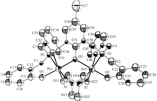
The structureof 1 (Fig. 2) reveals discrete diiron
com-plex anions and ammonium counterions. The iron atoms in the anion of 1 are in a pseudo-octahedral environment in
which the two terminal phenolate oxygen atoms coordinate
trans to the bridging phenolate group. This arrangement of
the ligand around the Fe(3) centers is very similar to those observed in the closely related [Fe2III(bbpmp) (CH3COO)2]
ClO4.H2O5,7, 2 and [Fe2III(bbpmp)(O2P(OPh)2)2]ClO4.
H2O10 [OP(OPh)2 = diphenylphosphate] complexes, but
with some significant differences in their bond lengths and angles. The Fe-O average distances within the bridging phenolate group increase from 2.055(8) Å in 2 to 2.07(2)
Å in 1 to 2.13(2) Å in Krebs’s complex and are significantly
larger than the corresponding Fe(III)-O(phenolate) bond
length observed in the mixed-valence [FeIIFeIII(bpmp)
(OPr)2] [BPh4]2 complex11 (1.943(2) Å) where bpmp is the
anion of 2,6-bis[bis(2-pyridylmethyl)aminomethyl]-4-methylphenol. This is a reflection of the short terminal Fe3-O
(phenolate) (av. 1.93(2), 1.855(2), and 1.853(2) Å) bond
lengths observed in 1, 2, and in the diphenylphosphate
complex, respectively, which are coordinated in trans
po-sitions relative to the bridging phenolate group. Further-more, it is important to note that the Fe1-O3-Fe2 bridging angle of 121.7(9)° in 1 is somewhat larger than the 118.3(4)
value observed for 2, but it is significantly smaller than the
128.2(4)° detected in [Fe2III(bbpmp)(O2(OPh)2)2] ClO4.
H2O10. Consequently, the Fe1...Fe2 distance of 3.624(6) Å
for 1 is slightly longer (0.096 Å) when compared to that
observed for 2, but is significantly shorter (0.21 Å) than in
the diphenylphosphate complex10. These facts can be
cor-related with the increasing O...O separation in phosphate and sulfate compared to the carboxylate bridging ligands. Similar data were reported by Wieghardt et al.12 for the
monophenylphosphate, sulfate and acetate diiron(III) com-plexes with N,N’,N’’-trimethyl-1,4,7-triazacyclononane as ligand. To our knowledge, 1 represents the first example
of an structurally characterized FeIII(µ-phenolate)(µ
-SO4)2FeIII unity with a biologically relevant N,O-donor
dinucleating ligand.
The oxidation states of the iron centers in 1 are
sup-ported by the Mössbauer spectrum at 115 K and the follow-ing parameters: isomer shift (relative to metallic iron with the source at room temperature) δ, 0.51 mm/s and quadru-pole splitting, ∆EQ, 0.87 mm/s which indicates the presence
of high-spin Fe2III centers13.
444 Brito et al. J. Braz. Chem. Soc.
Figure 1. Spectral change during the conversion of [FeIIFeIII(bbpmp) (CH3COO)2], in CH3CN-H2O/(NH4)2S2O8 solution, into [Fe2III(bbpmp)
The magnetic data for a powder sample of 1 were
collected in the temperature range of 5.1 to 300 K and indicate a weak antiferromagnetic coupling interaction for the two Fe3 ions in the complex. The data were fitted by
using the expression for the molar susceptibility vs.
tem-perature from the spin-exchange Hamiltonian H = -2JS1S2
(S1 = S2 = 5/2)14 and the following parameters: g = 2.00
(fixed); % imp = 5.5; θ = -3.5 K; TIP = 400 x 10-6 cm3/ mol; J = -6.4 cm -1 . This J value is very similar to those detected
for the complexes [Fe2III(bbpmp)(CH3COO)2]ClO4.
H2O5,7 and [Fe2III(bbpmp)(O2P(OPh)2)2]ClO4.H2O10,
de-spite the structural differences detected in these complexes. The electrochemical properties of 1 were investigated
by cyclic voltammetry in acetonitrile with [Bu4N][PF6] as
the supporting electrolyte. A quasi-reversible wave is ob-served at -1.29 V vs. Fc/Fc which is ascribed to the
Fe2III/FeIIFeIII redox couple. The corresponding couple in 2, is observed to occurs at -0.57 V vs. Fc+/Fc5,7 and, as
expected, the substitution of two acetate by sulfate groups,
shifts the redox couple to a more negative potential. We have synthesized and characterized 1 to serve as a
synthetic analogue for PAPox-sulfate complexes but, to our
knowledge, there are very few informations in the literature on the corresponding PAPox complexes to make further
comparisons1,3. On the other hand, due to the presence of
two terminal phenolate groups in 1 and based on the redox
potential reported for uteroferrin (Eo’ = -0.03 V vs. Fc/Fc at
pH 5)15 , one should expect a less negative redox potential
for the PAPo-sulfate complex compared to 1.
Finally, further preparative, structural and physico-chemical studies on the XO42- (X = Cr, Mo) complexes are
in progress in our laboratory, and will be the subject of a full paper.
Supplementary Material
The following tables are available from the authors on request: complete table of crystal data, positional parame-ters, bond distances, bond angles, hydrogen atoms coordi-nates, displacement parameters (12 pages), and list of observed and calculated structure factors (79 pages).
Acknowledgments
This work was supported by grants from PRONEX, CNPq, PADCT, FINEP (Brazil) and KFA (Germany).
References
1. Doi, K.; Antanaitis, B.C.; Aisen, P. Struct. Bonding (Berlin)1988, 70, 1.
Vol. 8, No. 5, 1997 New Di-µ-sulfate Complex as a Model of Purple Acid Phosphatase-Sulfate Complexes 445
2. Vincent, J.B.; Crowder, M.W.; Averill, B.A.; Bio-chemistry1992, 31, 3033.
3. Dietrich, M.; Münstermann, D.; Suerbaum, H.; Witzel, H. Eur. J. Biochem. 1991, 199, 105.
4. Neves, A.; Erthal, S.M.D.; Drago, V.; Griesar, K.; Haase, W. Inorg. Chim. Acta 1992,197, 121.
5. Neves, A.; de Brito, M.A.; Vencato, I.; Drago, V.;
Griesar, K.; Haase, W.; Mascarenhas, Y.P. Inorg.
Chim. Acta 1993, 214, 5.
6. Neves, A.; de Brito, M.A.; Drago, V.; Griesar, K.; Haase, W. Inorg. Chim. Acta 1995, 237, 131.
7. Neves, A.; de Brito, M.A.; Vencato, I.; Drago, V.; Griesar, K.; Haase, W. Inorg. Chem. 1996, 35, 2360.
8. de Brito, M.A.; Neves, A.; Zilli, L.R. Química Nova,
1997, 20(2), 154.
9. Fair, C.K. In MOLEN. An Interactive Intelligent Sys-tem for Crystal Structure Analysis. Enraf-Nonius,
Delft, Netherlands, 1990.
10. Krebs, B.; Schepers, K.; Bremer, B.; Henkel, G.; Althaus, E.; Müller-Warmurth, W.; Griesar, K.; Haase, W. Inorg. Chem.1994, 33, 1907.
11. Borovik, A.S.; Papaefthymiou, V.; Taylor, L.F.; An-derson, O.P.; Que Jr., L. J. Am. Chem. Soc. 1989, 111,
6183.
12. Drüke, S.; Wieghardt, K.; Nuber, B.; Weiss, J.; Fleischhauer, H-P.; Gehring, S.; Haase, W. J. Am. Chem. Soc. 1989, 111, 8622. Wieghardt, K.; Drüke,
S.; Chaudhuri, P.; Flörke, U.; Haupt, H-J.; Nuber, B.; Weiss, J. Z. Naturforsch. 1989446, 1093. Hartman,
J.R.; Rardin, R.L.; Chaudhuri, P.; Pohl, K.; Wieghardt, K.; Nuber, B.; Weiss, J.; Papaefthymiou, G.C.; Frankel, R.B.; Lippard, S.J. J. Am. Chem. Soc.
1987, 109, 7387.
13. Greenwood, N.N.; Gibb, T. C. Mössbauer Spectros-copy, Chapman and Hall, London, 1971, 113-168.
14. O’Connor, C.J. Prog. Inorg. Chem. 1982, 29, 203.
15. Wang, D.L.; Holz, R.C.; David, S.S.; Que Jr., L.; Stankovich, M.T. Biochemistry1991, 30, 8187.
![Figure 1. Spectral change during the conversion of [Fe II Fe III (bbpmp) (CH 3 COO) 2 ], in CH 3 CN-H 2 O/(NH 4 ) 2 S 2 O 8 solution, into [Fe 2 III (bbpmp) (SO 4 ) 2 ] - ( 1 ) with time intervals of 90 s.](https://thumb-eu.123doks.com/thumbv2/123dok_br/18987803.459500/2.918.99.444.824.1030/figure-spectral-change-conversion-bbpmp-solution-bbpmp-intervals.webp)