© 2014 Sociedade Brasileira de Hemodinâmica e Cardiologia Intervencionista. Published by Elsevier Editora Ltda. All rights reserved.
Very Late Stent Thrombosis by Angiography.
A Case of Atherosclerosis Progression by OCT
Antonio de Castro-Filho, Daniel Chamié, Alexandre Abizaid
ABSTRACT
The progressive nature of coronary atherosclerotic disease is often neglected in patients submitted to percutaneous coronary intervention. Very late (> 1 year) myocardial infarctions affect-ing the treated myocardial territory are usually attributed to device related complications. We report the case of a patient with acute inferior wall ST-elevation myocardial infarction, who had a thrombotic occlusion of a bare-metal stent implanted 8 years before. Despite the angiographic diagnosis of very late stent thrombosis, optical coherence tomography revealed that the acute myocardial infarction was caused by rupture of an atherosclerotic plaque outside of the previously stented segment.
DESCRIPTORS: Myocardial infarction. Percutaneous coronary intervention. Stents. Tomography, optical coherence. Coronary thrombosis.
Instituto Dante Pazzanese de Cardiologia, São Paulo, SP, Brazil. Correspondence to: Daniel Chamié. Serviço de Cardiologia Invasiva do Instituto Dante Pazzanese de Cardiologia. Avenida Dr. Dante Pazza-nese, 500 − Vila Mariana − CEP: 04012-180 − São Paulo, SP, Brazil E-mail: [email protected]
Received on: 9/1/2014 • Accepted on: 11/25/2014
RESUMO
Trombose Muito Tardia de Stent pela Angiografia. Um Caso de Progressão da Aterosclerose pela OCT
Em pacientes submetidos à intervenção coronária percutânea, a natureza progressiva da doença coronária aterosclerótica é frequentemente negligenciada. Geralmente, infartos muito tardios (> 1 ano) acometendo o território tratado são atribuídos a complicações relacionadas ao dispositivo. Apresentamos o caso de uma paciente com infarto agudo do miocárdio com supradesnivelamento do segmento ST na parede inferior, que apresentava oclusão trombótica de um stent não farmacológico implantado 8 anos antes. Apesar do diagnóstico angiográico de trombose muito tardia, a tomograia de coerência óptica revelou que a etiologia foi a ruptura de placa aterosclerótica no leito distal, fora do segmento previamente tratado.
DESCRITORES: Infarto do miocárdio. Intervenção coronária percutânea. Stents. Tomograia de coerência óptica. Trombose coronária.
T
he introduction of coronary stents, especially the drug-eluting, represented a great advance in the percutaneous treatment of coronary artery disease, making this intervention one of the therapeutic mo-dalities most often performed in medicine.1 However,in the mid-2000’s, reports of a higher incidence of late thrombosis after implantation of irst-generation drug-eluting stents gave rise to a series of debates on the long-term safety of these devices. In order to standardize the deinitions and provide consensus recommendations for the classiication of clinical out-comes after percutaneous coronary intervention (PCI), a document was published in 2007 by the Academic
Research Consortium (ARC).2 In brief, adverse events
occurring in a previously treated area are attributed to the implanted device, until another clear cause is documented. In the speciic case of stent thrombosis, the ARC provides the highest level of evidence for the diagnosis when there is angiographic documentation of thrombotic occlusion of a stent implanted in a vessel responsible for a territory under acute ischemia – the so-called deinite stent thrombosis.
In this report, the authors present a case of a patient with acute myocardial infarction (AMI) with ST-segment elevation, in which the angiographic diagnosis of very
June 2014, she was admitted to the emergency room of this hospital with typical chest pain, which had started at rest 2.5 hours before admission. Peripheral perfusion was adequate and there were no signs of pulmonary congestion. ECG showed ST-segment elevation in the inferior lateral and posterior walls (Figure 1).
The patient was treated with 300 mg acetylsalicylic acid, 600 mg of clopidogrel, and 5,000 units of un-fractionated heparin administered intravenously, being promptly referred to the cardiac catheterization laboratory.
Coronary angiography was performed through the right radial access route, using a 6-F hydrophilic arterial sheath. The left coronary artery showed discreet parietal irregularities, with no obstructions (Figures 2A to 2C). The RCA was dominant, showed a stent in the proximal third that was patent, with diffuse hyperplasia and 40% luminal obstruction in its narrowest point, followed by occlusion with contrast retention inside another stent located in the distal third (Figure 2D),demonstrating deinite and very late stent thrombosis.2 The left ventricle
showed moderate inferior hypokinesis (medio-basal) (Figures 2E and 2F).
After positioning the 0.014-inch guide wire on the RCA distal bed, the Export® thromboaspiration catheter
(Medtronic Inc., Minneapolis, USA) was passed multiple times, covering the entire distal segment of the vessel, removing large amounts of thrombus and restoring Thrombolysis in Myocardial Infarction (TIMI 3) distal coronary blood low. After administration of intracoro-nary nitroglycerin (200 μg), control angiography showed severe stenosis after the distal stent border, followed by
due to the presence of residual thrombus; (3) identify adequate reference sites for stent positioning; (4) ac-curately measure the target-vessel size for better selec-tion of devices to be implanted – the assessed segment showed large caliber disproportion between the ectatic and proximal and distal adjacent segments.
The OCT images were acquired using the commer-cially available Frequency Domain system (C7 XR®; St.
Jude Medical, St. Paul, United States) during intracoronary injection of Hexabrix® ionic low-osmolarity contrast
(Guerbet, Bloomington, United States) using a catheter guide with the help of a pump programmed to inject contrast at a constant speed of 3 mL/s for 3 seconds.
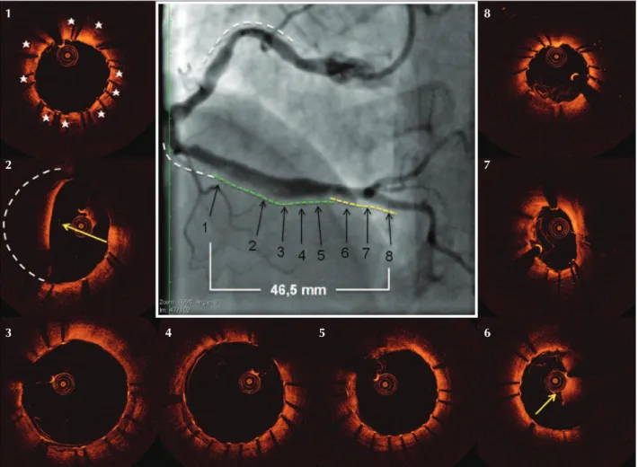
In the OCT images, adequate stent healing was observed, with complete coverage of the struts by tis-sue that had normal optical characteristics (Figure 3, panel 1). However, distal to the stent, a long segment with atherosclerotic involvement, of complex and heterogeneous composition, was observed. Eight mil-limeters after the stent distal border, it was observed the presence of ibrotic, focal plaque, promoting severe stenosis with minimal luminal area of 1.34 mm2 (Figure
3, panel 2). At the entrance of the ectatic segment, the presence of a lipid-rich plaque was observed, with ec-centric distribution and ibrous cap rupture (Figure 3, panel 3). The ectatic region had a mean diameter of 5 mm, almost normal morphology, and the presence of small eccentric ibrous plaque (Figure 3, panel 4). At the segments distal to the ectasia, the presence of a long lipid plaque that extended to the bifurcation was observed, with three additional sites of ibrous cap rupture (Figure 3, panels 5-7) and presence of
Figure 1 – Electrocardiogram at admission. ST-segment elevation is observed in the inferior, lateral, and posterior walls.
residual thrombus in the carina (Figure 3, panel 8). The bifurcation showed an eccentric ibrotic plaque with luminal area of 2.21 mm2 (Figure 3, panel 9), while
the proximal third of the posterior descending branch was normal (Figure 3, panel 10).
After intravenous bolus administration of abciximab (0.25 mg/kg), the authors chose to treat the entire af-fected segment, covering from the bifurcation to the distal edge of the stent. Based on the measurements obtained by OCT, a 2.5 × 18 mm zotarolimus-eluting Endeavor Sprint® stent (Medtronic Inc., Minneapolis,
United States) was implanted, positioned in the RCA towards the proximal third of the posterior descending branch, covering the bifurcation with the posterior ven-tricular branch (Figure 4A). This stent was post-dilated at its proximal portion using a 3.0 × 12 mm noncom-pliant balloon, inlated with high pressure (20 atm; Figure 4B), followed by post-dilation of the posterior ventricular branch with a 2.0 × 8 mm noncompliant balloon, inlated to 16 atm (Figure 4C). The implant optimization at the bifurcation was performed with simultaneous inlation of kissing-balloons (Figure 4D).
A second 4.0 × 30 mm Endeavor Sprint® stent was
im-planted proximally to obtain minimal overlap with the aforementioned stent and the previously existing stent (Figure 4E). Focal post-dilation with a 5.0 × 12 mm noncompliant balloon was performed only in the stent region positioned under the ectasia, aiming at adequate strut apposition (Figure 4F). Control angiography showed residual stenosis < 5%, hypertransparent image inside the treated segment, and TIMI 3 distal low (Figure 5).
OCT assessment (Figure 5) showed adequate ex-pansion and apposition of stent struts along the treated segment, including the ectatic region. However, the prolapse of a large amount of thrombotic material through the stent struts was observed in the region immediately proximal to the ectatic segment, affecting a short extension of 1.5 mm. Full coverage of the le-sion was also conirmed, with minimal overlap of the recently implanted 4.0 × 30 mm stent struts on those of the previously existing stent, as well as adequate opening of the bifurcation at the posterior ventricular branch level, and absence of dissections in the posterior descending branch borders.
Figure 2 – Coronary angiography. Left main coronary artery with no signiicant obstructions (A, B, and C). Right coronary artery shows a thrombus inside the stent positioned in the distal third (D). Left ventriculography (E and F) show moderate basal and mid-inferior wall hypokinesis. Dotted lines indicate the sites of previously implanted stents.
A
D
B
E
C
The electrocardiogram performed approximately 30 minutes after the PCI showed complete resolution of ST-segment elevation and absence of electrically inactive area. Glycoprotein IIb/IIIa inhibitor was maintained for 12 hours after the procedure. The echocardiography per-formed on the second day post-PCI showed an ejection fraction of 60% and mild hypokinesia of the lower and infero-lateral walls. The patient remained asymptomatic and was discharged on the 7º day after AMI.
DISCUSSION
The association of AMI with angiographic docu-mentation of thrombotic occlusion of a stent placed in a coronary artery responsible for the supply of a myocardial segment consistent with the topography of infarction constitutes a criterion for the so-called deinite stent thrombosis, as well as having the highest speciicity for the diagnosis of this phenomenon.2 In the
absence of angiographic documentation, an AMI that Figure 3 – Optical coherence tomography. The central panels show the right coronary artery before (A) and after (B) low restoration. Thrombi removed during thromboaspiration (C). The stent in the distal third showed struts with homogeneous tissue coverage. The stent and lumen areas, indicated by red and blue contours, deine the neointimal tissue with normal optical appearance (1). After the stent distal edge, stenotic ibrotic plaque can be observed (2). At the “shoulder” of the stenosis, a predominantly lipid plaque can be observed, with ibrous cap rupture and residual thrombus (3). The following ectatic segment is the vessel region with normal appearance (4). Distal to this segment, a lipid plaque, with multiple ibrous cap rupture sites, can be observed (5-7). At the bifurcation, there is large amount of thrombus (8), followed by eccentric ibrotic plaque (9). The proximal part of the posterior descending branch shows concentric intimal thickening (10).
Figure 4 – Percutaneous coronary intervention. A 2.5 × 18 mm stent was implanted in the distal segment (A). This stent was post-dilated in its proximal part with a 3.0 × 12 mm noncompliant balloon (B). After the guide wires were repositioned, the posterior ventricular branch ostium was post-dilated with a 2.0 × 8 mm non-compliant balloon (C), followed by simultaneous balloon inlation in the posterior descending and posterior ventricular branches (D). Another 4.0 × 30 mm stent was implanted proximally, with minimal overlap with the previously implanted stent (yellow line) and the existing stent (white line) (E). Focal stent post-dilation, under the ectatic region, was performed with a 12 × 5.0 mm noncompliant balloon (F).
3 4 5 6 7 8
is related to acute ischemia documentation in a previ-ously treated territory, regardless of the time elapsed after PCI, establishes the diagnosis of probable stent thrombosis.2 Although these criteria aim to increase
the detection of adverse events related to previously implanted coronary stents, they cannot differentiate whether the event occurrence is really related to the device or if it is associated with the atherosclerotic disease progression outside the treated segment.
In this report, the authors present the case of a patient who developed AMI on the inferior wall eight years after being treated with the implantation of two bare-metal stents in the RCA. Although the coronary angiography showed arterial occlusion with a thrombus inside the previously implanted stent, the diagnosis of very late thrombosis was challenged by the indings obtained at the invasive assessment with OCT, which
showed that coronary thrombosis was due to the de-stabilization of a long segment of lipid-rich plaques located distally to the treated segment – and not to an accident related to the stent.
It should be kept in mind that coronary atheroscle-rosis is a diffuse, progressive disease and the treatment of a certain coronary stenosis does not change the natural history of atherosclerotic disease. Non-obstructive stenosis and/or those not causing ischemia in sites that are distant from the treated one at the time of the index procedure can progress in the future into high-grade stenosis with recurrent angina or destabilize, causing thrombosis and AMI.3 In an analysis of 1,228 patients
treated with bare-metal stents, Cutlip et al.4 demonstrated
that stent-related events predominated in the irst year after implantation, becoming considerably rarer between the second and ifth years, when events related to the Figure 5 – Control angiography and optical coherence tomography. The previously implanted stents (white lines) and the newly implanted stents (yellow and green lines) show minimal overlap of the struts and complete segmental coverage. Panel 1 shows the location of the overlapping struts with the previous stent (asterisks). Thrombotic material prolapse can be observed, which explains the hypertransparent image on angiography (2). The focal post-dilation with a 5.0 × 12 mm balloon showed appropriate expansion and strut apposition in the ectatic segment (3 and 4). Outside the ectatic segment, the stent accompanies the size of the vessel (5). Next to the bifurcation, another point of thrombotic material prolapse is ob-served (6). Adequate stent expansion and apposition were attained at the bifurcation (7) and at its distal edge in the proximal third of the posterior descending branch (8).
1
2
3 4 5 6
with the Taxus™ stent (11.2% vs. 20.4%; p < 0.0001), target-vessel revascularization rates outside the previ-ously treated segment were relatively constant, of approximately 2% a year between the second and ifth years of follow-up, and not signiicantly different between patients treated with Taxus™ or bare-metal stents (3.3% vs. 3.8%; p = 0.21), more consistent with the natural disease progression than a speciic stent-related effect.5
Although the coronary angiography is suficient, in most cases, to differentiate in-stent restenosis from atherosclerosis progression outside the treated segment in stable patients, this case illustrates the dificulty of taining the angiographic diagnosis in the setting of acute coronary thrombosis. Even after obtaining the TIMI-3 low with thromboaspiration, the angiographic luminogram did not allow the authors to establish the coronary thrombosis origin. The documentation of thrombotic occlusion inside the stent, together with a long segment of distal atherosclerotic disease, with areas of ectasia and stenosis, together with the considerable residual thrombus burden, constituted an additional confounding factor and highlighted the importance of diagnostic complementation with an invasive imaging method.
In this context, the OCT has a critical role that is superior to existing imaging methods. In addition to being the only imaging method capable of accurately assessing the degree of stent struthealing,6,7 OCT also
allows for an accurate characterization of the athero-sclerotic plaque components and their morphometric aspects. Particularly in the setting of acute coronary syndrome, OCT has high sensitivity (94%) and specii-city (92%) for lipid plaque detection,8 and is the only in
vivo imaging method capable of precisely quantifying the ibrous capthickness9 and detecting the presence of
inlammation with macrophage aggregates,10
morphologi-cal aspects that are crucial for characterization of the thin-cap ibroatheroma – identiied as the “vulnerable plaque”, which most frequently leads to rupture and acute coronary occlusion.11,12 With the help of the OCT,
it was veriied that the stent was healed, with all its struts homogeneously covered by tissue with normal optical characteristics. Nonetheless, a long (~ 30 mm) segment of lipid-rich plaques with thin ibrous cap and multiple rupture points along its trajectory was identiied, causing the formation of a large thrombotic burden, which accumulated upstream, occluding the coronary
stent borders. It is important to mention that the as-surance of adequate and consolidated healing of the stent implanted eight years before allowed the team to treat only the distal vessel segment, without covering the previous stent with a new stent, a practice that possibly would not have been performed depending only on the angiographic assessment.
During primary PCI in AMI, stent implantation with low/moderate inlation pressures and less use of post-dilation are common practices, used to minimize the occurrence of the no-relow phenomenon, which justiies, in part, the higher frequency of poor acute apposition in AMI, in comparison with PCI in stable patients.15 In this case,
CONFLICTS OF INTEREST
The authors declare no conlicts of interest.
FUNDING SOURCES
None declared.
REFERENCES
1. Windecker S, Kolh P, Alfonso F, Collet JP, Cremer J, Falk V, et al. 2014 ESC/EACTS Guidelines on myocardial revascular-ization: The Task Force on Myocardial Revascularization of the European Society of Cardiology (ESC) and the European Association for Cardio-Thoracic Surgery (EACTS) Developed with the special contribution of the European Association of Percutaneous Cardiovascular Interventions (EAPCI). Eur Heart J. 2014;35(37): 2541-619.
2. Cutlip DE, Windecker S, Mehran R, Boam A, Cohen DJ, van Es GA, et al. Clinical end points in coronary stent trials: a case for standardized deinitions. Circulation. 2007;115(17):2344-51. 3. Pisterer ME, Zellweger MJ, Gersh BJ. Management of stable
coronary artery disease. Lancet. 2010;375(9716):763-72. 4. Cutlip DE, Chhabra AG, Baim DS, Chauhan MS, Marulkar S,
Massaro J, et al. Beyond restenosis: ive-year clinical outcomes from second-generation coronary stent trials. Circulation. 2004;110(10):1226-30.
5. Leon MB, Allocco DJ, Dawkins KD, Baim DS. Late clinical events after drug-eluting stents: the interplay between stent-related and natural history-driven events. JACC Cardiovasc Interv.2009;2(6):504-12.
6. Murata A, Wallace-Bradley D, Tellez A, Alviar C, Aboodi M, Sheehy A, et al. Accuracy of optical coherence tomography in the evaluation of neointimal coverage after stent implantation. JACC Cardiovasc Imaging. 2010;3(1):76-84.
7. Templin C, Meyer M, Muller MF, Djonov V, Hlushchuk R, Dimova I, et al. Coronary optical frequency domain imaging (OFDI) for in vivo evaluation of stent healing: comparison with light and electron microscopy. Eur Heart J. 2010;31(14):1792-801. 8. Yabushita H, Bouma BE, Houser SL, Aretz HT, Jang IK, Schlendorf
KH, et al. Characterization of human atherosclerosis by optical coherence tomography. Circulation. 2002;106(13):1640-5. 9. Kume T, Akasaka T, Kawamoto T, Okura H, Watanabe N, Toyota
E, et al. Measurement of the thickness of the ibrous cap by optical coherence tomography. Am Heart J. 2006;152(4):755 e1-4.
10. Tearney GJ, Yabushita H, Houser S, Aretz HT, Jang IK, Schlendorf KH, et al. Quantiication of macrophage content in atherosclerotic plaques by optical coherence tomography. Circulation.2003;107(1):113-9.
11. Virmani R, Kolodgie FD, Burke AP, Farb A, Schwartz SM. Lessons from sudden coronary death: a comprehensive morphological classiication scheme for atherosclerotic lesions. Arterioscler Thromb Vasc Biol. 2000;20(5):1262-75.
12. Virmani R, Burke AP, Farb A, Kolodgie FD. Pathology of the unstable plaque. Prog Cardiovasc Dis. 2002;44(5):349-56. 13. Little WC, Constantinescu M, Applegate RJ, Kutcher MA,
Bur-rows MT, Kahl FR, et al. Can coronary angiography predict the site of a subsequent myocardial infarction in patients with mild-to-moderate coronary artery disease? Circulation. 1988;78(5 Pt 1):1157-66.
14. Falk E, Shah PK, Fuster V. Coronary plaque disruption. Circula-tion. 1995;92(3):657-71.