Jose Geraldo MillI Karina PintoII
Rosane Härter GriepIII Alessandra GoulartIV Murilo FoppaV Paulo LotufoIV Marcelo K MaestriVI Antonio Luiz RibeiroVII Rodrigo Varejão AndreãoVIII Eduardo Miranda DantasI Ilka OliveiraIX
Sandra C FuchsVI Roberto de Sá CunhaI Isabela M BensenorIV
I Departamento de Ciências Fisiológicas. Centro de Ciências da Saúde. Universidade Federal do Espírito Santo. Vitória, ES, Brasil II Instituto de Saúde Coletiva. Universidade
Federal da Bahia. Salvador, BA, Brasil III Laboratório de Educação em Ambiente e
Saúde. Instituto Oswaldo Cruz. Fundação Oswaldo Cruz. Rio de Janeiro, RJ, Brasil IV Centro de Pesquisa Clínica e
Epidemiológica. Hospital Universitário, Universidade de São Paulo. São Paulo, SP, Brasil
V Programa de Pós-Graduação em Ciências Cardiovasculares, Faculdade de Medicina, Universidade Federal do Rio Grande do Sul. Porto Alegre, RS, Brasil
VI Hospital de Clínicas de Porto Alegre. Universidade Federal do Rio Grande do Sul. Porto Alegre, RS, Brasil
VII Hospital das Clínicas. Departamento de Clínica Médica. Faculdade de Medicina. Universidade Federal de Minas Gerais. Belo Horizonte, MG, Brasil
VIII Coordenadoria de Engenharia Elétrica. Instituto Federal do Espírito Santo. Vitória, ES, Brasil
IX Departamento de Radiologia. Faculdade de Medicina. Universidade de São Paulo. São Paulo, SP, Brasil
Correspondence: José Geraldo Mill
Departamento de Ciências Fisiológicas Centro de Ciências da Saúde Universidade Federal do Espírito Santo Av. Marechal Campos, 1468 Maruípe 29042-755 Vitória, ES, Brasil E-mail: [email protected] Received: 10/17/2011 Approved: 7/12/2012
Article available from: www.scielo.br/rsp
Medical assessments and
measurements in ELSA-Brasil
ABSTRACT
The article describes assessments and measurements performed in the Brazilian Longitudinal Study for Adult Health (ELSA-Brasil). Some assessments including anthropometric assessment, casual blood pressure measurement, and ankle-brachial index have an established clinical application while others including pulse wave velocity, heart rate variability, and carotid intima-media thickness have no established application and do not have reference values for healthy Brazilian population but may be important predictors of cardiovascular outcomes. Blood pressure measurement following postural change maneuver was included in the ELSA-Brasil because it has not been much tested in epidemiological studies. Innovative approaches were developed for assessing the ankle-brachial index using an automatic device instead of the mercury column to measure blood pressure and for assessing the anterior-posterior diameter of the right lobe of the liver by ultrasound for quantitative assessment of nonalcoholic fatty liver disease. All ELSA-Brasil subjects were younger (35 years or more) than those included in other cohorts studying subclinical atherosclerosis. The inclusion of younger individuals and a variety of assessments make the ELSA-Brasil a relevant epidemiology study nationwide and worldwide.
DESCRIPTORS: Diagnostic Techniques and Procedures. Diagnostic
The Longitudinal Study for Adult Health (ELSA) is a prospective cohort study designed to assess the incidence of cardiovascular diseases and diabetes as well as their biological and social determinants. The study originally included 15,105 subjects aged 35–74 years (2008–2010) for long-term follow-up.3 The ELSA was designed based
on the cohort model: subjects are required to attend visits at the Investigation Center (IC) for the assess-ment of clinical or subclinical parameters at baseline including diseases of interest. They all undergo a wide range of measures to assess cardiovascular diseases and diabetes. Some assessments including anthropometric assessment, casual blood pressure (BP) measure, and ankle-brachial index (ABI) have established clinical application.7,8,10,16,27,30 Although BP measure following
postural change maneuver has an established clinical application, it has not been much applied in epide-miological studies.2,21 Other assessments including pulse
wave velocity (PWV), heart rate variability (HRV), and carotid intima-media thickness (IMT) have no established clinical application but have been applied in cohort studies9,19,30,31 and may be important predictors of
cardiovascular outcomes. However, there are no refer-ence values in the general population, especially in the Brazilian population. One of the purposes of the ELSA is to estimate reference values for these measurements in healthy individuals, which can be a major epide-miological contribution in Brazil. Innovative approaches were developed for assessing the ABI using an automatic device instead of the mercury column to measure blood pressure and for assessing the anterior-posterior diameter of the right lobe of the liver by ultrasound for quantitative assessment of nonalcoholic fatty liver disease (NAFLD). Standard sample collections and readings in special-ized laboratories are key strategies for ensuring quality measurements. The issue of quality measurements in the ELSA is detailed elsewhere in this supplement.28
This article summarizes the ELSA-Brasil assessment protocols focusing on those with innovative approaches that could be incorporated into medical practice and on those that do not have established clinical applications. Thus, it is required to estimate reference values for healthy populations, one of ELSA’s goals. The assess-ments are here categorized according to their established clinical application, established application in epidemio-logical studies and innovative approaches. Table 1 lists all assessments according to this categorization and Table 2 lists cohort studies using similar approaches.
ASSESSMENTS WITH ESTABLISHED CLINICAL APPLICATION
Anthropometric assessment
Anthropometric measures included weight and height measures, waist and hip circumference, sitting height INTRODUCTION
and neck circumference taken following standard
techniques.20 The body mass index (BMI) was weight
(kg) divided by height squared (m2).
Casual blood pressure
BP was taken using a validated oscillometric device (Omron HEM 705CPINT) after a 5-minute rest with the subject in a sitting position in a quiet,
temperature-controlled room (20–24°C).22 Three measurements
were taken at one-minute intervals. The mean of the two latest BP measurements was casual BP.
Ankle-brachial index
ABI is a noninvasive test recommended for assess-ment of peripheral arterial disease.27 Three systolic BP
measurements were taken with the subject in the supine position at each ankle, alternating between right and left ankles every two minutes. Then three BP measures were taken at the right arm every two minutes (Omron HEM 705CPINT). The total time for all measurements was 20–25 minutes. The ABI at each leg was calculated by dividing the systolic BP measured at the ankles by the highest pressure obtained at the right arm. Measures lower than 0.9 require clinical investigation while those lower than 0.5 are suggestive of severe obstructive
vasculopathy.27 An innovative approach was applied
in the present study: ABI was measured using an auto-mated device rather than the mercury column. The use of mercury-containing devices have been subjected to restrictions due to environmental safety concerns in Brazil and the use of mercury columns was not allowed at the São Paulo study site (IC-SP) following the Brazilian Ministry of Labor requirements. The original technique was thus adapted for use with an automated device, which reduced the duration of the test by fi ve minutes.
Conventional electrocardiogram
Conventional -lead electrocardiograms (ECG) were
performed using a digital device (Atria 6100, Burdick, Cardiac Science Corporation, USA) with automated readings of heart rate, duration, amplitude, and axis of P wave, QRS complex and T wave in addition to QT, QTc, and QT dispersion. All precordial electrodes were positioned after identifying the location for V4 electrode with a square. The Electrocardiogram Reading Center (ERC) at the IC-MG provided all ECG readings in the ELSA following the Minnesota
ECG Coding.25
Blood pressure measurement following postural change maneuver
evaluation of patients with heart or neurological conditions, few studies have included this measure as a predictor of cardiovascular disease.11 Two studies −
the Honolulu Heart Program and the Atherosclerosis
Risk in Communities (ARIC) − investigated this
relationship and showed that postural hypotension was positively associated with hypertension, ischemic heart disease, and stroke.21,26,30 In the ELSA, BP after
postural change was measured following the ARIC technique and after ABI measures were taken while
the patient was at rest in the supine position. BP in the right arm was fi rst measured in the supine position (Omron HEM 705CPINT) and the subject was then asked to stand up at once (with the evaluator’s help as needed). BP was reassessed within two, three, and
fi ve minutes of standing. Orthostatic hypotension was defi ned as ≥ 20 mm Hg decrease in systolic BP and/or
≥ 10 mm Hg decrease in diastolic BP within 3 minutes of orthostasis.11,21
Table 1. Classifi cation of assessments and measurements. ELSA-Brasil. Assessments with established clinical application used in cohort studies
No technical innovation Anthropometric measurements Casual blood pressure measurement Conventional electrocardiogram Technical innovation
Ankle-brachial index
Assessments with established clinical application rarely used in cohort studies Blood pressure measurements following postural change maneuver Assessments with no established clinical application
Pulse wave velocity (PWV)
Carotid intima-media thickness (IMT) Abdominal wall fat thickness Heart rate variability (HRV) Retinography
Assessments with an innovative approach
Measurement of anteroposterior diameter of the right lobe of the liver
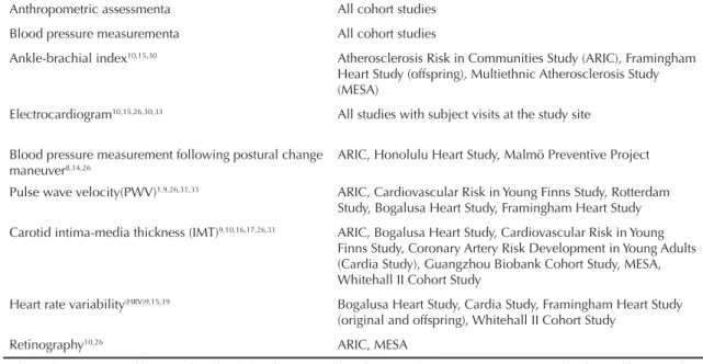
Table 2. Assessments/measurements included in the ELSA-Brasil and other cohort studies with similar methods Anthropometric assessmenta All cohort studies
Blood pressure measurementa All cohort studies
Ankle-brachial index10,15,30 Atherosclerosis Risk in Communities Study (ARIC), Framingham Heart Study (offspring), Multiethnic Atherosclerosis Study (MESA)
Electrocardiogram10,15,26,30,33 All studies with subject visits at the study site
Blood pressure measurement following postural change maneuver8,14,26
ARIC, Honolulu Heart Study, Malmö Preventive Project
Pulse wave velocity(PWV)1,9,26,31,33 ARIC, Cardiovascular Risk in Young Finns Study, Rotterdam Study, Bogalusa Heart Study, Framingham Heart Study
Carotid intima-media thickness (IMT)9,10,16,17,26,31 ARIC, Bogalusa Heart Study, Cardiovascular Risk in Young Finns Study, Coronary Artery Risk Development in Young Adults (Cardia Study), Guangzhou Biobank Cohort Study, MESA, Whitehall II Cohort Study
Heart rate variability(HRV)9,15,19 Bogalusa Heart Study, Cardia Study, Framingham Heart Study (original and offspring), Whitehall II Cohort Study
Retinography10,26 ARIC, MESA
ASSESSMENTS WITH NO ESTABLISHED CLINICAL APPLICATION USED IN OTHER COHORT STUDIES
In this group there were included assessments that were likely to provide other relevant input related to cardio-vascular disease and diabetes by evaluating structural and functional parameters of the heart and blood vessels and central fat accumulation. Some assessments (PWV, HRV, IMT) have been used in other cohort studies but have no established clinical application. This is mostly because many parameters obtained in these assessments including PWV, IMT, density of the liver, ABI and temporal and spectral parameters of HRV do not have well-defi ned normal ranges in healthy populations in general and in the Brazilian population in particular.
Assessment of structure and function of blood vessels
Atherosclerosis is the most prevalent cardiovascular condition worldwide causing cardiovascular events of great impact such as myocardial infarction and
stroke.18 The ELSA protocol included assessments
to evaluate large artery stiffness (PWV), subclinical atherosclerosis (IMT), and arteriolosclerosis of retinal blood vessels.
1. Pulse wave velocity
Large arteries act as a pressure reservoir during systole due to the predominance of elastic fi bers over collagen
fi bers in their walls. A decrease in arterial elasticity with aging is a result of both fragmentation and disor-ganization of elastic fi bers in the vascular wall.5 Artery
stiffness has been used as a marker of arterial aging but pressure inside the blood vessel and endothelial dysfunction can also cause stiffness.24,32 Sudden
disten-tion of the aortic root during systole generates a wave that propagates through the arterial wall at a speed ten times greater than that of blood. According to the Moens-Korteweg equation, PWV is proportional to the elastic modulus of the medium:
PWV = √(Eh/2ρR)
where:
E = Young’s modulus
h = vascular wall thickness
R = radius of the vessel
ρ = blood density
Thus, the single effective determinant of PWV in the arterial vascular network is the elastic modulus as all other factors can be roughly considered constant. Therefore, increased carotid-femoral PWV indicates loss of elasticity of the aortic wall.
The carotid-femoral PWV was measured using a vali-dated automated device (Complior, Artech Medicale, France) with the subject in the supine position in a temperature-controlled room (20°C–24ºC).4 First, BP
was taken in the right arm with the subject in the supine position using an oscillometric device (HRM Onrom 705 CP). The distance from the sternal furcula to the right femoral pulse was measured with a metric tape regardless of abdominal curvature. Pulse sensors were positioned in the right carotid and femoral arteries so that pulse waves were recorded and visualized on a computer screen. A computer program that can adequately detect and record pulse waves was used. PWV is calculated by dividing the distance from the sterna furcula to the femoral pulse by the difference between the rise delay of carotid and femoral pulses. A subject’s PWV was the arithmetic average of readings obtained in ten consecutive cardiac cycles at regular heart rate.
PWV is affected not only by stiffness of large arteries (mainly aorta), but also by the pressure inside the artery which determines elastic and collagen fi bers that are recruited.5,23 PWV must be adjusted for BP using the
pressure measure that is closest to the stiffness measure. All PWVs were measured and recorded at study sites and sent to the central cardiovascular physiology laboratory (IC-ES) for analysis and validation. ELSA is the largest study where PVW data was recorded and analyzed centrally generating robust data to construct normal references ranges and on the effect of biological and environmental factors on arterial aging.
2. Carotid intima-media thickness
The etiology of atherosclerosis is not yet fully understood, but its development and progression are dependent on the interaction between genetic and lifestyle factors.15,18,33 Atherosclerosis is a systemic
disease that involves different arterial territories with varying degrees of severity.33 Atherosclerotic plaques
are slowly progressive, starting with the transport of low density lipoproteins across the endothelium into the subendothelial space. Endothelial dysfunction is characterized by increased permeability of the endo-thelium to lipoproteins and may initiate the process of atherosclerotic plaque formation. Oxidation of LDL-cholesterol would be the next step in plaque formation triggering a local infl ammatory reaction.18
The degree of fi broblast proliferation and migration of smooth muscle cells from the medial layer to the subendothelial space can be inferred by IMT. IMT has been used in epidemiological research as a marker of subclinical atherosclerosis and an independent risk factor for ischemic heart disease.1,9,10,23 In ELSA,
over a length of 1 cm, starting 1 cm below the carotid
bifurcation.17 They were collected and recorded at
study sites and send to the central ultrasound imaging laboratory (SS-SP) for analysis. Image data of three cardiac cycles were analyzed centrally using an automated computer program (MIA™), and IMT was calculated by averaging measures taken in the right and left carotids.
3. Retinography
The retina is a unique body site where the micro-circulation can be imaged directly, providing an opportunity to detecting vascular diameter changes, microaneurysms, micro-hemorrhages, among others. The finding of arteriolar narrowing is associated with the development of diabetes, hypertension and dyslipidemia and can be correlated with myocardial infarction, stroke, and cardiovascular mortality.10,14,18,34
The ELSA is an excellent opportunity to assess retinal vascular changes and their association with large artery changes. Data from cohort follow-up will also allow to prospectively assessing the association of retinal changes with cardiovascular outcomes in coronary and cerebral circulations.
Retinography was performed using a nonmydriatic retinograph (CR-1, Canon, Japan) with a 10-mega-pixel digital camera (Canon EOS 40 D). The subjects had their pupils naturally dilated (about four minutes in a dark room), and for each eye two 45º fundus images were obtained, one centered on the optical disk and the second one on the macula. The central retinography laboratory (IC-RS) developed standard-ized image acquisition and reading protocols and DICOM (~30MB) and JPEG (~3MB) images were acquired. DICOM images were transferred and stored in the ELSA’s Picture Archiving and Communication System (PACS); JPEG images were recorded on CD/ DVD at study sites and mailed to the central retinog-raphy laboratory.
All retinal images were processed using an imaging program (Canon CR-1 Retinal Imaging Control Software, Canon, USA). First, medical cues were identifi ed using Image J (Java-Based Image Processing and Analysis Program, National Health Institute, USA) and then vascular caliber was measured using a measurement system for retinal blood vessels (IVAN, Nicola J. Ferrier, College of Engineering, Fundus Photography Reading Center, University of Wisconsin-Madison, USA), provided by the University of Wisconsin (USA).14 The Retinal Assessment System
(RAS), developed at the Universidade Federal do Rio Grande do Sul, will be used to assess a selected group of subjects.24 The severity of retinal changes will be
rated for microvascular complications in subgroups of subjects such as diabetics.
Assessment of heart structure and function
Echocardiograms were performed to assess heart struc-ture and function and a 10-minute continuous ECG recording to determine HRV.
1. Echocardiogram
The echocardiogram is an ultrasound test performed to assess heart structure and function and can provide diagnostic and prognostic parameters. It can detect the development and impact of maladaptive processes in cardiovascular diseases and common pathophysi-ological mechanisms of cardiovascular and metabolic conditions. Echocardiograms were performed in all ELSA study sites, giving priority to tthis exam in a random subsample of the cohort (10% of the sample) and in subjects older than 55 years. All tests were performed by echocardiographers following a standard acquisition protocol developed in line with current
research recommendations.13
Echoimages were obtained using a device (Aplio XG, Toshiba) with a 2.5 MHz sector transducer. They were sent to ELSA PACS and staff at the central echocardio-gram laboratory (IC-RS) performed all readings at their end (ComPACS, Medimatic, Srl, Italy). The readings consisted of qualitative analysis of echocardiographic
fi ndings and measurements of quantitative parameters to defi ne outcomes of interest including left ventricle (LV) geometry and size, left atrial size, LV systolic and diastolic function, segmental LV dysfunction, valvular heart disease, and fi brocalcifi c degeneration and epicardial fat thickness.
2. Heart rate variability
HR is continuously modulated by the degree of sympa-thetic and parasympasympa-thetic nerve discharge to the
sinus node. HR fl uctuations provide information on
autonomic balance to the heart, which depends on the integration of efferent signals to brainstem nuclei from carotid pressoreceptors, chemoreceptors, and atrium volume and pulmonary artery sensors, among others. The predominance of vagal control increases HRV, which is associated with better prognosis in patients with heart failure, myocardial infarction, and other cardiovascular diseases. In contrast, increased sympa-thetic activity is associated with decreased HRV and poor prognosis in patients with these same conditions.29
performed in a computer program that eliminated artifacts and selected RR intervals lasting 0.5 to 2.0 seconds and then temporal and spectral analysis of HRV was performed using an autoregressive model to identify very low-frequency (VLF, 0 to 0.04 Hz), low-frequency (LF, 0.04 to 0.1 Hz) and high-frequency
spectral bands (HF, 0.1 to 0.4 Hz). HF amplitude29
relates to vagal modulation of HR while LF band
depends on both sympathetic and vagal modulation.29
A 10-minute standing ECG was also obtained in about 5% of the subjects because there is a major increase of LF energy and increased sympathetic control of HR in the standing position.12
Abdominal ultrasound imaging
Abdominal fat accumulation is strongly correlated with chronic diseases including cardiovascular diseases and diabetes. It has been postulated that abdominal fat accumulation induces a chronic proinfl ammatory state leading to the development of hypertension, diabetes, and atherosclerosis. Thus, one of ELSA’s focus of interest was to measure fats deposits in the abdominal wall and omentum.
Like in NAFLD, visceral mesenteric and omental fat has been associated to endocrine, metabolic, and cardio-vascular risk factors. Ultrasound imaging has been used in recent years for biometric profi ling abdominal fat distribution. Central obesity in ELSA subjects was assessed by measuring fat layers in the abdominal wall (subcutaneous and preperitoneal fat) and intra-abdominal fat (visceral fat) through ultrasound images obtained in a Toshiba Aplio XG™ scanner with a 7.5 MHz linear transducer. Three lines were traced in two abdominal wall images (xiphoid and umbilical) to measure subcutaneous and preperitoneal fat layers below the xiphoid process. All images were saved and sent to the central ultrasound imaging laboratory
(IC-SP) for analysis using the MIA™ program. ELSA
data will allow correlating omental fat with standard anthropometric measurements (waist circumference and waist-to-hip ratio) that have been used as indicators of abdominal obesity.18,20
ASSESSMENT WITH AN INNOVATIVE APPROACH
NAFLD is the most common liver disease associated with insulin resistance.6 It is usually quantitatively
assessed by medical imaging specialists through increased echogenicity in liver ultrasound examinations . These assessments, however, are evaluator-dependent. An innovative quantitative approach was proposed in ELSA to assess NAFLD: measurement of the antero-posterior (AP) diameter of the right lobe of the liver. Subjects with NAFLD are expected to have increased AP diameters. Liver images were obtained using
standard equipment (Toshiba SSA-770A Aplio, Japan) and a broadband, convex transducer (PVT-375BT) at a center frequency of 3.5 MHz (2.5 to 5.5 MHz). The transducer was placed in the intercostal space and the gallbladder and the inferior vena cava were used as anatomical landmarks for gain control settings, which allowed to making adjustments according to the subject biotype. Steatosis was diagnosed by liver parenchymal echogenicity and increased ultrasound attenuation. The semi-quantitative assessment of attenuation of the acoustic beam was based on the visualization of the diaphragm: good visualization (normal); partial visualization; and non-visualization.
Static images of the right lobe of the liver in axis oblique sections including anatomical structures aligned in the AP axis − the anterior surface of right liver lobe, gallbladder, inferior vena cava and diaphragmatic angle of the liver − were acquired and saved on CD/DVD. If the gallbladder was not visualized inferior vena cava
blood fl ow was used as an anatomical landmark for
gain control settings.
The saved images were sent to the central ultrasound imaging laboratory (IC-SP) and then analyzed using
TomTec’s Image-Arena. It was measured by drawing
a straight line perpendicular to the skin in the AP axis of the right lobe of the liver from the surface to the posterior diaphragmatic angle of the liver. All images were assessed for three sonographic parameters: echo-genicity, echotexture and acoustic beam attenuation. Increased echogenicity is seen when liver parenchyma is brighter than usual. Heterogeneous echotexture occurs when there are coarse echoes compared to the usual pattern. Abnormal acoustic beam attenuation occurs when visualization of intrahepatic vessels or the posterior diaphragm is reduced, partial (mild) or absent (severe).
FINAL CONSIDERATIONS
The ELSA-Brasil protocol included assessments with well-established clinical and epidemiological research application that can provide data that are relevant and comparable with those already available from other populations. There were also included assessments/ measurements applied in other cohorts to assess whether they have predictive ability for cardiovascular diseases and diabetes in the Brazilian population. The inclusion of subjects from age 35, i.e., younger compared to other cohort studies with similar goals to the ELSA,10,30 was
Suitable spaces at each study site were required to conduct all assessments and specialized teams were trained in data collection centrally. All ELSA-Brasil procedures are detailed elsewhere.7 Furthermore, the
ELSA took an innovative approach by assessing ABI using an automated BP device successfully. Another novel approach was the proposed quantitative assess-ment for NAFLD, which is being validated using computed tomography as the gold standard. If the results are satisfactory, it would provide an easier to perform diagnostic tool that is evaluator-independent. Finally, the centralized readings of imaging assessments
required the implementation of internet transmission of DICOM images collected in the ELSA study sites to the study’s central laboratories. These laboratories provided training and certifi cation of data collection teams and developed algorithms for standard readings of all assessments.
Some of the assessments included in the ELSA such as PWV, IMT, and spectral indices of HRV do not have normal reference values for general population. Since this cohort study comprises mostly healthy subjects, reference values in apparently healthy Brazilian popula-tion can be established.
1. Aatola H, Hutri-Kähönen N, Juonala M, Viikari JS, Hulkkonen J, Laitinen T, et al. Lifetime risk factors and arterial pulse wave velocity in adulthood: the cardiovascular risk in young Finns study. Hypertension. 2010;55(3):806-11. DOI:10.1161/HYPERTENSIONAHA.109.145102
2. Alagiakrishnan K, Masaki K, Schatz I, Curb JD, Blanchette P. Postural hypertension in elderly men: the Honolulu Heart Program. Hawaii Med J. 2000;59(2):48-50.
3. Aquino EM, Barreto SM, Benseñor IM, Carvalho MS, Chor D, Duncan BB, et al. Brazilian Longitudinal Study of Adult Health (ELSA-Brasil): objectives and design. Am J Epidemiol. 2012;175(4):315-24. DOI:10.1093/aje/kwr294
4. Asmar R, Benetos A, Topouchian J, Laurent P, Pannier B, Brisac AM, et al. Assessment of arterial distensibility by automatic pulse wave velocity measurement: validation and clinical application studies. Hypertension. 1995;26(3):485-90. DOI:10.1161/01.HYP.26.3.485
5. Avolio A, Jones D, Tafazzoli-Shadopour M.
Quantifi cation of alterations in structure and function of elastin in the arterial media. Hypertension. 1998;32(1):170-5. DOI:10.1161/01.HYP.32.1.170
6. Bellentani S, Scaglioni F, Marino M, Bedogni G. Epidemiology of non-alcoholic fatty liver disease. Dig Dis. 2010;28(1):155-61. DOI:10.1159/000282080
7. Bensenor IM, Griep RH, Pinto MA, Faria CP, Felisbino-Mendes M, Caetano EI, et al. Rotinas de organização de exames e entrevistas no centro de investigação ELSA-Brasil. Rev Saude Publica. 2013;47(Supl 2): 45-55.
8. Berglund G, Eriksson KF, Israelsson B, Kjellström T, Lindgärde F, Mattiasson I, et al. Cardiovascular risk groups and mortality in an urban Swedish male population: the Malmö Preventive Project. J Intern Med. 1996;239(6):489-97.
9. Bhuiyan AR, Srinivasan SR, Chen W, Paul TK, Berenson GS. Correlates of vascular structure and function measures in asymptomatic young adults: the Bogalusa Heart Study. Atherosclerosis. 2006;189(1):1-7. DOI:10.1016/j.atherosclerosis.2006.02.011
10. Bild DE, Bluemke DA, Burke GL, Detrano R, Diez Roux AV, Folsom AR, et al. Multi-ethnic study of atherosclerosis: objectives and design. Am J Epidemiol. 2002;156(9):871-81. DOI:10.1093/aje/kwf113
11. Consensus Committee of the American Autonomic Society; American Academy of Neurology. Consensus statement on the defi nition of orthostatic hypotension, pure autonomic failure, and multiple system atrophy.
Neurology. 1996;46(5):1470.
12. Dantas EM, Gonçalves CP, Silva ABT, Rodrigues SL, Ramos MS, Andreão RV, et al. Reproducibility of heart rate variability parameters measured in healthy subjects at rest and after a postural change maneuver. Braz J Med Biol Res. 2010;43(10):982-8. DOI:10.1590/S0100-879X2010007500101
13. Douglas PS, DeCara JM, Devereux RB, Duckworth S, Gardin JM, Jaber WA, et al. Echocardiographic Imaging in clinical trials: American Society of Echocardiography Standards for echocardiography core laboratories: endorsed by the American College of Cardiology Foundation. J Am Soc Echocardiogr. 2009;22(7):755-65. DOI:10.1016/j.echo.2009.05.020
14. Hubbard LD, Brothers RJ, King WN, Clegg LX, Klein R, Cooper LS, et al. Methods for evaluation of retinal microvascular abnormalities associated with hypertension/sclerosis in the Atherosclerosis Risk in Communities Study. Ophthalmology. 1999;106(12):2269-80.
15. Hubert HB, Eaker ED, Garrison RJ, Castelli WP. Life-style correlates of risk factor change in young adults: an eight-year study of coronary heart disease risk factors in the Framingham offspring. Am J Epidemiol. 1987;125(5):812-31.
16. Jiang CQ, Xu L, Lam TH, Lin JM, Cheng KK, Thomas GN. Smoking cessation and carotid atherosclerosis: the Guangzhou Biobank Cohort Study-CVD. J
Epidemiol Community Health. 2010;64(11):1004-9.
DOI:10.1136/jech.2009.092718
17. Kanters SD, Algra A, Leuween MS, Banga JD. Reproducibility of in vivo carotid intima-media thickness measurements: a review. Stroke. 1997;28(3):665-71. DOI:10.1161/01.STR.28.3.665
1 8. Kuller LH. Prevention of cardiovascular disease and the future of cardiovascular disease epidemiology.
The Brazilian Longitudinal Study for Adult Health (ELSA-Brazil) was funded by the Brazilian Ministry of Health (Division of Science and Technology [DECIT] of the Department of Science and Technology) and the Brazilian Ministry of Science and Technology (FINEP and CNPq) (protocol number 01 06 0010.00 RS, 01 06 0212.00 BA, 01 06 0300.00 ES, 01 06 0278.00 MG, 01 06 0115.00 SP, 01 06 0071.00 RJ)
This manuscript was peer reviewed as any other manuscripts submitted to this publication. Anonymity is guaranteed for authors and reviewers.
Editors and reviewers declare no confl icts of interest that could affect the peer-review process. The authors declare no confl icts of interest.
Int J Epidemiol. 2001;30(Suppl 1):S66-72.
DOI:10.1093/ije/30.suppl_1.S66
19. Lauer MS, Anderson KM, Levy D. Separate and joint infl uences of obesity and mild hypertension on left ventricular mass and geometry: the Framingham Heart Study. J Am Coll Cardiol. 1992;19(1):130-4. DOI:10.1016/0735-1097(92)90063-S
20. Lohman TG, Roche AF, Martorell R, editors. Anthropometric standardization reference manual. Champaign (IL): Human Kinetics Publications; 1988.
21. Masaki KH, Schatz IJ, Burchfi el CM, Sharp DS, Chiu D, Foley D, et al. Orthostatic hypotension predicts mortality in elderly men: the Honolulu Heart Program. Circulation. 1998;98(21):2290-5. DOI:10.1161/01.CIR.98.21.2290
22. O’Brien E, Mee F, Atkins N, Thomas M. Evaluation of three devices for self-measurement of blood pressure according to the revised British Hypertension Society Protocol: The Omron HEM-705CP, Philips HP 5332, and Nissei DS-175. Blood Pr Monitor.
1996;1(1):55-61.
23. Olivier JJ, Webb DJ. Noninvasive assessment of arterial stiffness and risk of atherosclerotic events.
Atheroscler Thromb Vasc Biol. 2003;23(4):554-66.
DOI:10.1161/01.ATV.0000060460.52916.D6
24. Pakter HM, Fuchs SC, Maestri MK, Moreira LB, Dei Ricardi L, Pamplona VF, et al. Computer-assisted methods to evaluate retinal vascular caliber: what are they measuring? Invest Ophthalmol Vis Sci. 2011;52(2):810-5. DOI:10.1167/iovs.10-5876
25. Prineas RJ, Crow RS, Zhang ZM. The Minnesota code manual of electrocardiographic fi ndings. 2.ed. New York: Springer; 2009.
26. Rose KM, Figenbrodt ML, Biga RL, Couper DJ, Light KC, Sharret AR, et al. Orthostatic hypotension predicts mortality in middle-aged adults: the Atherosclerosis Risk in Communities
(Aric) Study. Circulation. 2006;114(7):630-6. DOI:10.1161/CIRCULATIONAHA.105.598722
27. Sacks D, Bakal CW, Beatty PT, Becker GJ, Cardella JF, Raabe RD, et al. Position statement on the use of the ankle-brachial index in the evaluation of patients with peripheral vascular disease. J Vasc Interv Radiol. 2002;13(4):353.
28. Schmidt MI, Griep RH, Passos VMA, Luft VC, Goulart AC, Menezes GMS, et al. Estratégias e desenvolvimento de garantia e controle de qualidade no ELSA-Brasil. Rev
Saude Publica. 2013;47(Supl 2):113-20.
29. Task Force of the European Society of Cardiology; North American Society of Pacing and
Electrophysiology. Heart rate variability standards of measurement, physiological interpretation and clinical use. Circulation. 1996;93(5):1043-65. DOI:10.1161/01.CIR.93.5.1043
30. The Aric Investigators. The Atherosclerosis Risk in Communities (Aric) Study: design and objectives. Am J
Epidemiol. 1989;129(4):687-702.
31. Van Popele NM, Grobbee DE, Bots ML, Asmar R, Topouchian J, Reneman VS, et al. Association between arterial stiffness and atherosclerosis: the Rotterdam Study. Stroke. 2001;32(2):454-60. DOI:10.1161/01.STR.32.2.454
32. Wilkinson IB, Webb DJ, Cockcroft JR. Aortic pulse wave velocity. Lancet. 1999;354(9194):1996-7. DOI:10.1016/S0140-6736(05)76767-2
33. Wilson PW, Kannel WB, Silbershatz H, D’Agostino RB. Clustering of metabolic factors and coronary heart disease. Arch Intern Med. 1999;159(10):1104-9. DOI:10.1001/archinte.159.10.1104