582
Revista da Sociedade Brasileira de Medicina Tropical 40(5):582-584, set-out, 2007
RELATO DE CASO/CASE REPORT
Oral cavity lymphoma as secondary AIDS-defining neoplasm
in a patient on HAART with immune reconstitution
HAART with immune reconstitution
Linfoma da cavidade oral como neoplasia secundária determinante da AIDS
em um paciente com HAART e reconstituição imune
Marcelo Corti
1, Rubén Solari
1, Diana Cangelosi
1, Luis De Carolis
1,
Ricardo Schtirbu
2and Daniel Lewi
3ABSTRACT
Lymphomas of the oral cavity are a rare complication of advanced HIV/AIDS disease. The clinical appearance of these neoplasms includes masses or ulcerative lesions that involve the oral soft tissue and the jaw as the predominant manifestation. We report the case of a patient with AIDS who developed diffuse large B-cell non-Hodgkin’s lymphoma of the oral cavity during highly active antiretroviral therapy, with undetectable plasma viral load and immune reconstitution.
Key-words: Oral cavity lymphoma. AIDS. HIV. Immune reconstitution.AIDS. HIV. Immune reconstitution.
RESUMO
Os linfomas da cavidade oral são uma complicação rara da AIDS/HIV avançada. A aparência clínica dessas neoplasias inclui massas ou lesões ulcerativas que envolvem o tecido mole oral e da mandíbula como manifestação predominante. Relatamos um caso de um paciente com AIDS que desenvolveu um linfoma não Hodgkin de células B difuso e extenso da cavidade oral durante a terapia antiretroviral altamente ativa com carga viral plasmática indetectável e reconstituição imune.
Palavras-chaves:Linfoma da cavidade oral. AIDS. HIV. Reconstituição imunológica.AIDS. HIV. Reconstituição imunológica.
1. Division of HIV/AIDS disease, F. J. Muñiz Infectious Diseases Hospital, Buenos Aires, Argentina. 2. Histopathology Laboratory, Infectious Diseases F. J. Muñiz Hospital, Buenos Aires, Argentina. 3. Oncology Section, Acute General Hospital J.A. Fernandez, Buenos Aires, Argentina.
Address to: Dr. Marcelo Corti. HIV/AIDS Division, Infectious Diseases F.J. Muñiz Hospital, Puán 381 2ºC 1406 CQG, Buenos Aires, Argentina. Tel: +00 54 11 4432- 3762
e-mail: [email protected] Recebido para publicação em 10/1/2007 Aceito em 24/9/2007
Non-Hodgkin’s lymphoma (NHL) is the second most frequent malignancy among AIDS patients. More than 90% of HIV-associated NHL cases are derived from B cells and the majority are high-grade. Extranodal presentation is most frequent in HIV-seropositive patients and occurs in 70% to 80% of the cases4 6.
The oral cavity is a rare site for presentation of NHL and only 3% of the cases involve primarily the oral cavity9. We report a case of
primary oral cavity lymphoma as a secondary defining neoplasm in an AIDS patient undergoing highly active antiretroviral therapy (HAART) with immune reconstitution.
CASE REPORT
A 38-year-old man with history of intravenous drug abuse who was infected with the human immunodeficiency virus (HIV) and hepatitis C virus (HCV) was admitted to our HIV/AIDS division with a one-month history of mild fever, weight loss and night sweating. The patient also reported odynodysphagia and oral pain. He had
been diagnosed with AIDS because he had developed cerebral toxoplasmosis and a single lesion of oral Kaposi’s sarcoma three months prior to the present hospitalization. At this time, his plasma viral load was over 500,000 copies/ml (log10 5.23) and his CD4 T-lymphocyte count was 23 cell/µl.
One month after being diagnosed with these two AIDS-defining illnesses he was started on HAART, based on zidovudine plus lamivudine plus efavirenz. The oral Kaposi’s sarcoma retrograded without the need for systemic chemotherapy, and a computed tomography (CT) scan showed that the cerebral mass lesions caused by Toxoplasma gondii had resolved.
583
Corti M et alFigure 1 - Large ulcerative lesion of the oral cavity in the region of the left hard palate.
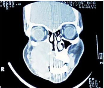
Figure 2 - Computed tomography scan of the maxillofacial area showing a large and heterogeneous mass along the left hard palate, with osseous erosion of the ethmoid sinus and left jaw.
diagnosis of primary NHL of the oral cavity was made and an incisional tissue biopsy was performed. The biopsy smears showed diffuse and aggressive lymphoid infiltration with replacement of the normal tissue by a proliferation of large cells with hyperchromatic central round nuclei, several nucleoli near the basal membrane and abundant eosinophilic cytoplasm.
Immunostaining with monoclonal antibodies demonstrated that the atypical cells showed intensive reactivity to anti-CD20 (PAN B). The T-cell marker CD3 was negative. These histopathological findings confirmed the diagnosis of diffuse large B-cell NHL. In situ hybridization and immunohistochemical tests to detect the Epstein-Barr (EBV) virus genome were negative. The nested polymerase chain reaction (PCR) test in tumor cells, for human herpes virus-8 (HHV-8) DNA, was also negative. A thoracoabdominal-pelvic CT scan was normal and a bone marrow biopsy was negative for atypical cells. At this time, the CD4 T-cell count was 198 cell/µl and the plasma viral load was undetectable.
The patient was sustained with the same scheme of HAART and was started on combined chemotherapy, comprising
cyclophosphamide, doxorubicin, vincristine and prednisone (CHOP), every 21 days and up to a total of six cycles, supporting by granulocyte-stimulating colony factor, with a complete remission of the neoplasm (Figure 3).
Figure 3 - Complete remission of the neoplasm after six cycles of chemotherapy plus HAART.
DISCUSSION
The lymphomas associated with the human immunodeficiency virus include high-grade B-cell lymphomas (which are the vast majority), Burkitt’s lymphoma, diffuse large B-cell lymphoma with two subtypes (centroblastic and immunoblastic) and, finally, two uncommon lymphomas that occur specifically in HIV-positive patients, namely primary effusion lymphoma (PEL) and plasmablastic lymphoma of the oral cavity4. AIDS-associated
B-cell lymphomas have commonly been described as having atypical morphology, extranodal locations and an aggressive clinical course. The most frequent extranodal anatomical sites involved in these tumors are the central nervous system and the gastrointestinal tract6.
Oral cavity lymphomas (OCL) are rare tumors that have a frequency of less than 1% in the general population9. However,
patients with immunodeficiency-associated diseases present a high risk of developing oral lymphomas. Moreover, a new histopathological form of oral cavity lymphoma has been described in patients with HIV/AIDS and has been named plasmablastic lymphoma of the oral cavity3. Primary oral cavity lymphoma is
defined as a neoplasm that involves the oral soft tissue or the jaw as the predominant expression of the disease, and in which the diagnostic smear was obtained from the oral lesion. We observe both of these two conditions, thereby confirming the diagnosis of primary oral cavity lymphoma in our patient.
Cattaneo et al1analyzed 18 cases of OCL: nine cases among
584
Revista da Sociedade Brasileira de Medicina Tropical 40(5):582-584, set-out, 2007
(34 versus 47.5 years, p = 0.0118). Intravenous drug use is an independent risk factor for the development of lymphomas in the HIV/AIDS population1. As in our patient, in Cattaneo’s series the
most frequent risk factor for HIV infection was intravenous drug abuse (seven out of nine patients).
Clinically, OCL in AIDS patients involve the hard palate, gingiva and jaw, as we could see in our patient, and it presents as masses or ulcers. Gingival involvement is characteristic in HIV-seropositive patients, whereas tongue involvement is more frequent among immunocompetent patients1. Plasmablastic lymphoma of the oral
cavity always involves the hard palate.
Patients with a diagnosis of AIDS-associated lymphomas generally present advanced HIV/AIDS, with CD4 T-cell counts lower than 100 cells/µl10. In Cattaneo’s series1, the median CD4 T-cell
count at the time of diagnosis was 39.5 cells/µl and no patient was receiving HAART at the time of diagnosing the neoplasm. In contrast, the patient that we reported was receiving HAART and had a good clinical, virological and immunological response to the lymphoma diagnosis.
We believe that, in the patient we have presented, development of NHL of the oral cavity can be considered to be an expression of the clinical entity named immune reconstitution inflammatory syndrome (IRIS). The Medline, Embase and Cochrane databases were searched to identify articles on lymphomas associated with IRIS in AIDS patients. This search was performed using the following key words: HIV, AIDS, lymphoma and IRIS. We only found one article, by Powles et al.8, in which the authors report
on three cases of IRIS following a diagnosis of AIDS-related NHL that resembled lymphoma relapse.
EBV is strongly associated with the pathogenesis of NHL in AIDS patients, and EBV expression by atypical cells is frequent in AIDS-associated lymphomas2 5. However, we were unable
to demonstrate the presence of the EBV genome in the atypical cells by two techniques: immunohistochemical analysis for latent membrane protein-1 and in situ hybridization for EBER-1 RNA. Furthermore, the PCR test to detect HHV-8 DNA was also negative.
The significantly improved survival of patients with HIV-NHL is associated with the positive impact of HAART on their survival,
especially among those with the centroblastic and immunoblastic subtypes of NHL7.
Recognition of OCL is very important because most cases are clinically aggressive and have a poor prognosis. Early diagnosis followed by chemotherapy and HAART improve the prognosis and survival of this kind of patient. Moreover, NHL should be considered to be an expression of IRIS in patients with AIDS who develop the neoplasm after starting on HAART with a good clinical, virological and immunological response.
REFERENCES
1. Cattaneo C, Facchetti F, Re A, Borlenghi E, Majorana A, Bardellini E, Casari S, Tucci A, Conti G, Rossi G. Oral cavity lymphomas in immunocompetent and human immunodeficiency virus infected patients. Leukemia & Lymphoma 46: 77-81, 2005.
2. Corti M, Villafañe F, Trione N, Schtirbu R, Yampolsky C, Narbaitz M. Primary central nervous system lymphomas in AIDS patients. Enfermedades Infecciosas Microbiología Clínica 22:332-336, 2004.
3. Delecluse HJ, Anagnostopoulos F, Dallenbach M, Hummel M, Marafioti T, Schneider U, Huhn D, Schmidt-Westhausen A, Reichart PA, Gross U, Stein H. Plasmablastic lymphomas of the oral cavity: a new entity associated with the human immunodeficiency virus infection. Blood 89:1413-1420, 1997. 4. Gandhi MK, Khanna R. Viruses and lymphoma. Pathology 37:420-423, 2005. 5. Hamilton-Dutoit SJ, Raphael M, Audouin J, Diebold J, Lissel Pedersen C,
Oksenhendler E, Marelle L, Pallesen G. In situ demonstration of Epstein-Barr virus small RNAs (EBER 1) in acquired immunodeficiency syndrome-related lymphomas: correlation with tumor morphology and primary site. Blood 82:619-624, 1993.
6. Levine AM. Acquired immunodeficiency syndrome-related lymphoma. Blood 80:8-20, 1992.
7. Navarro JT, Ribera JM, Oriol A, Tural C, Millá F, Feliú E. Improved outcome of AIDS-related lymphoma in patients with virologic response to highly active antiretroviral therapy. Journal of Acquired Immune Deficiency Syndrome 32: 347-348, 2003.
8. Powles T, Thirlwell C, Nelson M, Bower M. Immune reconstitution inflammatory syndrome mimicking relapse of AIDS-related lymphoma in patients with HIV-1 infection. Leukemia & lymphoma 44:1417-1419, 2003.
9. Takahashi H, Fujita S, Okabe H, Tsuda N, Tezuka F. Immunophenotypic analysis of extranodal non-Hodgkin’s lymphomas in the oral cavity. Pathology, Research and Practice 189: 300-311, 1993.