369
RELATO DE CASO CASE REPORTPrimeira submissão em 12/02/07 Última submissão em 11/06/07 Aceito para publicação em 19/06/07 Publicado em 20/10/07
Primary diffuse large B-cell lymphoma of the oral cavity
Linfoma difuso de grandes células B primário de boca
Bruno Correia Jham1, 2; Eliza Carla Barroso Duarte1; Anacélia Mendes Fernandes1; Aline Cristina Batista Rodrigues Johann1; Maria Cássia Ferreira Aguiar3; Ricardo Santiago Gomez3; Ricardo Alves Mesquita3
Lymphomas arising within the oral cavity account for only 3.5% of all oral malignancies. Diffuse large B-cell lymphoma is a non-Hodgkin lymphoma subtype characterized by diffuse proliferation of large neoplastic B lymphoid cells. This paper reports a case of diffuse large B-cell lymphoma affecting the oral cavity of a Brazilian woman, along with its clinical, microscopical, immunohistochemical, and molecular features.
resumo
abstract
Linfomas correspondem a 3,5% de todos os casos de lesões malignas de boca. O linfoma difuso de grandes células B é um subtipo de linfoma não-Hodgkin caracterizado pela proliferação difusa de células linfóides B. Este artigo relata um caso de linfoma difuso de grandes células B localizado na cavidade bucal de uma mulher brasileira, incluindo os achados clínicos, microscópicos, imuno-histoquímicos e moleculares.
key words
unitermos
Diffuse large B-cell lymphoma
Non-Hodgkin lymphoma Oral cancer
Linfoma difuso de grandes células B
Linfoma não-Hodgkin Câncer bucal
J Bras Patol Med Lab • v. 43 • n. 5 • p. 369-372 • outubro 2007
1. Graduate student at the Department of Oral Surgery, Oral Medicine, and Oral Pathology, School of Dentistry, Universidade Federal de Minas Gerais (UFMG). 2. Graduate student at the Department of Diagnostic Sciences and Pathology, University of Maryland Dental School.
3. Professor at the Department of Oral Surgery, Oral Medicine, and Oral Pathology, School of Dentistry, UFMG.
This work was conducted in the Department of Oral Surgery, Oral Medicine, and Oral Pathology, School of Dentistry, UFMG. The study was sponsored by Fundação de Amparo à Pesquisa de Minas Gerais (FAPEMIG CDS895/05) and by Conselho Nacional de Desenvolvimento Cientíico e Tecnológico (CNPq 484974/2006-8). M.C.F. Aguiar, R.S. Gomez and R.A. Mesquita are fellow researchers of CNPq. B.C. Jham gratefully acknowledges CNPq-Brazil doctorate scholarship (201590/2006-9).
Background
Diffuse large B-cell lymphoma (DLBCL) is a non-Hodgkin lymphoma (NHL) subtype characterized by diffuse proliferation of large neoplastic B lymphoid cells with nuclear size equal to or exceeding normal macrophage nuclei, or more than twice the size of a normal lymphocyte(7).
Affected patients are usually in their seventh decade of life, and the clinical aspect is typically a rapidly enlarging, often symptomatic mass(7). The most commonly affected site of NHL of the oral cavity and maxillofacial region is the Waldeyer’s ring: tonsils, nasopharynx, base of the tongue, and palatine tonsil(8). Currently, the treatment
of DLBCL consists of radiotherapy, chemotherapy or both. Importantly, DLBCL may be cured in a signiicant percentage of patients, depending on the initial characteristics of the tumor and the host(1).
Here we report a case of primary DLBCL affecting the oral cavity of a Brazilian woman, along with clinical, microscopic, immunohistochemical, and molecular features.
Case report
370
BCL-2 gene and the ampliication of the c-rel locus on chromosome 2p. In this tumor subtype, oncogenesis is related to BCL-6 expression. In contrast, ABC-like tumors demonstrate trisomy 3, gains of 3q and 18q21-q22, and losses of 6 q21-22; in this case, oncogenesis is associated with expression of NF-κB target genes resulting from a constitutive activity of inhibitor kappa B (IκB) kinase(11).
Lymphomas arising within the oral cavity, such as in our case, account for only 3.5% of all oral malignancies. In the oral cavity, lymphomas usually present as an extranodal, soft-elastic, asymptomatic lesion(4). One study reviewed forty cases of oral lymphomas and veriied that in 66% of the cases, the lesion arose from soft tissues, and 77% of that had been increasing in size for the past six months.
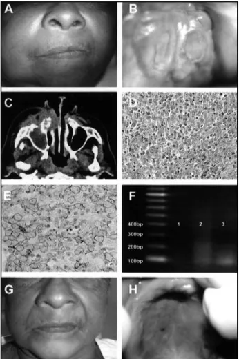
Extra-oral examination revealed asymmetry and swelling of the right side of the face (Figure 1A). Intra-oral exam showed a soft mass on the right side of the palate, containing two 3-cm eroded areas (Figure 1B). Computed tomography revealed destruction of the maxilla and zygomatic bone and invasion of soft tissue on the right side (Figure 1C), as well as invasion of the nasal cavity and maxillary sinus of the opposite side. There were no signs of the disease elsewhere in the body. Clinical diagnoses were mucoepidermoid carcinoma or maxillary sinus carcinoma. An incisional biopsy was performed, with histology showing neoplastic sheets of lymphoid cells with a solid growth pattern. Individually, cells showed scarce cytoplasm and large nucleus (Figure 1D). Sections were submitted to immunohistochemical evaluation, and the malignant cells were immunopositive for LCA and CD20 (Figure 1E). AE1/AE3, CD3, CD10, CD45RO, and S-100 were immunonegative. Following extraction of the genomic DNA, heavy immunoglobulin gene rearrangement (IgH) analysis was performed(3, 6, 10). The obtained DNA showed a discrete and homogeneous band on electrophoresis (Figure 1F, line 3). Final diagnosis was diffuse large B-cell lymphoma. The treatment consisted of dexamethasone, cyclophosphamide, vincristine, etoposide, carboplatin, adriamycin, and iphosphamide. Initially, the patient showed signiicant clinical improvement (Figures 1G and 1H). However, after being submitted to ive cycles of chemotherapy, she unfortunately passed away.
Discussion
Clinical, microscopic, immunohistochemical and molecular features of the reported case confirm the diagnosis of primary DLBCL of the oral cavity. Following squamous cell carcinoma, lymphomas are the second most common neoplasm of the head and neck(8). Nevertheless, the etiology of DLBCL remains unknown. They may originate de novo or represent progression from a less aggressive lymphoma, such as follicular lymphoma or small lymphocytic lymphoma. Underlying immunodeiciency is a signiicant risk factor, and DLBCL in the setting of immunodeiciency is more often Epstein-Barr virus-positive than sporadic DLBCL(3, 7). In accordance to the analysis of global gene expression employing DNA microarrays, DLBCL was broadly divided into normal germinal center B-cells (GC-like DLBCL) or activated peripheral blood B-cells (ABC-like DLBCL). Cytogenetically, GC-like lymphomas present t(14;18)(q32;q21) translocations involving the
JHAM, B. C., et al. Primary diffuse large B-cell lymphoma of the oral cavity • J Bras Patol Med Lab • v. 43 • n. 5 • p. 369-372 • outubro 2007
Figure 1 – A: Asymmetry and swelling of the right side of the face; B: tumoral swelling in the palate with two 3-cm eroded areas; C: computed tomography demonstrated invasion of maxillary sinus, nasal cavity and soft tissue of the right side; D: neoplastic cells are represented by centroblasts (large cell with two or more nucleolus) and immunoblasts (large with one nucleolus) (haematoxilin and eosin, original magnification 400x); E: immunoblasts and centroblasts are CD20-positive (streptoavidin-biotin technique; Dako Corporation, Carpinteria, CA, USA; clone L26; dilution 1:50, antigen retrieval with 0.01 M citric acid, 95oC, 30 min); F: IgH analysis demonstrated a discrete and homogenous
371
these were seen in the upper jaw(14). Likewise, our patient showed a soft, asymptomatic lesion on the upper jaw. The lesion was mainly located in the palate; however, it was clinically dificult to precisely determine its limits and to deine whether or not it extended into maxillary alveolar ridge.
The diagnosis of oral lymphomas may be challenging because frequently there is a low index of clinical suspicion, leading to misdiagnosis and/or delayed treatment. For instance, in a supericial biopsy specimen, superimposed inlammatory changes can result in a lymphoma being mistaken for a reactive or infectious process(12). Thus, biopsies should be carefully performed to obtain adequate specimens. On the other hand, recent advances have made it possible to investigate lymphomas for the expression of a wide range of antigens in ixed tissues. Although standard hematoxylin- and eosin-stained sections still provide the basis for all lymphoma diagnosis, immunophenotyping (IP) is by far the most common ancillary technique used today. IP is useful both for distinguishing benign from malignant processes as well as for accurate subclassiication with high sensibility and speciicity. In addition, IP allows diagnosis on small biopsies that would have previously been deemed inadequate for diagnosis(5). Furthermore, detection of IgH at the DNA level by use of polymerase chain reaction (PCR) to assess monoclonality has become a routine technique in the initial diagnosis of lymphoproliferative disorders. Clonality assays are useful for distinguishing reactive or benign lymph nodes from neoplastic lymphoid iniltrates
1. COIFFIER, B. State-of-the-art therapeutics: diffuse large
B-cell lymphoma. J Clin Oncol, v. 23, n. 26, p.
6387-93, 2005.
2. de FARIA, P. R. et al. Clinical presentation of patients with oral squamous cell carcinoma when first seen by dentists or physicians in a teaching hospital in Brazil.
Clin Oral Investig, v. 7, n. 1, p. 46-51, 2003.
3. DUARTE, E. C. B. et al. Plasmablastic lymphoma of oral
mucosal type: a case report. Oral Oncol Extra, v. 41,
n. 6, p. 121-4, 2005.
4. EPSTEIN, J. B. et al. Characteristics of oral and paraoral
malignant lymphoma: a population-based review of 361
cases. Oral Surg Oral Med Oral PatholOral Radiol
Endod, v. 92, n. 5, p. 519-25, 2001.
5. HIS, E. D.; YEGAPPAN, S. Lymphoma immunophenotyping:
Referências
a new era in paraffin-section immunohistochemistry.
Adv Anat Pathol, v. 8, n. 4, p. 218-39, 2001.
6. ISOLA, J. et al. Analysis of changes in DNA sequence copy number by comparative genomic hybridization
in archival paraffin-embedded tumor samples. Am J
Pathol, v. 145, n. 6, p. 1301-8, 1994.
7. JAFFE, E. S. et al. World Health Organization classification of tumors. Pathology and genetics of tumors of haematopoietic and lymphoid tissues. Lyon, France: IARC Press, 2001.
8. KOLOKOTRONIS, A. et al. Localized B-cell non-Hodgkin’s
lymphoma of oral cavity and maxillofacial region: a clinical study. Oral Surg Oral Med Oral Pathol Oral Radiol Endod, v. 99, n. 3, p. 303-10, 2005.
9. KRAFFT, A. E. et al. Enhanced sensitivity with a novel
in most cases. In addition, the inclusion of T-cell antigen receptors in the assessment of lymphomas results in a signiicant increase in the sensitivity of clonality detection(9). Furthermore, microarrays are powerful tools for discovery and hypothesis generation, allowing researchers to obtain an unbiased survey of gene expression in lymphoma samples. However, their direct clinical application for individualized diagnosis has been questioned(11). In the reported case, immunohistochemistry and IgH analysis were essential for diagnosis and classiication.
NHLs are usually staged according to the Ann Harbor system, being classiied into four stages depending on the extent of disease(8). Patients who are in stage I on admission have a better prognosis than those in stages II to IV, with 5-year overall survival rates ranging from 26% to 73%(12, 13). Unfortunately, oral cancer patients frequently take too
long to seek care, what results in more dificult treatment and worse prognosis, as occurred in the reported case. This most likely occurs due to lack of information(2). Therefore, a major requirement to increase lymphoma survival rates is providing more information on oral cancer to the population. Also, the addition of rituximab to combination chemotherapy in the treatment regimen has increased the proportion of cured patients(12).
In our case report of primary DLBCL of the oral cavity we concluded that efforts should be made to diagnose this disease as rapidly as possible, since prognosis is directly related to disease staging on the patient’s admission. Both health professionals and patients should be aware of the
372
Mailing address
Ricardo Alves Mesquita Universidade Federal de Minas Gerais
Faculdade de Odontologia, Disciplina de Patologia Bucal Av. Antônio Carlos, 6.627, sala 3.204 – Pampulha CEP 31270-901 – Belo Horizonte-MG Fax: (31) 3499-2472
e-mail: [email protected]
TCRgamma PCR assay for clonality studies in 569 formalin-fixed, paraffin-embedded (FFPE) cases. Mol Diagn, v. 4, n. 2, p. 119-33, 1999.
10. MESQUITA, R. A. et al. Avaliação de três métodos
de extração de DNA de material parafinado para amplificação de DNA genômico pela técnica PCR. Pesq Odontol Bras, v. 15, n. 4, p. 314-9, 2001.
11. MORGENSZTERN, D. et al. Gene expression profiling in
diffuse large B-cell lymphoma. Leuk Lymphoma,v. 48,
n. 4, p. 669-82, 2007.
12. PAZOKI, A. et al. Primary non-Hodgkin’s lymphoma of the
jaws: report of 4 cases and review of the literature. J Oral Maxillofac Surg, v. 61, n. 1, p. 112-7, 2003.
13. SEHN, L. H.; CONNORS, J. M. Treatment of aggressive non-Hodgkin’s lymphoma: a North American perspective. Oncology (Williston Park), v. 19, n. 4, p. 26-34, 2005.
14. van der WAAL, R. I. et al. Characteristics of 40 primary
extranodal non-Hodgkin lymphomas of the oral cavity in perspective of the new WHO classification and the International Prognostic Index. Int J Oral Maxillofac
Surg, v. 34, n. 4, p. 391-5, 2005.