Correlation between flow cytometry and histologic findings:
ten year experience in the investigation
of lymphoproliferative diseases
Correlação entre citometria de luxo e anatomia patológica: experiência de dez
anos na investigação de doenças linfoproliferativas
Alanna Mara Pinheiro Sobreira Bezerra1, Denise da Cunha Pasqualin1, João Carlos de Campos Guerra2, Marjorie Paris
Colombini3, Elvira Deolinda Rodrigues Pereira Velloso2, Paulo Augusto Achucarro Silveira2, Cristovão Luis Pitangueira
Mangueira1, Ruth Hissae Kanayama3, Sonia Tsukasa Nozawa3, Rodolfo Correia3, Ana Carolina Apelle1, Welbert de
Oliveira Pereira3, Rodrigo Gobbo Garcia4, Nydia Strachman Bacal5
ABSTRACT
Objective: To demonstrate the advantages of correlating flow cytometry immunophenotyping with the pathology/ immunohistochemistry of lymph nodes or nodules in the diagnosis of lymphoproliferative diseases. Methods: A retrospective study was carried out of 157 biopsy or fine-needle aspiration lymph nodes/ nodule specimens taken from 142 patients, from 1999 and 2009. The specimens were simultaneously studied with flow cytometry and pathology at Hospital Israelita Albert Einstein. The specimens were prepared in hematoxylin/eosin, Giemsa, or monoclonal antibody stained slides for detecting specific antibodies for the purposes of pathology/immunohistochemical analysis. The samples were hemolyzed and marked with different monoclonal antibody panels for different antigens in flow cytometry immunophenotyping. Results:
The diagnostic results of pathology/immunohistochemical studies and flow cytometry immunophenotyping agreed in 115 patients (81%), corresponding to 127 specimens, as follows according to the pathologic diagnosis: 63 patients with non-Hodgkin’s B-cell lymphoma; 26 patients with reactive lymphoid hyperplasia; 5 patients with non-Hodgkin’s T-cell lymphoma; 4 patients with atypical lymphoid proliferation; 5 patients with a chronic granulomatous inflammatory process; 5 patients with a non-hematologic diagnosis; 2 patients with granulocytic sarcoma; 2 patients with thymoma; 1 patient with byphenotypic leukemia; 1 patient with kappa plasmocytoma; 1 patient with Hodgkin’s lymphoma. Subtypes of lymphomas could be classified by associating the two techniques: 19 patients with
follicular lymphoma; 15 patients with diffuse large B-cell lymphoma; 7 patients with small lymphocytic B-cell lymphoma/chronic lymphocytic leukemia; 3 patients with mantle cell lymphoma; 1 patient with Burkitt’s lymphoma; 1 patient with MALT type lymphoma; 1 patient with post-transplant lymphoproliferative disease; 2 patients with high grade Hodgkin’s B-cell lymphoma; 1 patient with low grade non-Hodgkin’s B-cell lymphoma not otherwise specified; 1 patient with Hodgkin’s lymphoma; and 12 patients with B-cell non-Hodgkin’s lymphoma not otherwise specified. Conclusion: Flow cytometry adds to the results of morphologic and immunohistochemical studies, facilitating a rapid and accurate diagnosis of lymphoproliferative diseases.
Keywords: Lymphoma; Lymphoproliferative disorders/diagnosis; Immunophenotyping; Flow cytometry; Immunohistochemistry
RESUMO
Objetivo: Evidenciar as vantagens da correlação entre imunofenotipagem por citometria de fluxo e exame anatomopatológico/ imunoistoquímico de adenomegalias e/ou nódulos no diagnóstico de doenças linfoproliferativas. Métodos: Estudo retrospectivo no qual foram avaliadas 157 amostras de biópsias ou punções aspirativas de gânglios ou nódulos de 142 pacientes, durante o período de 1999 a 2009. As amostras tinham sido encaminhadas simultaneamente para os Serviços de Citometria de Fluxo e Anatomia Patológica do Hospital
Study carried out at Hospital Israelita Albert Einstein - HIAE - São Paulo (SP), Brazil.
1 Pathological Anatomy - Department of Clinical Pathology, Hospital Israelita Albert Einstein - HIAE - São Paulo (SP), Brazil. 2 Hematology, Department of Clinical Pathology, Hospital Israelita Albert Einstein - HIAE - São Paulo (SP), Brazil. 3 Clinical Pathology Department, Hospital Israelita Albert Einstein - HIAE - São Paulo (SP), Brazil.
4 Department of Imaging, Hospital Israelita Albert Einstein - HIAE - São Paulo (SP), Brazil.
5 Division of Flow Cytometry, Department of Clinical Pathology, Hospital Israelita Albert Einstein - HIAE - São Paulo (SP), Brazil.
Corresponding author: Nydia Strachman Bacal - Division of Flow Cytometry, Department of Clinical Pathology, Hospital Israelita Albert Einstein - HIAE - Avenida Albert Einstein, 627/701 - Morumbi - CEP 05651-901 - São Paulo (SP), Brasil - e-mail: [email protected]
Israelita Albert Einstein, em São Paulo. Para a análise na anatomia patológica, as amostras foram preparadas em lâminas e coradas com hematoxilina-eosina, Giemsa, ou marcadas com anticorpos monoclonais para detecção de antígenos específicos. Para a análise por imunofenotipagem por citometria de fluxo, as amostras foram hemolisadas e marcadas com diferentes painéis de anticorpos monoclonais para detecção dos diferentes antígenos. Resultados:
Foram concordantes os diagnósticos entre a anatomopatológico e imunofenotipagem por citometria de fluxo em 115 (81%) pacientes, o que correspondeu a 127 amostras distribuídas da seguinte forma, conforme o diagnóstico anatomopatológico: 63 pacientes com linfoma não Hodgkin de células B; 26 pacientes com hiperplasia linfoide reacional; 5 pacientes com linfoma não Hodgkin de células T; 4 pacientes com proliferação linfoide atípica; 5 pacientes com processo inflamatório crônico granulomatoso; 5 pacientes com diagnósticos não hematológicos; 2 pacientes com sarcoma granulocítico; 2 pacientes com timoma; 1 paciente com leucemia bifenotípica; 1 paciente com plasmocitoma Kappa; e 1 paciente com linfoma de Hodgkin. A correlação entre os resultados das duas técnicas permitiu a classificação dos subtipos de linfomas da seguinte forma: 19 pacientes com linfoma folicular; 15 pacientes com linfoma difuso de grandes células B; 7 pacientes com linfoma linfocítico de pequenas células B/leucemia linfocítica crônica; 3 pacientes com linfoma de células do manto; 1 paciente com linfoma de Burkitt; 1 paciente com linfoma do tipo MALT (tecido linfoide associado à mucosa); 1 paciente com doença linfoproliferativa pós-transplante; 2 pacientes com linfoma não Hodgkin de células B de alto grau; 1 paciente com linfoma não Hodgkin de células B de baixo grau; 1 paciente linfoma de Hodgkin; e 12 pacientes com linfoma não Hodgkin de células B, sem outra especificação. Conclusão: A imunofenotipagem por citometria de fluxo complementa os achados do estudo anatomopatológico/ imunoistoquímico, permitindo um diagnóstico hematopatológico rápido e preciso das doenças linfoproliferativas.
Descritores: Linfoma; Transtornos linfoproliferativos/diagnóstico; Imunofenotipagem; Citometria de fluxo; Imunoistoquímica
INTRODUCTION
Lymph node neoplasms often affect lymph nodes, the spleen, mucosa-associated lymphoid tissues, the skin, or non-lymphoid solid organs, resulting in tumors and enlarged organs. The diagnosis of lymphoproliferative diseases is made by pathology and immunohistochemistry of lymph nodes, which are the gold standard. These approaches demonstrate tissue structure and the classiication of the type of lymphoma. These procedures, however, are time consuming and invasive; furthermore, some tissues are dificult to access.
Fine needle aspiration biopsy (FNAB) is often the irst investigation (screening) for the differential diagnosis among benign/reactional and malignant tumors in cases of enlarged lymph nodes. It is a fast, simple, safe, and only slightly invasive technique for gathering well-preserved cells for studies.
FNAB may be used not only in tissue biopsies for pathology, but also as a technique for obtaining tissue specimens for other studies, including low cytometry immunophenotyping,(1,2) because of its high positive
predictive value. Several studies have been proposed for establishing the reliability of FNAB in the diagnosis of lymphoproliferative diseases(3-9) (Chart 1).
Immunological marker analysis of lymph nodes based on low cytometry immunophenotyping has progressed rapidly in the last few decades, going from restricted use in research to routine use in laboratory diagnoses. This has become possible mainly by the wide availability of low cytometers and the signiicant amount of monoclonal antibodies for rapidly detecting membrane and intracellular antigens in different cell suspensions(10,11).
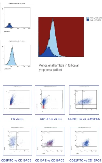
The immunophenotyping study of lymphoproliferative processes is used for distinguishing benign reactions and malignancies; it identiies monoclonality – mainly of B cells – where there is restriction of one of the light immunoglobulin chains (Figure 1). Besides diagnosis, low cytometry immunophenotyping is also applied for classifying the types and subtypes of lymphoproliferative diseases(12-14).
The current classiication of lymphoproliferative diseases (World Health Organization)(14) emphasizes
histologic, clinical, cytologic, immunophenotypic, and genotypic aspects for diagnosing and deining the prognosis of lymphoproliferative diseases. Thus, new highly speciic markers are described on an ongoing basis to improve the diagnosis and to yield information about the prognosis of these diseases; monoclonal antibody panels may include these new markers depending of the needs of clinical investigation(15-17).
High quality smears are useful, since a differential diagnosis may be made based on the nature of cells (monomorphic; polymorphic; small, medium or large
Advantages Disadvantages
Biopsy Adequate study of histological architecture Traumatic procedure High relative cost Limited access in some
tissues Time for results (between 10
and 15 days)
FNAB Minimum trauma Loss in observation of
architectural pattern Low cost
Fast diagnosis
Chart 1. Advantages and disadvantages of fine needle aspiration biopsy and pathological examination by lymph node biopsy in diagnosis of lymphoproliferative diseases
the diagnosis. A κkappa/λlambda proportion below 0.5 or over 3.0 suggests the presence of a clonal B cell population in peripheral blood, bone marrow, lymph nodes, the spleen, or other tissues with larger numbers of mature B lymphocytes(18).
OBJECTIVE
The purpose of this study was to demonstrate the advantages of correlating low cytometry immunophenotyping and pathology/immunohistochemistry of enlarged lymph nodes and/or nodules in the diagnosis of lymphoproliferative diseases.
METHODS
A retrospective study was made of 157 biopsy or ine needle aspiration specimens of lymph nodes or nodules obtained from 142 patients from 1999 to 2009; the specimens were sent simultaneously to the Flow Cytometry Unit and the Pathology Unit of the Israelita Albert Einstein Hospital, São Paulo, SP.
Pathology/immunohistochemistry and cytology
Biopsies or FNAB of lymph nodes or nodules were done in all patients for histologic and immunohistochemical diagnosis. The hematologic and laboratory routine practices were not altered or interfered with for this study.
Pathologists at our hospital usually have three moments to evaluate fresh tissue samples: during ultrasonography for FNABs, during computed tomography for guided needle biopsies, and during intraoperative freeze sections in the surgical theater.
Pathologists promptly examined FNABs
specimens of lymph node/masses to establish cell representativeness; before tissue ixation of the specimens in 95% alcohol), the material was transferred to a tube containing ethylenediaminetetraacetic acid (EDTA) and 2 mL of a RPMI culture medium (RPMI 1640, developed at the Roswell Park Memorial Institute). Flow cytometry immunophenotyping took place within 6 hours of obtaining the specimen, which precluded the need for ixation.
For the biopsies, pathologists selected representative samples of fresh specimens for tissue ixation (10% formaldehyde). The specimens were then transferred to a tube containing EDTA, and low cytometry immunophenotyping was done similarly to the FNAB cases.
In the pathology laboratory, smears and cell centrifugates of FNAB specimens were routinely prepared Figure 1. Flow cytometry analysis of axillary lymphadenomegaly of patient with
follicular lymphoma.
Monoclonalida de Lambda em Paciente com Linfoma Folicular
Monoclonal lambda in follicular lymphoma patient
FS vs SS CD19PC5 vs SS CD20FITC vs CD19PC5
CD5FITC vs CD19PC5 CD10PE vs CD19PC5 CD22FITC vs CD19PC5
sized). Attention should be given to signiicantly hemodiluted materials, because proliferative cases may be mistakenly diagnosed as reactional.
The immediate morphological evaluation of specimens after ine needle aspiration may lead to a second FNAB or to a lymph node biopsy to obtain a specimen with more adequate cellularity (1,6,16,18,19).
B cells comprise about 40% and T cells comprise about 55% of normal lymph nodes. The subtype CD4 predominates among CD3+ cells, and the CD4/CD8 ratio is over 4. The frequency of natural killer cells in normal lymph nodes is very low (about 1%). On the other hand, the tonsils are lymphoid organs in which B cells (CD19+) predominate; the remaining cells are CD3+ with a predominance of the CD4 subtype, as in lymph nodes(20,21).
B cell lymphomas are the majority among non-Hodgkin lymphomas; in such cases, establishing cell clonicity – by restriction of one of the κ (kappa) or λ
for cytology; the Papanicolaou and Giemsa ixation were used. Biopsies went through routine histologic preparation and the slides were hematoxylin-eosin stained.
Immunohistochemistry consisted of placing the specimens on glass slides previously prepared with a poly-D-Lysine adhesive (Sigma, St. Louis, MO, US, code P7886) and kept in an oven at 60oC for 4 hours. Deparafining was
done with repeated xylol baths, absolute ethyl alcohol, and washing with a buffered saline solution, a phosphate buffer solution (PBS), and blockage of endogenous activity with a 3% H2O2 solution. Antigenic recovery was attained by heat or the enzyme method. After recovery of the epitopes, the slides were incubated with the primary antibodies for 12 to 18 hours at 4oC at appropriate dilutions
for each antibody. The slides were then washed again with PBS and incubated for 60 minutes with the respective secondary antibodies. Polymer detection systems were then applied. The slides were processed by a treatment with 3,3’-diaminobenzidine (DAB, Sigma, St. Louis, MO, US, code. D5637), H2O2 (inal concentration = 0.2%), Mayer hematoxylin counterstained, and mounted with histologic resin. Pathologists evaluated all assays with common microscopy; the immunohistochemical reaction controls were positive.
An initial panel consisted of the following primary antibodies: CD20 (clone L26), CD3 (polyclonal), CD10 (clone 56C6), Bcl-2 (clone Bcl-2-100), Bcl-6 (clone lymph node22), CD5 (clone RTB-CD5), CD23 (clone 1B12), cycline D1 (clone SP4), Ki-67 (clone SP6), CD30 (clone Ber-H2), Epstein Barr virus (EBV – clone CS.1-4), and CD15 (clone BY87). This panel was increased by adding the following antibodies, as needed: CD138 (clone MI15), Kappa (polyclonal), Lambda (polyclonal), CD4 (clone 1F6), CD8 (clone C8/144B), CD43 (clone DF-T1), CD56 (clone 123C3), myeloperoxidase (polyclonal), granzyme B (polyclonal), TIA-1 (clone C-20), multiple myeloma-1/ interferon regulatory factor-4 (MUM1/IRF4 – clone MUM 1P), and terminal deoxynucleotidyl transferase (TdT – polyclonal), cytokeratins (clone AE1/AE3), Melan A (clone M27C10), protein S-100 (polyclonal), and HMB45 (clone HMB45).
Pathologists carried out the inal histologic evaluation under a common light microscope, based on the 2001 and 2008 tumor classiication systems of the World Health Organization (WHO), as recommended in the literature(14).
Flow cytometry immunophenotyping
FNAB samples were placed in a collecting medium (Vitrocell), and lymph node/mass samples were imbibed in a saline solution or a collecting medium (RPMI, Vitrocell). Cells were irst counted in a Neubauer chamber. The slides were prepared in Cytospin and
colored with a Rosenfeld dye for cytomorphology. The 7-AAD cell viability assay was used. After the morphologic analysis, the specimens were pipette in 12 x 75 mm tubes depending on the sample volume and the number of cells. The specimens were then PBS (phosphate buffer) washed before marking with monoclonal antibodies. These were obtained from several manufacturers: Beckman Coulter (BC), Becton Dickinson (BD), IQ Products (IQP), Immunotech (IM).
Basic screening of the phenotypic proile of the specimens consisted of applying a panel with the following antibodies: anti-CD2(BC), anti-CD3(BC), CD4(BC), CD8(BC), CD14(BC), CD15(IM), CD19(BC), CD30(IM), anti-CD34(IM), anti-CD45(IM), anti-Kappa (Dako), and anti-Lambda (Dako)(22).
If clonality was present, a complete antibody panel was used, including a panel for the B cell proliferative disease: CD2(BC), CD3(BC), CD5(IM), CD7(BC), CD10(IM), CD11c(IM), CD20(IM), CD22(IM), CD23(Dako), CD25(BC), CD38(IM), CD79b(IM), CD103(IQP), FMC-7(IM), HLA-DR(IM), IgM(Dako), IgD(Dako), and IgG(Dako).
Other panels were used, as follows:
- panel for T cell lymphoproliferative disease: CD1a(IM), CD2(BC), CD3(BC), CD5(IM), CD7(BC), CD10(IM), CD20(IM), CD38(IM), CD56((IM), TCR Alfa/Beta(IM), TCR Gamma/Delta(IM);
- panel for multiple myeloma and associated diseases: CD19(PC5), CD20(CD5), CD33(IM), CD38(IM), CD45(IM), CD56(IM), CD117(IM), HLA-DR(IM), and intracytoplasmatic markers for Kappa, Lambda, IgM, IgG, IgD, and IgA.
After marking with monoclonal antibodies, cells were incubated during 15 minutes at room temperature and away from light. A hemolytic buffer (ammonium chloride) was applied during 15 minutes at room temperature for lysis of red blood cells.
Specimens were washed three times with PBS and fetal bovine serum, and incubated during one hour in a water bath at 37oC for surface marking into light
and heavy chain immunoglobulins. The IntraPrep kit (Beckman Coulter) was used for intracytoplasmatic marking. Data gathering and analysis was done in an EPICS XL-MCL and FC-500 (Beckman Coulter) low cytometer. Analyses were interpreted based on the resulting histograms together with cytomorphology of the specimens, according to the tumor classiication system of the WHO( 2001 and 2008) or others recommended previously in the literature (14).
immunophenotyping. Sensitivity and specificity were used as parameters for assessing the performance of flow cytometry immunophenotyping relative to pathology (the gold standard).
Sensitivity was calculated to assess the proportion of diseases subjects that tested positive, and speciicity was calculated to assess the proportion of disease-free subjects that tested negative. The positive predictive value was calculated to assess the probability of a subject having the disease when tested positive, and the negative predictive values was calculated to assess the probability of a subject not having the disease when tested negative.
RESULTS
There were 157 specimens of 142 patients during the period from 1999 to 2009, of which 75 were male and 67 were female; the mean age was 55 years (ranging from 4 to 92 years).
The procedures for obtaining the specimens consisted of biopsies in 119 patients, FNAB in 16 patients, and FNAB followed by biopsy in 7 patients.
The sites for 145 lymph node specimens were the neck, inguinal, axillary, mediastinal, peripancreatic, paraaortic, and juxtacarotid regions; the sites of 12 tumor mass samples were the spleen, kidney, small intestine, lung, ischium, parotid, scalp, and nasopharynx. There were more than one specimens in 12 patients because of different procedures (for instance, FNAB specimens followed by biopsy specimens), different sites obtained at the same time, different years in a single patient, or duplication of specimens (Table 1).
To investigate the eficacy of associating low cytometry immunophenotyping with pathology for accurate diagnoses, we assessed the agreement percentage between the two techniques for each disease group in the study (Figure 2).
Pathological diagnosis Number of patients Patients % Number of FNAB Number of FNAB + biopsy Number of biopsies
B-NHL 73 51.41 5 4 64
Reactional 26 18.31 4 0 22
T-NHL 7 4.93 0 0 7
Atypical lymphoid proliferation 5 3.52 3 0 2
Chronic granulomatous inlammation 5 3.52 4 0 1
Non-hematological 5 3.52 0 0 5
Granulocytic sarcoma 2 1.41 0 0 2
Thymoma 2 1.41 0 0 2
Biphenotypic 1 0.70 0 0 1
Plasmocytoma 1 0.70 0 0 1
HL 15 10.56 0 3 12
Total 142 100 16 7 119
Table 1. List of patients per pathological diagnosis and types of specimen collection
FNAB: ine needle aspiration biopsy; B-NHL: B-cell non-Hodgkin lymphoma; T-NHL: T-cell non-Hodgkin lymphoma; HL: Hodgkin’s lymphoma.
Figure 2. Percentage of diagnosis with agreement in pathological examination and flow cytometry immunophenotyping per studied group.
Diagnosis with agreement (%)
Pathological diagnosis
Chronic granulomatous inflammation
Non-hematologicalGranulocytic sarcoma Thymoma
Biphenotypic Plasmocytoma
HL
Atypical lymphoid proliferation T-NHL Reactional B-NHL 100.0
90.0
80.0
70.0
60.0
50.0
40.0
30.0
20.0
10.0
0.0 86.3
80.0
100.0 100.0
71.4
6.7 100.0 100.0 100.0 100.0 100.0
The 142 study patients were classiied according to the diagnosis of the disease (Table 1). The bars show the percentage of agreement in diagnosis by each technique (Figure 2).
Interestingly, agreement was above 80% in 9 of 11 disease groups; it was above 70% in one group. Only the diagnosis of Hodgkin’s lymphoma (LH) was mostly discordant, which has been predicted in the literature, as will be discussed below.
study). The agreement between pathology and low cytometry immunophenotyping was 80% in the ive patients with atypical lymphoid proliferation. Flow
cytometry immunophenotyping was completely
effective in the diagnosis of reactional hyperplasia, granulomatous inlammation, non-hematologic cancer, granulocytic sarcoma, thymoma, and individual cases of biphenotypic leukemia and plasmacytoma (100% agreement with pathology).
Figure 3 shows the diagnoses of patients according to pathology.
- post-transplant lymphoproliferative disease (PTLD): 1 patient;
- high grade B cell non-Hodgkin’s lymphoma: 2 patients;
- low grade B cell non-Hodgkin’s lymphoma: 1 patient;
- B cell non-Hodgkin’s lymphoma, not otherwise speciied: 12 patients;
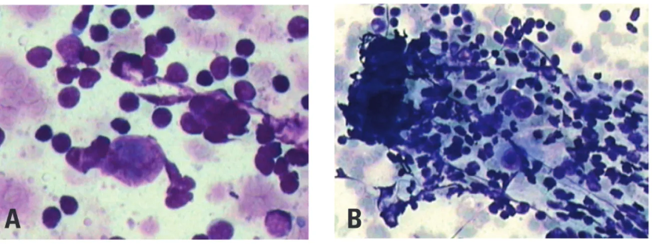
- Hodgkin’s lymphoma: 1 patient (Figure 5A and 5B).
Discordant diagnoses between pathology and low cytometry immunophenotyping comprised 27 patients (19.0%) in 30 specimens (19.1%), as shown on table 2.
P
ercentage of patients
FL
B-NHL , NO
S
CLL/ SLL: MCL
High-grade B
-NHL
Burkitt ’s lymphoma
Low-grade B
-NHL
Hodgkin ’s lymphoma
LDGCB MAL
T
PTLD
20
15
10
5
0
Figure 3. Distribution of diagnosis agreement in percentage of patients per subtype of lymphoma.
Source: Department of Pathology - Hospital Israelita Albert Einstein
The distribution according to subtypes in 63 concordant cases of non-Hodgkin’s lymphoma was as follows:
- follicular lymphoma (LF): 19 patients (Figure 4A and 4B);
- large B cell diffuse lymphoma: 15 patients;
- lymphocytic lymphoma/chronic lymphocytic
leukemia (LLC/ LL): 7 patients; - mantle cell lymphoma: 3 patients; - Burkitt’s lymphoma: 1 patient;
- MALT lymphoma (mucosa-associated lymphoid tissue): 1 patient;
Figure 4. Immunohistochemical panel for diagnosis of follicular lymphoma. (A) Histological features of follicular lymphoma with identification of neoplastic follicle architecture (200x, hematoxylin-eosin); (B) Bcl-2 positivity in immunohistochemical examination in a neoplastic follicle (400X).
A
B
Source: Departament of Hematology - Hospital Israelita Albert Einstein
Diagnoses per pathology Patients (%) Samples (%)
Hodgkin’s lymphoma 14 (51.85) 17 (56.7)
B-cell non-Hodgkin lymphoma 10 (37) 10 (33.3)
T-cell non-Hodgkin lymphoma 02 (7.4) 2 (6.7)
Reactional lymphoid hyperplasia 01 (3.7) 01 (3.3)
Table 2. General distribution of non-agreed diagnoses per type of definite pathological examination.
The following subtypes were found when the diagnoses of non-Hodgkin’s lymphoma made with low cytometry immunophenotyping and pathology did not agree:
- B cell lymphoma rich in T cells and histiocytes: 2 patients;
- large B cell diffuse lymphoma: 6 patients;
- precursor B cell lymphoblastic lymphoma / lymphoblastic leukemia: 1 patient;
- marginal zone (MALT) B cell non-Hodgkin’s lymphoma with focal involvement of lymph nodes: 1 patient
standard) was 0.77; the specii city value was 0.97. The positive predictive value was 0.77, and the negative predictive value was 1.00.
DISCUSSION
The traditional technique of choice for diagnosing lymph node diseases has been histopathology of parafi n-included tissues. Immunohistochemistry is an important tool for analyzing biopsies of lymph nodes and other tissues; cell morphology and tissues architecture are preserved, and immunophenotypic analysis of histological sections are possible(23,24).
Detecting specii c antigens in lymphoid cells is fundamental for classifying tumors, assessing the outcome, and identifying specii c targeted therapy(13,14).
However, pathology of immunohistochemical results in routine laboratory work has its limits: the analysis may be subjective, reproducibility is limited, and the process is time-consuming. Inter- and intra-observer variability is high because so many factors may interfere with the processing of specimens and interpretation of results. Thus, lack of consensus in quantifying antigen expression and dei ning positive and negative results in poor reproducibility. Cytomorphological assays and l ow cytometry immunophenotyping overcome some of these hurdles by providing faster diagnoses, quantitative and qualitative analysis of cell antigens, and multiparametric analyses(2,7,17,18). However, l ow cytometry also has its
limits: variability in antigenic signature expression; loss of cells during the pre-analytical process; specimen preparation issues; work with fresh specimens; and availability of sufi cient neoplastic cells. Large cell lymphoma cells may be lost in the preparation process
because of cell frailty. According to the literature, a negative result does not exclude malignancy(11,18,19,23).
FNAB is a minimally invasive procedure for which cytomorphological analysis combined with l ow cytometry immunophenotyping are important tools, which are able to rapidly differentiate lymphoproliferative diseases from reactional lymphoid hyperplasia in most cases of enlarged lymph nodes(20,23,25).
In our study, the diagnosis by l ow cytometry immunophenotyping and pathology differed in 27 cases; this occurred more often when diagnosing Hodgkin’s lymphoma in 14 patients out of 15 patients with positive pathology for this disease (51.85% of the total number of discordant cases), in 17 specimens (56.7%).
Although l ow cytometry is useful for diagnosing several hematopoetic neoplasms, and may often detect small cell populations (< 0.01% of leukocytes), it is a limited technique for the diagnosis of Hodgkin’s lymphoma involving lymph nodes. Many studies on l ow cytometry immunophenotyping in Hodgkin’s lymphoma have shown changes in reactional lymphocytes, such as the CD4/CD8 ratio in T cells; however, this technique fails to detect Reed Sternberg cells, especially because of their large volume(26). In 2009, Wood described a highly
sensitive and specii c technique based on l ow cytometry using nine colors and three lasers for diagnosing classic Hodgkin’s lymphoma (27).
Flow cytometry failed to diagnose non-Hodgkin’s B lymphoma in ten patients of our sample. In these cases, there was partial distribution of anomalous cells in lymphoid tissues in two patients, which affected neoplastic cell representation. In two cases, the i nal diagnosis was large B cell diffuse lymphoma rich in T cells/histiocytes, where those few detected Figure 5. Fine needle aspiration biopsy showing, in cytology, cells with voluminous nuclei and evident nucleoli (A), sometimes multinucleated (B) diagnostic of Hodgkin lymphoma (400x and 200x, respectively, hematoxylin-eosin).
Source: Department of Pathology - Hospital Israelita Albert Einstein.
neoplastic cells are spread out in a rich background of T lymphocytes and histiocytes; furthermore, the cells are large and more fragile compared to other lymphocytes. Thus, they are not well represented, which may mask low cytometry analysis. Meda et al.(28) and Verstovsek et al.(1) also reported this inding.
They are often wrongly characterized as a polyclonal population because of signiicant contamination by residual normal cells. Therefore, a low cytometry immunophenotyping result showing no evidence of malignancy did not exclude a cancer; in these situations, a detailed cytomorphological exam is required, as demonstrated in the literature(9,29).
According to Meda et al.(28) and Zardawi et al.,(18) if κ
and λ light chains are not restricted, further evidence of clonal proliferation may be investigated, such as major antigen proliferation (CD19, CD20) in speciic tissues (over 85%), CD10 ≥> 18% or CD20+CD5+ ≥> 35%.
Among other studies, Martins et al.(7) conducted a
retrospective study of 627 lymph node FNAB specimens and underlined the importance of cytomorphological analysis for the diagnosis of large cell non-Hodgkin’s lymphoma.
In three of our patients, the specimens consisted of necrotic material, which was frail or sparse for low cytometry immunophenotyping; the specimen with sparse material was obtained in a bone biopsy, and was not representative of the neoplasm. Representativeness of malignancy was lost in three patients, and low cytometry immunophenotyping diagnosis was not possible (in one, the PCR technique for B clonality was used to supplement the study); furthermore no distinct representative IgH loci monoclonal rearrangement band was found, which demonstrated the paucity of cells in the specimen. The presence of necrosis, accelerated tumor growth, and bone, affected pre-analytical processing, thereby interfering with cell viability for analysis in the cytometer; this again has been reported in the literature(19,23).
In T cell lymphomas, cell clonality may only be characterized if antigenic expression of a T lineage marker is absent. A demonstration of T clonality may be done using PCR, the Southern Blot molecular biology technique, or low cytometry for clonality detection in the Vβ family with simultaneous analysis of more than 20 monoclonal antibodies – a technique that is not available in Brazil.
In our study, low cytometry was more speciic than sensitive in diagnostic agreement, a result of poor agreement in patients with Hodgkin’s lymphoma,(14,15)
and in ten patients with large cell diffuse lymphoma, as described previously(10). These factors also affected the
global positive predictive value.
Immunohistochemistry is an important tool in biopsies of lymph nodes and other tissues; it is possible
to analyze the immunophenotype of histological sections where identiication of the tissue architecture is also possible(30). For example, identifying CD5 antigen
expression in some lymphomas, such as between the small cell lymphocytic lymphoma/chronic lymphocytic leukemia (LL/CLL) and the mantle cell lymphoma (ML), which are two types of lymphoproliferative diseases that progress differently, thereby requiring a correct deinition. These cases require investigating and charactering the D1 cyclin marker, which is found in 70 to 80% of mantle cell lymphoma cases(15) - a technique that
is done successfully only with immunohistochemistry. Detection is also possible of the typical t(11:14) of the mantle cell lymphoma in classic cytogenetics, luorescent in situ hybridization (FISH), and reverse transcription polymerase chain reaction (RT-PCR).
In mature B cell lymphomas, the main differential diagnosis to be made among those that are positive for the CD10 marker is between the follicular lymphoma, the large cell diffuse lymphoma, and Burkitt’s lymphoma. Demonstrating histologically the Bcl-2 marker helps identify neoplastic follicles in follicular lymphomas and differentiate them from reactional follicles in follicular lymphoid hyperplasia. Identifying Bcl-2 is dificult in low cytometry, which however may differentiate follicular lymphomas from lymphoid hyperplasia by testing clonality in the κ and λ ratio(16,21,31).
Diagnostic centers that currently provide these technologies as supplementary diagnostic tools reduce the limitations of each method, add speed, and further choices for staging and deining the most appropriate treatment.
CONCLUSION
We have been able to show that in several situations, for many hematologic diseases, low cytometry associated with cytomorphology and immunohistochemistry made it possible to diagnose and differentiate reactional processes from neoplasms, and to subclassify lymphoproliferative diseases.
In our experience with the majority of suspected cases of lymphoproliferative diseases, low cytometry data supplemented the indings of cytomorphology, immunohistochemistry (FNABs) and biopsy specimens.
REFERENCES
1. Verstovsek G, Chakraborty S, Ramzy I, Jorgensen JL. Large B-cell lymphomas: ine-needle aspiration plays an important role in initial diagnosis of cases which are falsely negative by low cytometry. Diagn Cytopathol. 2002;27(5):282-5.
3. Nicol TL, Silberman M, Rosenthal DL, Borowitz MJ. The accuracy of combined cytopathologic and low cytometric analysis of ine-needle aspirates of lymph nodes. Am J Clin Pathol. 2000;114(1):18-28.
4. Caraway NP. Strategies to diagnose lymphoproliferative disease disorders by ine-needle aspiration by using ancillary studies. Cancer. 2005;105(6):432-42 5. Sigstad E, Dong HP, Davidson B, Berner A, Tierens A, Risberg B. The role of low
cytometric immunophenotyping in improving the diagnostic accuracy in referred ine-needle aspiration specimens. Diagn Cytopathol. 2004;31(3):159-63. 6. Costa FPS, Pereira FG, Vassalo J, Freitas LLL, Lorand-Metze I. A utilidade da
citologia por punção com agulha ina aliada à imunofenotipagem no diagnóstico dos linfomas não-Hodgkin. Rev Bras Hematol Hemoter. 2005;27(1):16-20. 7. Martins MR, Santos GC. Fine-needle aspiration cytology in the diagnosis of
supericial lymphadenopathy: a 5-year Brazilian experience. Diagn Cytopathol. 2006;34(2):130-4.
8. Gupta R, Naseem S, Kashyap R, Paul L. Role of ine-needle aspirate immunophenotyping by low cytometry in rapid diagnosis of lymphoproliferative disorders. Diagn Cytopathol. 2007;35(7):381-5
9. Jorgensen, JL. State of the Art Symposium: low cytometry in the diagnosis of lymphoproliferative disorders by ine-needle aspiration. Cancer. 2005;105(6):443-51.
10. Szczepa ski T, van der Velden VH, van Dongen JJ. Flow cytometric immunophenotyping of normal and malignant lymphocytes. Clin Chem Lab Med. 2006;44(7):775-96.
11. Jennings CD, Foon KA. Recent advances in low cytometry: application to the diagnosis of hematologic malignancy. Blood. 1997;90(8):2863-92.
12. Wood BL, Arroz M, Barnett D, DiGiuseppe J, Greig B, Kussick SJ, et al. 2006 Bethesda International Consensus recommendations on the immunophenotypic analysis of hematolymphoid neoplasia by flow cytometry: optimal reagents and reporting for the flow cytometric diagnosis of hematopoietic neoplasia. Cytometry B Clin Cytom. 2007;72 Suppl 1:S14-22.
13. Harris NL, Jaffe ES, Diebold J, Flandrin G, Muller-Hermelink HK, Vardiman J, et al. World Health Organization classiication of neoplastic diseases of the hematopoietic and lymphoid tissues: report of the Clinical Advisory Committee Meeting. J Clin Oncol. 1999;17(12):3835-49.
14. Swerdlow SH, Campo E, Harris NL, Jaffe ES, Pileri SA, Stein H, et al. World Health Organization Classiication of Tumours of Haematopoietic and Lymphoid Tissues. 4th ed. Lyon: IARC Press; 2008.
15. Elnenaei MO, ,Jadayel DM, Matutes E, Morilla R, Owusu-Ankomah K, S et al. Cyclin D1 by low cytometry as a useful tool in the diagnosis of B-cell malignancies. Leuk Res. 2001;25(2):115-23.
16. Cornield DB, Mitchell DM, Almasri NM, Anderson JB, Ahrens KP, Dooley EO, et al. Follicular lymphoma can be distinguished from benign follicular hyperplasia by low cytometry using simultaneous staining of cytoplasmatic Bcl-2 and cell surface CD20. Am J Clin Pathol. 2000;114(2):258-63
17. Colorado M, Cuadrado MA, Insunza A, Mazorra F, Acinas O, Iriondo A. Simultaneous cytomorphologic and multiparametric flow cytometry
analysis on lymph node sample is faster than as valid as histopathologic study to diagnose most non-Hodgkin lymphomas. Am J Clin Pathol. 2010;133(1):83-91.
18. Zardawi IM, Jain S, Bennett G. Flow-cytometric algorithm on ine-needle aspirates for the clinical workup of patients with lymphadenopathy. Diagn Cytopathol. 1998;19(4):274-8.
19. Gong JZ, Williams DC Jr, Liu K, Jones C. Fine-needle aspiration in non-Hodgkin lymphoma: evaluation of cell size by cytomorphology and low cytometry. Am J Clin Pathol. 2002;117(6):880-8.
20. Küppers R. Mechanisms of B-cell lymphoma pathogenesis. Nat Rev Cancer. 2005;5(4)251-62.
21. Wood BL, Borowitz MJ. The low cytometric evaluation of hematopoietic neoplasia. In: McPherson RA, Pincus MR, editors. Henry’s Clinical Diagnosis and Management by Laboratory Methods. 21st ed. Philadelphia, PA: Saunders; 2007. p. 599-616.
22. Martínez A, Aymerich M, Castillo M, Colomer D, Bellosillo B, Campo E, et al. Routine use of immunophenotype by low cytometry in tissues with suspected hematological malignancies. Cytometry B Clin Cytom. 2003;56(1):8-15.
23. Mayall F, Dray M, Stanley D, Harrison B, Allen R. Immunolow cytometry and cell block immunohistochemistry in the FNA diagnosis of lymphoma: a review of 73 consecutive cases. J Clin Pathol. 2000;53(6):451-7.
24. Chan JK. Advances in immunohistochemistry:impact on surgical pathology practice. Semin Diagn Pathol. 2000;17(3):170-7.
25. Orfao A, Alameida J, Sanches ML, San Miguel JF. Immunophenotypic diagnosis of leukemic b-cell chronic lymphoproliferative disorders other than chronic lymphocytic leukemia. contemporary hematology. chronic lymphocitic leukemia: molecular, genetics, biology, diagnosis, and management. Totowa, NJ: G.B. Faguet. Human Press; 2004. p. 173-92.
26. Poppema S, Potters M, Emmens R, Visser L, van den Berg A. Immune reactions in classical Hodgkin’s lymphoma. Semin Hematol. 1999;36(3):253-9. 27. Fromm JR, Thomas A, Wood BL. Flow cytometry can diagnose classical
hodgkin lymphoma in lymph nodes with high sensitivity and speciicity. Am J Clin Pathol. 2009;131(3):322-32.
28. Meda BA, Buss DH, Woodruff RD, Cappellari JO, Rainer RO, Powell BL, et al. Diagnosis and subclassiication of primary and recurrent lymphoma. Am J Clin Pathol. 2000;113(5):688-99.
29. Sneige N, Dekmezian RH, Katz RL, Fanning TV, Lukeman JL, Ordoñez NF et al. Morphologic and immunocytochemical evaluation of 220 fine needle aspirates of malignant lymphoma and lymphoid hyperplasia. Acta Cytol. 1990;34(3):311-22.
30. Chan JK. Advances in immunohistochemistry:impact on surgical pathology practice. Semin Diagn Pathol. 2000;17(3):170-7.