http://dx.doi.org/10.1590/0037-8682-0266-2015
Major Article
INTRODUCTION
Corresponding author: Dr. Wander Rogério Pavanelli.
e-mail: [email protected] I [email protected]
Received 25 August 2015
Accepted 4 December 2015
Sodium nitroprusside has leishmanicidal
activity independent of iNOS
Natália Yoshie Kawakami
[1], Fernanda Tomiotto-Pellissier
[1],
Allan Henrique Depieri Cataneo
[1], Tatiane Marcusso Orsini
[1],
Ana Paula Fortes Dos Santos Thomazelli
[1], Carolina Panis
[2],
Ivete Conchon-Costa
[1]and Wander Rogério Pavanelli
[1][1]. Departamento de Ciências Patológicas, Centro de Ciências Biológicas, Universidade Estadual de Londrina, Londrina, Paraná, Brasil. [2]. Laboratório de
Mediadores Infl amatórios, Universidade Estadual do Oeste do Paraná, Francisco Beltrão, Paraná, Brasil.
ABSTRACT
Introduction: Leishmaniasis is a zoonotic disease caused by protozoa of the genus Leishmania. Cutaneous leishmaniasis is the most common form, with millions of new cases worldwide each year. Treatments are ineffective due to the toxicity of existing
drugs and the resistance acquired by certain strains of the parasite. Methods:We evaluated the activity of sodium nitroprusside
in macrophages infected with Leishmania (Leishmania) amazonensis. Phagocytic and microbicidal activity were evaluated by
phagocytosis assay and promastigote recovery, respectively, while cytokine production and nitrite levels were determined by
ELISA and by the Griess method. Levels of iNOS and 3-nitrotyrosine were measured by immunocytochemistry. Results: Sodium
nitroprusside exhibited in vitro antileishmanial activity at both concentrations tested, reducing the number of amastigotes and
recovered promastigotes in macrophages infected with L. amazonensis. At 1.5µg/mL, sodium nitroprusside stimulated levels
of TNF-α and nitric oxide, but not IFN-γ. The compound also increased levels of 3-nitrotyrosine, but not expression of iNOS,
suggesting that the drug acts as an exogenous source of nitric oxide. Conclusions:Sodium nitroprusside enhances microbicidal
activity in Leishmania-infected macrophages by boosting nitric oxide and 3-nitrotyrosine.
Keywords: Leishmaniasis. Nitric oxide. 3-nitrotyrosine. Leishmania amazonensis. Sodium nitroprusside.
Leishmaniasis, a group of infectious diseases found
worldwide, is caused by protozoa of the genus Leishmania.
Approximately 20 to 40 thousand deaths each year are attributed to this disease, which can manifest in various forms with different symptoms depending on the infecting species and the host immune response. The main forms of the disease are cutaneous, mucocutaneous, and visceral, of which the cutaneous form is the most common, with 0.7 to 1.2 million new cases
each year in 98 countries(1). Pentavalent antimonials are the
standard treatments, of which meglumine antimoniate and
sodium stibogluconate are the most frequently used(2).However,
these drugs require long treatment regimens and parenteral
or intralesional administration(3), and cause numerous side
effects, including pancreatitis, hepatitis, and cardiotoxicity(3)(4).
In addition, some strains of the parasite have acquired resistance to these drugs(5).
Macrophages mount various mechanisms to combat
parasites, including oxidative burst, acidifi cation of vesicles, and expression of inducible nitric oxide synthase (iNOS)(6).
iNOS synthesizes nitric oxide, a highly reactive,
membrane-diffusible molecule used to control various pathogens(7). Nitric
oxide reacts with reactive oxygen species to generate reactive
nitrogen species such as peroxynitrite(8), which damage DNA,
inhibit enzymes, and peroxidize lipids(9).
Leishmania spp. has evolved several mechanisms to evade
macrophage activity. For instance, parasites suppress nitric oxide production by taking up L-arginine, a required substrate for iNOS, as well as by inhibiting the release of IFN-γ and TNF-α, proinfl ammatory cytokines that stimulate expression
of this enzyme(10). These observations suggest that exogenous
sources of nitric oxide may potentially be used to control leishmaniasis.
Indeed, studies have shown that such sources are active against Leishmaniain vitro(11) and in vivo(12), as well as other
microorganisms, including Strongyloides venezuelensis(13),
Paracoccidioides brasiliensis(14), and Trypanosoma cruzi(15). One
METHODS
an inorganic compound(16) active against promastigotes and
axenic amastigotes of Leishmania (Leishmania) amazonensis(17).
In this report, we demonstrate that sodium nitroprusside
enhances the microbicidal activity of Leishmania-infected
macrophages by enhancing production of nitric oxide and
3-nitrotyrosine.
Leishmania (Leishmania) amazonensis
Promastigotes of L. amazonensis (MHOM/BR/1989/166MJO)
were maintained in 199 medium pH 7.18-7.22 (Invitrogen-GIBCO)
supplemented with 10% fetal bovine serum (Invitrogen-GIBCO),
10mM HEPES, 0.1% human urine, 0.1% L-glutamine, 10U/mL
penicillin, 10µg/mL streptomycin (Invitrogen-GIBCO), and 10%
sodium bicarbonate. Cultures were grown in 25cm2 fl asks at 24°C.
Culture of peritoneal macrophages and phagocytosis assay
Macrophages were obtained from the peritoneal cavity
of BALB/c mice, re-suspended in RPMI 1640 medium pH
7.2 (Gibco BRL), and incubated for 2h at 37ºC and 5% CO2
in 24-well plates (5 × 10⁵ cells per well) with 13mm glass
coverslips with 200µL RPMI 1640 medium. Adherent cells
were infected for 2h with L. amazonensis promastigotes at a
ratio of 1:5, washed with phosphate-buffered saline to remove
non-phagocytized parasites, and treated for 24h at 37ºC and
5% CO2 with RPMI 1640 (control) or with 0.5 and 1.5µg/mL
sodium nitroprusside. Subsequently, cells were stained with Giemsa, and 200 cells per sample were imaged at 1,000× under
a CX31RBSFA light microscope (Olympus Optical Co. Ltd.,
Tokyo, Japan) to quantify the number of infected macrophages and the average number of amastigotes per macrophage.
Promastigote recovery
Promastigote recovery was performed as previously
described(18). Briefl y, peritoneal macrophages (5 × 10⁵ cells)
were infected with L. amazonensis, treated for 24h at 37ºC and
5 % CO2 with 0.5 and 1.5µg/mL sodium nitroprusside, washed
with phosphate-buffered saline, and incubated at 24°C with 199 medium to induce differentiation of intracellular amastigotes into promastigotes. Promastigotes recovered were counted daily in a Neubauer chamber for three days after infection.
Cytokine levels
Supernatants were collected from cultures of infected macrophages 24h after treatment with sodium nitroprusside, centrifuged at 460 ×g for 7 min at 4°C, and stored at -80°C
until analysis. TNF-α, IFN-γ, and IL-12-p70 were determined
by enzyme-linked immunosorbent assay (ELISA) according to the manufacturer's instructions (eBiosciences®, USA). Plates
were read at 450nm using a plate reader (Thermo-TP-Reader).
Nitrite levels
Nitrite was also determined in the supernatant of cultures of infected macrophages treated with sodium nitroprusside. Nitric oxide was measured with the Griess reagent according to published methods(19). Briefl y, aliquots were diluted in 45g/L
glycine pH 9.7, and treated for 10 min with cadmium granules
previously activated with 5mM CuSO4. Subsequently, 200µL
of this mixture reacted for 10 min at room temperature with an equal volume of Griess reagent. Tubes were then centrifuged
at 10,845 ×g for 2 min at 25°C, and transferred to 96-well
microplates in triplicate. Absorbance at 550nm was determined in a microplate reader. A calibration curve was constructed using dilutions of NaNO2.
Immunocytochemistry for iNOS and 3-nitrotyrosine
Slides with adherent macrophages were prepared in triplicate as described for the phagocytic assay, and labeled
by the streptavidin-biotin method (Universal Dako LSAB®+ System-HRP Kit, DAKO Japan, Kyoto, Japan) without microwave pretreatment. Slides were then treated with
10% Triton-X for 15 min, washed with phosphate-buffered saline, and incubated in 1% fetal bovine serum for 30 min. Subsequently, slides were probed overnight at 4 ºC with 1:300
dilutions of rabbit polyclonal primary antibodies against iNOS
and 3-nitrotyrosine (Santa Cruz Biotechnology), and then
with biotinylated anti-rabbit, anti-mouse, and anti-goat IgG (LSAB+ System-HRP, DAKO, Japan, Kyoto, Japan) for 2h at room temperature. Negative controls were performed omitting the primary antibodies. Horseradish peroxidase activity was
visualized with H2O2 and 3,3'-diaminobenzidine for 5 min, and
cells were counterstained with Harris hematoxylin (Merck).
Finally, slides were digitally imaged in color at 400× using a BX41 photomicroscope (Olympus Optical Co. Ltd., Tokyo, Japan). Representative fi elds of view from 10 images of each replicate were scored semi-quantitatively using color deconvolution in ImageJ (NIH, USA). Pixels with intensity 0-255 were categorized as strongly positive (3+, intensity 0-60), positive (2+, intensity 61-120), weakly positive (1+, intensity 121-170), and negative (0, intensity 171-230), as previously
described(20). Slides that were not probed with primary antibody
were used as negative control.
Statistical analysis
Data are reported as mean ± standard error of the mean. Duplicate datasets from three independent experiments with three animals per experiment were analyzed in Prism GraphPad 5.00 (GraphPad Software, Inc., USA). Data were found to
be normally distributed by Kolmogorov-Smirnov test, and variances were found to be homogeneous by F test. Treatments were compared by Student’s t-test or analysis of variance
followed by Tukey’s test for multiple comparisons. Differences
RESULTS Ethics statement
Female BALB/c mice weighing approximately 25-30g and aged 6-8 weeks were housed in pathogen-free conditions
according to protocols approved by the Institutional Animal Care and Use Committee at Londrina State University. This study was approved by the Londrina State University Ethics Committee
for Animal Experimentation (33064.2012.42).
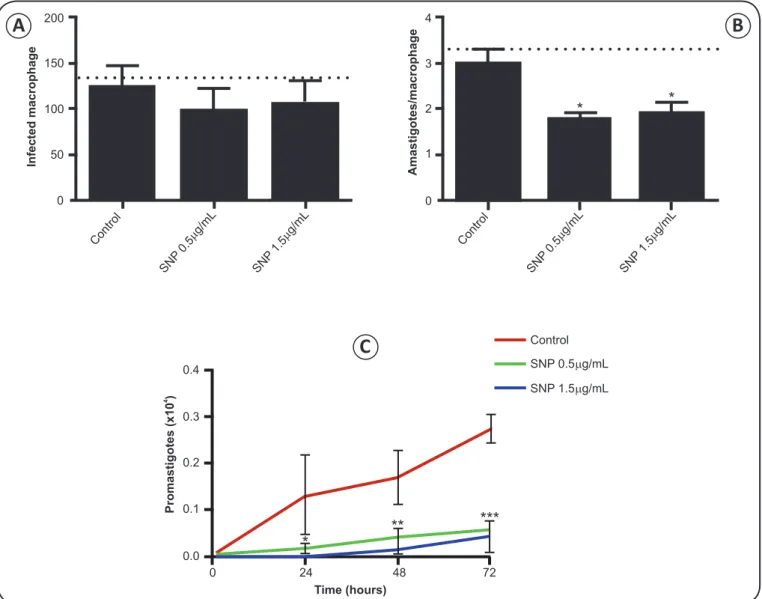
Sodium nitroprusside alters phagocytic capacity and increases microbicidal activity
To characterize the impact of sodium nitroprusside on phagocytic and microbicidal activity, macrophages were treated
Infected macrophage
Promastigotes (x10
)
4
Amastigotes/macrophage
200 4
3
2
1
0
Control
0.4
0.3
0.2
0.1
0.0
Time (hours)
0 24 48 72
SNP 0.5 g/mLµ
SNP 1.5 g/mLµ 150
100
50
0
Contro l
Control
SN P0.5
g/mL µ
SN P1.5
g/mL µ
SN P0.5
g/mL µ
SN P1.5
g/mL µ
*
** ***
* *
with different concentrations of the compound for 24h after infection. We found that exposure to sodium nitroprusside for 24h
did not signifi cantly affect the number of infected macrophages.
However, the number of amastigotes per macrophage was
signifi cantly reduced in cells treated with 0.5µg/mL (p = 0.0188) and 1.5µg/mL (p = 0.0409) sodium nitroprusside (Figure 1A
and 1B). Accordingly, sodium nitroprusside also reduced the
number of promastigotes recovered, with p < 0.0001 for both
concentrations 72h after exposure (Figure 1C). The data indicate
that treatment with sodium nitroprusside for 24h enhanced leishmanicidal activity in macrophages.
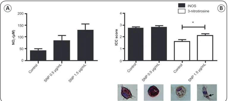
Sodium nitroprusside increases nitric oxide and favors formation of 3-nitrotyrosine
Exposure to 1.5µg/mL sodium nitroprusside for 24h
signifi cantly increased nitric oxide (p = 0.0304, Figure 2A)
FIGURE 1 – Peritoneal macrophages from BALB/c mice were infected in vitro with Leishmania amazonensis and treated for 24h with 0.5 and
1.5μg/mL sodium nitroprusside (SNP). A) Number of infected macrophages. B) Number of amastigotes per macrophage. Dashed line indicates the number of (A) infected macrophages and (B) the amount of internalized parasites after infection period (2 h). C) Leishmania amazonensis promastigotes recovered over three days after infection. *p < 0.05; **p < 0.01; ***p < 0.001 vs. control, by one-way ANOVA followed by Tukey test.
A
B
in macrophages infected with L. amazonensis. Accordingly,
there was increased immunostaining for 3-nitrotyrosine (p = 0.0467, Figure 2B), implying that sodium nitroprusside acts
as an exogenous source of nitric oxide. Notably, 3-nitrotyrosine
colocalized with the parasite in some cells.
Sodium nitroprusside does not induce iNOS expression Semi-quantitative immunocytochemistry demonstrated that sodium nitroprusside exposure was not associated with elevated
expression of iNOS (Figure 2B), indicating that the increase in
nitric oxide was exogenous.
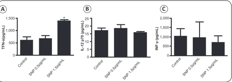
Sodium nitroprusside increases TNF-α
Treatment of infected macrophages for 24h with 1.5µg/mL
sodium nitroprusside increased TNF-α (p = 0.0065, Figure 3A),
but not IFN-γ and IL-12 (Figure 3B and 3C).
200
NO
-(
M)
2
μ
150
100
50
0
Contro l
Contro l
Control
SN P0.5
g/mL μ
SN P0.5
g/mL μ
SNP 1.5
g/mL μ
SNP 1.5
g/mL μ
4
iNOS 3-nitrotirosine
ICC score
3
2
1
0
*
FIGURE 2 - A) Nitrite levels in peritoneal macrophages isolated from BALB/c mice, infected in vitro with Leishmania amazonensis, and treated for
24h with 0.5 and 1.5μg/mL sodium nitroprusside (SNP). B) iNOs and 3-nitrotyorsine, as measured by immunocytochemistry, in BALB/c peritoneal
macrophages infected with L. amazonensis, and treated with 1.5µg/mL sodium nitroprusside for 24h. Pixel intensities (ICC score) were scored as strongly
positive (3+), positive (2+), weakly positive (1+), and negative (0). Sodium nitroprusside did not affect expression of iNOS, but increased 3-nitrotyrosine, which colocalized with the parasite (arrow). *, p < 0.05 vs. control, by t test or one-way ANOVA followed by Tukey test.
DISCUSSION
Nitric oxide is well known to be a key effector in clearing
Leishmania(21), although the parasite is capable of suppressing
nitric oxide production via several mechanisms(22) (23). Thus,
drugs that release nitric oxide, including sodium nitroprusside(24),
may enhance leishmanicidal activity in macrophages. The pharmacological characteristics of these drugs were established in 1955(25), and clinical application has since widened(26) (27). Indeed,
sodium nitroprusside remains in use due to its effectiveness and rapid action(26) (28), despite reports of cyanide toxicity.
A previous study in vitro demonstrated that sodium
nitroprusside decreased the number of L. amazonensis
promastigotes and axenic amastigotes in a dose-dependent
manner(17). In accordance with this result, we observed that
the drug reduced the number of intracellular amastigotes, and, consequently, the number of promastigotes recovered (Figure 1B and 1C). Thus, we investigated the mechanism by which sodium nitroprusside enhanced leishmanicidal activity. We found that nitric oxide levels increased in the supernatant of cultured macrophages exposed to 1.5µg/mL sodium
nitroprusside (Figure 2A). Consequently, 3-nitrotyrosine was
generated, indicating enhanced formation of reactive nitrogen
radicals(29). Notably, 3-nitrotyrosine and nitrated proteins
accumulated in phagosomes and in intracellular parasites (Figure 2B), reinforcing the idea that parasite clearance depends
on reactive nitrogen species. Indeed, 3-nitrotyrosine peaks early in infection in leishmaniasis-resistant C57BL6 mice, presaging a subsequent decline in parasitosis. In contrast, 3-nitrotyrosine peaks late in leishmaniasis-susceptible BALB/c mice(30), implying that 3-nitrotyrosine formation is a relevant indicator
of antiparasitic activity.
In addition to 3-nitrotyrosine, the outcome of Leishmania
infection is critically dependent on the host immune response.
In mice, protective immunity depends on the ability of IL-12 to trigger Th1 activity and release of IFN-γ(22)(31) (32). Indeed,
macrophages stimulated with IL-12 secrete IFN-γ(33), although
FIGURE 3 - TNF-α (A), IL-12 (B), and IFN-γ (C), as measured by ELISA, in BALB/c peritoneal macrophages infected in vitro with Leishmania amazonensis
and treated with 0.5 and 1.5μg/mL sodium nitroprusside (SNP) for 24h. *p < 0.01 vs. control, by one-way ANOVA followed by Tukey test.
this cytokine is primarily produced by natural killer cells, CD4+,
and CD8+ T lymphocytes(34). We note that Leishmania is capable
of suppressing IL-12 expression(35) (36). Importantly, we found that IL-12 production (Figure 3B) and IFN-γ secretion were
not affected by sodium nitroprusside (Figure 3C).
In contrast, sodium nitroprusside stimulated levels of TNF-α
(Figure 3A), a key cytokine involved in macrophage expression
of iNOS(37). However, the increase in TNF-α did not stimulate iNOS expression (Figure 2B), in line with published data demonstrating that exogenous sources of nitric oxide suppress
expression of this enzyme(38) (39). Thus, we conclude that the
antileishmanial activity of sodium nitroprusside depends on its properties as a source of nitric oxide(40) (41).
In summary, we have demonstrated in vitro that sodium
nitroprusside enhances leishmanicidal activity in macrophages
infected with L. amazonensis via release of nitric oxide.
Even though the drug has some toxicity and is challenging to administer, the results provide a rationale for further studies
in vivo, in light of the serious limitations of current therapies,
which have limited effi cacy and signifi cant toxicity, and require
long treatment regimens.
ACKNOWLEDGMENTS
FINANCIAL SUPPORT
REFERENCES
The authors declare that there is no confl ict of interest. CONFLICT OF INTEREST
Dr. A. Leyva helped with English translation and editing.
This work was financially supported by Secretaria de
Ciencia e Tecnologia do Paraná, Conselho Nacional de Desenvolvimento e Pesquisa (CNPq: 82195/2013-4), and State
University of Londrina.
1. Alvar J, Vélez ID, Bern C, Herrero M, Desjeux P, Cano J, et al.
WHO Leishmaniasis Contro Team. Leishmaniasis worldwide and global estimates of its incidence. PLoS One 2012; 7:e35671.
2. Palumbo E. Treatment stra tegies for mucocutaneous leishmaniasis. J Glob Infect Dis 2010; 2:147-150.
3. Frézard F, Demicheli C. New delivery strategies for the old pentavalent antimonial drugs. Expert Opin Drug Deliv 2010; 7:1343-1358.
4. Sundar S, Chakravarty J. Leishmaniasis: an update of current
pharmacotherapy. Expert Opin Pharmacother 2013; 14:53-63.
5. Kaur G, Rajput B. Comparative analysis of the omics technologies used to study antimonial, amphotericin B, and pentamidine resistance in leishmania. J Parasitol Res 2014; 2014:726328. 6. Cunningham AC. Parasitic adaptive mechanisms in infection by
leishmania. Exp Mol Pathol 2002; 72:132-141.
7. Qadoumi M, Becker I, Donhauser N, Röllinghoff M, Bogdan C. Expression of inducible nitric oxide synthase in skin lesions of patients with american cutaneous leishmaniasis. Infect Immun
2002; 70:4638-4642.
8. Rosselli M, Keller PJ, Dubey RK. Role of nitric oxide in the biology, physiology and pathophysiology of reproduction. Hum Reprod
Update 1998; 4:3-24.
9. James SL. Role of nitric oxide in parasitic infections. Microbiol Rev
1995; 59:533-547.
10. Perrella-Balestieri FM, Queiroz AR, Scavone C, Costa VM,
Barral-Netto M, Abrahamsohn IA. Leishmania (L.) amazonensis-induced inhibition of nitric oxide synthesis in host macrophages. Microbes
Infect 2002; 4:23-29.
C
B
A
1,500 25
20
15
10
5
0
2,000
1,500
1,000
500
0 1,000
500
0
TFN-(pg/mL)
α
INF-(pg/mL)
γ
IL-12 p70 (pg/mL)
SN P0.5
g/mL μ
SN P0.5
g/mL μ
SNP 0.5
g/mL μ Contro
l
Control Contro
l
SN P1.5
g/mL μ
SN P1.5
g/mL μ
SNP 1.5
g/mL μ
11. Tfouni E, Truzzi DR, Tavares A, Gomes AJ, Figueiredo LE, Franco DW.
Biological activity of ruthenium nitrosyl complexes. Nitric Oxide 2012; 26:38-53.
12.Miranda MM, Panis C, Cataneo AH, da Silva SS, Kawakami NY, Lopes LG, et al. Nitric oxide and Brazilian propolis combined accelerates tissue repair by modulating cell migration, cytokine production and collagen deposition in experimental leishmaniasis.
PLoS One 2015; 10:e0125101.
13. Ruano AL, López-Abán J, Fernández-Soto P, de Melo AL, Muro A.
Treatment with nitric oxide donors diminishes hyperinfection by Strongyloides venezuelensis in mice treated with dexamethasone.
Acta Trop 2015; 152:90-95.
14. Pavanelli W, da Silva JJ, Panis C, Cunha TM, Costa IC, de Menezes MC, et al. Experimental Chemotherapy in Paracoccidioidomycosis
Using Ruthenium NO Donor. Mycopathol 2011; 172:95-107.
15. Silva JJN, Guedes PMM, Zottis A, Balliano TL,
Nascimento-Silva FO, França Lopes LG, et al. Novel ruthenium complexes as
potential drugs for Chagas's disease: enzyme inhibition and in vitro/ in vivo trypanocidal activity. Br J Pharmacol 2010; 160:260-269. 16. Bates JN, Baker MT, Guerra Jr R, Harrison DG. Nitric oxide
generation from nitroprusside by vascular tissue. Evidence that reduction of the nitroprusside anion and cyanide loss are required.
Biochem Pharmacol 1991; 42:S157-S165.
17. Genestra M, Soares-Bezerra RJ, Gomes-Silva L, Fabrino DL,
Bellato-Santos T, Castro-Pinto DB, et al. In vitro sodium nitroprusside-mediated toxicity towards Leishmania amazonensis promastigotes
and axenic amastigotes. Cell Biochem Funct 2008; 26:709-717.
18. Da Silva SS, Thomé GS, Cataneo AHD, Miranda MM, Felipe I, Guadalupe C, et al. Brazilian propolis antileishmanial and immunomodulatory effects. Evid Based Complement Alternat Med
2013; 2013:673058.
19. Panis C, Mazzuco TL, Costa CZ, Victorino VJ, Tatakihara VL,
Yamauchi LM, et al. Trypanosoma cruzi: Effect of the absence of
5-lipoxygenase (5-LO)-derived leukotrienes on levels of cytokines, nitric oxide and iNOS expression in cardiac tissue in the acute phase
of infection in mice. Exp Parasitol 2011; 127:58-65.
20.Chatterjee S, Malhotra R, Varghese F, Bukhari AB, Patil A, Budrukkar A, et al. Quantitative Immunohistochemical Analysis Reveals Association between Sodium Iodide Symporter and
Estrogen Receptor Expression in Breast Cancer. PLoS ONE 2013;
8:e54055.
21. Horta MF, Mendes BP, Roma EH, Noronha FSM, Macêdo JP,
Oliveira LS, et al. Reactive oxygen species and nitric oxide in cutaneous leishmaniasis. J Parasitol Res 2012; 2012:203818.
22.Isnard A, Shio MT, Olivier M. Impact of Leishmania metalloprotease
GP63 on macrophage signaling. Front Cell Infect Microbiol 2012;
2:72.
23.Shio MT, Hassani K, Isnard A, Ralph B, Contreras I, Gomez MA,
et al. Host cell signalling and leishmania mechanisms of evasion.
J Trop Med 2012; 2012:819512.
24.Lockwood A, Patka J, Raninovich M, Wyatt K, Abraham P. Sodium
nitroprusside-associated cyanide toxicity in adult patients - fact or fi ction? A critical review of the evidence and clinical relevance. J Clin Trials - Dovepress 2010; 2:133-148.
25. Page IH, Corcoran AC, Dustan HP, Koppanyi T. Cardiovascular actions of sodium nitroprusside in animals and hypertensive
patients. Circulation 1955; 11:188-198.
26. Hottinger DG, Beebe DS, Kozhimannil T, Prielipp RC, Belani
KG. Sodium nitroprusside in 2014: A clinical concepts review. J Anaesthesiol Clin Pharmacol 2014; 30:462-471.
27. Taylor TH, Styles M, Lamming AJ. Sodium nitroprusside as a
hypotensive agent in general anaesthesia. Br J Anaesth 1970; 42:859-864.
28. Friederich JA, Butterworth JF. Sodium nitroprusside: twenty years
and counting. Anesth Analg 1995; 81:152-162.
29. Schopfer FJ, Baker PR, Freeman BA. NO-dependent protein
nitration: a cell signaling event or an oxidative infl ammatory response? Trends Biochem Sci 2003; 28:646-654.
30. Linares E, Giorgio S, Mortara RA, Santos CX, Yamada AT, Augusto O. Role of peroxynitrite in macrophage microbicidal mechanisms
in vivo revealed by protein nitration and hydroxylation. Free Radic
Biol Med 2001; 30:1234-1242.
31. Heinzel FP, Rerko RM, Hatam F, Locksley RM. IL-2 is necessary
for the progression of leishmaniasis in susceptible murine hosts.
J Immunol 1993; 150:3924-3931.
32. Sypek JP, Chung CL, Mayor SE, Subramanyam JM, Goldman SJ, Sieburth DS, et al. Resolution of cutaneous leishmaniasis: interleukin 12 initiates a protective T helper type 1 immune
response. J Exp Med 1993; 177:1797-1802.
33. Gessani S, BelardellI F. IFN-gamma expression in macrophages and its possible biological signifi cance. Cytokine Growth Factor Rev 1998; 9:117-123.
34. Farrar MA, Schreiber RD. The molecular cell biology of interferon-gamma and its receptor. Annu Rev Immunol 1993; 11:571-611. 35. Lapara NJ, Kelly BL. Suppression of LPS-induced infl ammatory
responses in macrophages infected with Leishmania. J Infl amm
(Lond) 2010; 7:8.
36. Murphy TL, Cleveland MG, Kulesza P, Magram J, Murphy KM. Regulation of interleukin 12 p40 expression through an NF-kappa B half-site. Mol Cell Biol 1995; 15:5258-5267.
37. Fonseca SG, Romão PR, Figueiredo F, Morais RH, Lima HC, Ferreira SH, et al. TNF-alpha mediates the induction of nitric oxide
synthase in macrophages but not in neutrophils in experimental
cutaneous leishmaniasis. Eur J Immunol 2003; 33:2297-3306. 38. Chang K, Lee SJ, Cheong I, Billiar TR, Chung HT, Han JA, et al.
Nitric oxide suppresses inducible nitric oxide synthase expression
by inhibiting post-translational modifi cation of IkappaB. Exp Mol Med 2004; 36:311-324.
39. Colasanti M, Persichini T, Menegazzi M, Mariotto S, Giordano E, Calderena CM, et al. Induction of nitric oxide synthase mRNA expression. Suppression by exogenous nitric oxide. J Biol Chem
1995; 270:26731-26733.
40. Emre A, Bayram O, Salman B, Ercan S, Anadol Z, Akin O. Sodium Nitroprusside as a Nitric Oxide Donor in a Rat Intestinal Ischemia-Reperfusion Model. Clinics 2008; 63:91-96.
41. William M, Vien J, Hamilton E, Garcia A, Bundgaard H, Clarke RJ, et al. The nitric oxide donor sodium nitroprusside stimulates the