2255-4823/$ – see front matter © 2013 Elsevier Editora Ltda. All rights reserved.
7PMVNFt/ÞNFSPt/PWFNCSP%F[FNCSPt*44/t*44/ 0OMJOF
www.ramb.org.br
ARTIGOS
ARTIGOS ORIGINAIS _____________Qualidade da informação da internet disponível para pacientes em páginas em português ___________________________________________________________645 Acesso a informações de saúde na internet: uma questão de saúde pública? ______650 Maus-tratos contra a criança e o adolescente no Estado de São Paulo, 2009_______659 Obesidade e hipertensão arterial em escolares de Santa Cruz do Sul – RS, Brasil ___666 Bone mineral density in postmenopausal women with and without breast cancer ___673 Prevalence and factors associated with thoracic alterations in infants born prematurely __________________________________________________679 Análise espacial de óbitos por acidentes de trânsito, antes e após a Lei Seca, nas microrregiões do estado de São Paulo ___________________________________685 Sobrevida e complicações em idosos com doenças neurológicas em nutrição enteral ______________________________________________________691 Infliximab reduces cardiac output in rheumatoid arthritis patients without heart failure ______________________________________________________698 Análise dos resultados maternos e fetais dos procedimentos invasivos genéticos fetais: um estudo exploratório em Hospital Universitário _______________703 Frequência dos tipos de cefaleia no centro de atendimento terciário do Hospital das Clínicas da Universidade Federal de Minas Gerais __________________709 ARTIGO DE REVISÃO ______________Influência das variáveis nutricionais e da obesidade sobre a saúde e o metabolismo __714
EDITORIAL Conclusão: como exibir a cereja do bolo 633 PONTO DE VISTA
Os paradoxos da medicina contemporânea 634 IMAGEM EM MEDICINAObstrução duodenal maligna: tratamento
endoscópico paliativo utilizando prótese metálica autoexpansível 636 Gossypiboma 638
DIRETRIZES EM FOCO Hérnia de disco cervical no adulto: tratamento cirúrgico 639 ACREDITAÇÃOAtualização em perda auditiva: diagnóstico radiológico 644
SEÇÕES ____________________________
www.ramb.org.br
Revista da
ASSOCIAÇÃO MÉDICA BRASILEIRA
Original article
Laser therapy in oral mucositis control:
a meta-analysis
q
André Luiz Peixoto Figueiredo
a, Liliane Lins
a,b,*,
Ana Carolina Cattony
a, Antônio Fernando Pereira Falcão
ca Escola Bahiana de Medicina e Saúde Pública, Salvador, BA, Brazil
b Medicine School of Bahia, UFBA, Salvador, BA, Brazil
c Faculty of Dentistry, Universidade Federal da Bahia (UFBA), Salvador, BA, Brazil
A RT I C L E I N F O
Article history:
Received 25 August 2012 Accepted 19 August 2013
Keywords:
Laser therapy Oral mucositis Prevention Meta-analysis
q Study conducted at the Escola Bahiana de Medicina e Saúde Pública, Salvador, BA, Brazil.
* Corresponding author.
E-mail: [email protected]; [email protected] (L. Lins).
A B S T R A C T
Objective: To perform a systematic review and meta-analysis of the effectiveness of laser
therapy (LT) in the prevention of oral mucositis (OM) in patients undergoing oncotherapy.
Methods: A search was conducted in the MEDLINE, LILACS, and Cochrane databases using the
keywords “laser therapy” and “oral mucositis” in order to perform this systematic review and meta-analysis. The case-control studies included were submitted to odds ratio (OR) analysis, whose cut-off for statistic calculation was OM grade ≥ 3. Calculations were performed with the BioEstat program, release 5.0, using DerSimonian-Laird’s random effects statistical analysis.
Results: In this systematic review, twelve studies were included; the meta-analysis of seven
of them demonstrated that LT in patients undergoing oncotherapy is approximately 10 times more effective in the prevention of OM grade ≥ 3 than in patients without laser treatment (OR: 9.5281; 95% CI: 1.447-52.0354; p = 0.0093).
Conclusion: The data demonstrated the significant prophylactic effect of OM grade ≥ 3 in
patients undergoing LT. Further studies, with larger sample sizes, are needed for better evaluation of LT’s prophylactic effect on OM grade ≥ 3.
© 2012 Elsevier Editora Ltda. All rights reserved.
Laser terapia no controle da mucosite oral: um estudo de metanálise
R E S U M O
Objetivo: Realizar uma metanálise da eficácia da laser terapia (LT) na prevenção da mucosite
oral (MO) em pacientes submetidos à oncoterapia.
Métodos: Foi realizada uma busca nas bases de dados MEDLINE, LILACS e Cochrane, utilizando
as palavras-chave “laser therapy” e “oral mucositis”. Os estudos de caso-controle incluídos foram submetidos à análise do odds ratio (OR), cujo ponto de corte para a estatística foi MO
Palavras-chave:
grau ≥ 3. Os cálculos foram realizados com o programa BioEstat 5.0, utilizando a análise estatística de Efeito Aleatório de DerSimonian-Laird.
Resultados: Doze estudos foram incluídos na revisão sistemática. A metanálise de sete deles
evidenciou que a LT em pacientes submetidos à oncoterapia é aproximadamente 10 vezes mais eficaz na prevenção de MO grau ≥ 3 do que em pacientes sem o tratamento com laser (OR: 9,5281; intervalo de confiança de 95% 1,447-52,0354, p = 0,0093).
Conclusão: Esses dados demonstraram efeito profilático significativo de MO grau ≥ 3 nos
pacientes submetidos à LT. Estudos com maior tamanho amostral são necessários para melhor avaliação do efeito profilático de MO grau ≥ 3 por LT.
© 2012 Elsevier Editora Ltda. Todos os direitos reservados.
Introduction
Many patients with cancer are submitted to an initial therapy by radiotherapy (RT), surgery and chemotherapy (CT). RT is usually the treatment of choice in cases involving the head and neck, where the irradiation field involves the oral mucosa and salivary glands.1 Alone or combined with CT, RT has a good clinical response in the treatment of stage I and stage II cancer. However, cancer therapy is closely related to the location of the tumor, its staging, its histological type, as well as the patient’s status.2
Additionally, in cases of malignant and non-malignant hematological diseases, severe immunodeficiency, and bone marrow aplasia, one of the recommended treatments is hematopoietic stem cell transplantation3 (HSCT). Therefore, bone marrow transplantations require the continuous use of a conditioning regimen responsible for myeloablation, in order to create space in the recipient’s bone marrow.4 Therefore, immunosuppression and destruction of neoplastic cells are other effects of high doses of CT drugs, whether or not combined with RT.
Mucosal inflammation is a frequent acute complication in patients with malignancies undergoing oncotherapy. Among patients with head and neck cancer treated with RT, 90% to 97% have some degree of oral mucositis5 (OM). Literature indicates that the incidence of OM, in any degree, associated with oncotherapy for HSCT varies between 76.3% and 89%.6 However, some risk factors appear to be implicated in the pathogenesis of OM, such as the location of the radiation field, preexisting dental disease, poor oral hygiene, low saliva production, compromised immune function, and focus of local infection.7,8
The toxicity produced by the treatment causes alterations that manifest as mucositis, in light of its action on cells with high mitotic activity.9 Thus, there is an intense mucosal involvement, with a decrease in the capacity to overcome the natural exfoliation process, and consequent inflammation and edema.
Associated with a directly harmful effect on the mucous cells, pro-inflammatory cytokines play a role in the worsening of initial mucosal lesions. Tumor necrosis factor-a (TNF-a) and interleukin-1b, interleukin-11, and interleukin-6 appear to play an important role in tissue damage associated with oncotherapy.10 According to the literature,11 there are four
stages in the mucosal lesion process: (1) white patches, with intra- and extracellular edema; (2) appearance of erythematous areas in mucosa, in addition to dysphagia; (3) raised areas of the superficial layers of the mucosa, with reddish borders and re-covered by serofibrinous pseudomembrane, (4) when erythematous areas or areas with pseudomembrane are not re-covered in time, there is a loss of mucous lining, increase of the pain, and fever can occur, and oncotherapy interruption becomes necessary.
The inflammatory picture causes pain and discomfort, with impairment of speech, deglutition, and feeding, and ulcerating lesions can lead to dehydration and poor nutrition. Furthermore, the ulcerations bring a high risk of microbial invasion, causing predisposition to local or systemic infections.12 The increased severity of OM may cause fever, infection risk, need for total parenteral nutrition, need for intravenous analgesics, and mortality during the first 100 days.13
The severity of OM is commonly assessed by the Oral Toxicity Scale, a graduated scale established by the World Health Organization (WHO). This scale contains criteria such as the presence of erythema and ulceration, local pain, and deglutition capacity. When the score is 0 no abnormality has been detected; the presence of erythema without need for treatment characterizes a score of 1; a score of 2 indicates the presence of painful symptoms with no need for analgesics, with difficulty in feeding; a score of 3 indicates painful ulceration requiring the use of analgesics and preventing feeding; finally, a score of 4 indicates necrosis requiring parenteral nutrition.14
Tardieu et al. created the Quantitative Scale of Oral Mucositis by HSCT, used in some articles to assess OM.16
Despite a considerable range of studies performed in the last ten years regarding the prevention of OM due to cancer treatment, preventive measures have yet to be established for mucosal inflammation due to oncotherapy.17 The literature has recorded the use of over 20 preventive measures for oral mucositis caused by oncotherapy,18 of which the following may be cited: cryotherapy, chlorhexidine gluconate, oral hygiene, glutamine, benzydamine, sucralfate, vitamin E, and rinsing the mouth with salt and soda.19,20 However, there is a scarcity of scientific evidence regarding the clinical usage of agents such as benzydamine and cryotherapy.20 Currently, OM prevention is predominantly based on palliative care (oral rinses, anti-inflammatory drugs, and oral hygiene) and prevention of secondary infections.
Some studies, however, suggest other prophylactic measures, acting on the biological mechanisms involved in each phase of OM, such as the use of low-intensity laser.12
Laser therapy (LT) is known to stimulate biological effects, such as elimination of pain and inflammatory modulation. The ability to modulate a variety of metabolic events through photophysical and biochemical processes explains the effects of this therapy.21,22 The laser energy is absorbed only by a thin layer of surrounding tissue beyond the point affected by the radiation. For this reason, the use of lasers with low penetration power is currently recommended, with wave lengths of 640 nm to 940 nm, which should be used immediately after the lesion occurs.23 For comparison, the visible red light-emitting diode has less penetration power, being more suitable for tissue repair, while the diode with longer wavelength, which emits infrared laser, has a greater capacity to penetrate, being more indicated for analgesia. Low-intensity lasers increase cell metabolism by stimulating mitochondrial activity,24 acting as analgesic, anti-inflammatory, and reparative agents in mucosal lesions.25 They cause several biological events, such as epithelial and fibroblast proliferation as well as maturation, transportation, and transformation of fibroblasts into myofibroblasts.26
There are also cell and vascular alterations that depend, among other factors, on the laser wavelength. These include production of collagen, elastin, and proteoglycans; revascularization; wound contraction; increased phagocytosis by macrophages; and increased proliferation and activation of lymphocytes and higher tensile strength, accelerating the healing process.
Helium-neon (He-Ne) and gallium-aluminun-arsenic (Ga-Al-As) lasers have shown good results when used in cases of OM caused by oncotherapy. Although studies have suggested a significant role of LT in the treatment of OM due to oncotherapy,27 further studies are necessary to assess the efficacy of prophylactic LT use at low doses in severe inflammation of the oral mucosa.
Considering the need for further studies on the use of LT in patients with OM caused by oncotherapy, this study aimed to perform a systematic review and meta-analysis of the effectiveness of LT in preventing the development of OM ≥ 3 in patients undergoing cancer treatments.
Methods
Search strategy
From January to February of 2012, a search was performed in the LILACS, MEDLINE, and Cochrane electronic databases, with no restriction regarding the year of publication of the articles. The following key words used were in all databases: “laser therapy” and “oral mucositis”, aiming at standardizing the research. Initially, three researchers analyzed the titles and abstracts of studies listed in the databases. Then, the studies selected after evaluation of their abstracts were further analyzed by the researchers. Based on this assessment, the studies were submitted to the inclusion and exclusion criteria for the meta-analysis.
Criteria for inclusion and exclusion of studies
Considering the expected statistical analysis, studies were included when (1) the patients had a diagnosis of OM caused by oncotherapy, during or after treatment; (2) the treatment of the oral mucosa was performed with low-intensity laser, whose wavelength was set between 632 and 1,064 nm; and (3) the study design consisted of a randomized trial with a control group.
Data collection and study quality
The relevant data for the study were extracted by three researchers using a standardized form. The form was created based on the identification of the most relevant data for the study, containing information about the authors, year of publication, country of origin, and study design.
The data on the population of each study were also analyzed: sample, type of cancer, type of oncotherapy, gender, age of patients and controls, as well as the type of treatment used for the control group. The wavelength applied through the LT (in nm), type of laser used, laser power (in mW), dose (in J/cm2), irradiation time (in seconds), and number of sessions per week were also included in the standardization. To assess the methodological quality, the studies included in the analysis were analyzed according to the Jadad scale.28
Statistical analysis
analgesics and preventing feeding.11 The determination of this criterion is justified by the painful symptoms, food restriction, and discontinuation of treatment in the presence of severe inflammation of the mucosa.
Results
A literature search disclosed 149 studies with keywords “oral mucositis” and “laser therapy”. Forty-one studies considered potentially relevant were selected for detailed evaluation of the inclusion and exclusion criteria. Of these, five were review articles, four were case studies, four addressed LT only as a therapeutic measure, four did not have a control group, and two did not include oncotherapy in the analysis. In addition, two studies did not address LT and two were article comments and were excluded.
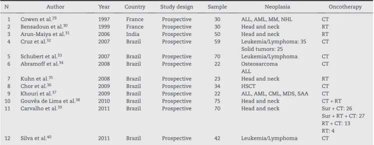
The final sample consisted of 12 prospective randomized studies, published between 1997 and 2011, with a total sample of 527 patients. The total number of patients undergoing LT included in the 12 studies was 276, whereas the control group consisted of 251 patients. Among the patients in the sample, the overall frequency of head and neck cancer was 47%, whereas 53% of the patients in the sample were treated for hematological malignancies (Table 1).
Study description
Of the articles included, two studies29,30 were performed in France, one in India,31 and nine studies were conduc-ted in Brazil.32-40 All studies included in the analysis were case-control studies, and three of them31,34,39 reported pros-pec tive follow-up. Among the selected studies, it is unclear whether there were methodological differences in patient
assessment, with varying inclusion criteria in the sample. In seven studies,31,32,34-37,40 at the beginning of the experiments, the group undergoing LT and the control group did not present OM. Four studies29,30,33,38 did not report the status of the oral mucosa at the beginning of their study.
Regarding the type of oncotherapy, in three studies30,31,36 patients were treated with RT alone, while in six others29,33,34,36,37,40 CT was the only anticancer treatment. One study subdivided the sample submitted to oncotherapy into a group treated with CT for HSCT, and a group where CT was the conventional treatment of solid tumors.32 Another article described the treatment of their patients restrictively with combined CT and RT.38 Similarly, other authors39 used four types of combinations of treatments that included surgery, CT, and RT.
Regarding demographic data, the group of patients submitted to LT consisted mostly of males.30,32,35,37-40 The mean age of patients undergoing LT was ≥ 50 years in four studies30,31,38,39 and among controls, in four as well.30,31,38,39 Two of the studies included in the analysis consisted of pediatric patients only.32,34
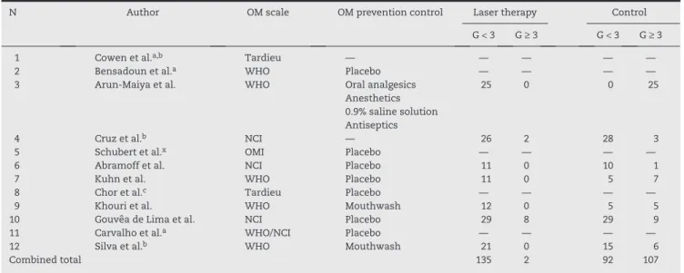
The WHO scale was used in four studies,30,37,39,40 whereas the NCI oral toxicity scale was used in three.32,34,38 One study33 assessed OM through the OMI15; however, this same study established a degree of comparison between the OMI and the scale recommended by WHO; thus, the numbers based on the latter were used by the present study. One study39 used the WHO and NCI scales, finding similar results, and the NCI classification showed a higher percentage of grade 3 OM than the WHO scale. Two studies29,36 evaluated OM using the Tardieu classification.16
The measure used for OM prevention in the control groups showed a significant variation. Eight studies30,33-36,38-40 used placebo laser, and one study39 used in the control group the same laser as in the study group, but with lower potency and
N Author Year Country Study design Sample Neoplasia Oncotherapy 1 Cowen et al.29 1997 France Prospective 30 ALL, AML, MM, NHL CT
2 Bensadoun et al.30 1999 France Prospective 30 Head and neck RT
3 Arun-Maiya et al.31 2006 India Prospective 50 Head and neck RT
4 Cruz et al.32 2007 Brazil Prospective 59 Leukemia/Lymphoma: 35 CT
Solid tumors: 25
5 Schubert et al.33 2007 Brazil Prospective 70 Leukemia/Lymphoma CT
6 Abramoff et al.34 2008 Brazil Prospective 22 Osteosarcoma CT
ALL
7 Kuhn et al.35 2008 Brazil Prospective 23 Head and neck RT
8 Chor et al.36 2009 Brazil Prospective 34 HSCT CT
9 Khouri et al.37 2009 Brazil Prospective 22 ALL, AML, CML, MDS, SAA CT
10 Gouvêa de Lima et al.38 2010 Brazil Prospective 75 Head and neck CT + RT
11 Carvalho et al.39 2011 Brazil Prospective 70 Head and neck Sur + CT: 26
Sur + RT + CT: 27 RT + CT: 13 RT: 4 12 Silva et al.40 2011 Brazil Prospective 42 Leukemia/Lymphoma CT
ALL, acute lymphoblastic leukemia; AML, acute myeloid leukemia; CML, chronic myeloid leukemia; CT, chemotherapy; HSCT hematopoietic stem cell transplant; MDS, myelodysplastic syndrome; MM, multiple myeloma; NHL, non-Hodgkin lymphoma; RT, radiotherapy; SAA, severe aplastic anemia; Sur, surgery.
dose. Conversely, the other placebo-controlled studies used the same laser device as the group submitted to LT, but the device was turned off during the use in the control group. One study31 used a combination of oral analgesics, anesthetics, 0.9% saline solution, and antiseptics as prophylactic tools in the control group. Information regarding the type of prophylactic measure chosen for the control group, the type of scale used to assess OM, and the cutoff used in the present study are shown in Table 2.
Regarding LT, five studies33-36,38 used only the Ga-As-Al laser, four studies29-31,39 used the He-Ne laser, one study40 used only indium-gallium-aluminum-phosphorus (In-Ga-Al-P) laser. and another37 used two types of lasers on alternate days, one emitting red visible light (660 nm [In-Ga-Al-P]) and another infrared (780 nm [–Ga-Al-As]) on alternate days. One article32 did not report the type of laser used in patients undergoing CT. Only one study33 subdivided the group submitted to LT with Ga-Al-As into two smaller groups: a subgroup submitted to a wavelength of 650 nm, with a 2 J dose and 40 mW power, and another submitted to 780 nm, with a 2 J dose and 60 mW power.
Four of the 12 studies did not observe losses in their samples,29,30,31,34 whereas three did not describe losses during the experiments.36,37,38 Two patients in one study33 died from complications related to OM. In one study,39 eight patients were excluded from the sample. Another study32 started with a sample of 62 patients, two of whom were excluded for not following the protocol established by the authors. Furthermore, at the end of the experiments, the researchers32 evaluated only 59 of the 60 patients included, providing no justification for the procedure. One study intended to evaluate 30 patients, but
only 23 completed the treatment with LT. Moreover, without giving any justification, the authors35 did not include patient number 12 in the sample reported in the publication. Another study40 intended to evaluate 56 patients; however, 14 subjects were excluded for not meeting the criteria established for inclusion in the experiment.
Study quality
Study assessment were performed according to the Jadad scale,28 showing similar methodology validations. Eight studies were considered as high quality,29,30,32-35,38,39 three were classified as moderate quality,31,37,40 and one as low quality.36
Meta-analysis
Only seven of the 12 studies included in this review provided sufficient data to classify successes (OM grade < 3) and failures (OM grade ≥ 3). The meta-analysis was performed based on the seven articles listed,31,32,34,35,37,38,40 with a total sample of 293 patients. The combined OR of the studies was 9.5, with p = 0.0093, contained in a 95% CI 1.447-52.0354, whose lower limit was > 1. The results rejected the null hypothesis and demonstrated that the effectiveness in preventing OM grade ≥ 3 by LT is approximately 10 times (OR: 9.5) higher than in those individuals not treated with LT (p = 0.0093).
The forest plot chart shows the analysis of the LT effect on the prevention of OM grade ≥ 3, compared with no treatment. Results to the right show the values of studies favorable to LT;
N Author OM scale OM prevention control Laser therapy Control G < 3 G ≥ 3 G < 3 G ≥ 3 1 Cowen et al.a,b Tardieu — — — — —
2 Bensadoun et al.a WHO Placebo — — — —
3 Arun-Maiya et al. WHO Oral analgesics 25 0 0 25 Anesthetics
0.9% saline solution Antiseptics
4 Cruz et al.b NCI — 26 2 28 3
5 Schubert et al.x OMI Placebo — — — —
6 Abramoff et al. NCI Placebo 11 0 10 1 7 Kuhn et al. WHO Placebo 11 0 5 7 8 Chor et al.c Tardieu Placebo — — — —
9 Khouri et al. WHO Mouthwash 12 0 5 5 10 Gouvêa de Lima et al. NCI Placebo 29 8 29 9 11 Carvalho et al.a WHO/NCI Placebo — — — —
12 Silva et al.b WHO Mouthwash 21 0 15 6
Combined total 135 2 92 107
G < 3, OM grade < 3; G ≥ 3, OM grade ≥ 3; Tardieu, Quantitative scale of oral mucositis associated with autologous bone marrow transplantation (Tardieu et al., 1996), daily index of mucositis (DIM); WHO, Oral Toxicity Scale of the World Health Organization (1979);
NCI, Toxicity Scale of the National Cancer Institute; RTOG, Oral Toxicity Scale of the Radiation Therapy Oncology Group; OMI, Oral Mucositis Index.
a Disclosed only the means of OM grades at the end of the study. b There was no treatment for the control group.
c Disclosed only the p-value in results.
the combined effect was represented by the diamond shape (Fig. 1). The heterogeneity test applied in the DerSimonian-Lair analysis was significant (p = 0.0018), statistically demonstrating the effect of LT on the prevention of OM grade ≥ 3. The Chi-squared analysis of the heterogeneity of the seven studies included in the meta-analysis31,32,34,35,37,38,40 showed p = 0.0006, with six degrees of freedom, which was significant, indicating the existence of heterogeneity among the studies and justifying the choice of the DerSimonian-Laird test.
All studies included in this meta-analysis investigated the possibility of adverse effects associated with LT. None of the included studies demonstrated any side effects resulting from their experiments.29-40
Discussion
The use of low-intensity laser in the oral cavity is capable of preventing the occurrence of OM grade ≥ 3 in patients undergoing oncotherapy, and, in individuals undergoing LT, this prophylaxis is approximately nine times more effective than the absence of LT in the control group (OR: 9.52). These results differ from some literature data; for instance, among the 12 studies included in this study, three did not observe, in a general manner, the effectiveness of LT in OM prevention in patients undergoing anticancer therapy.32,36,38
After a systematic review aiming to combine studies in a meta-analysis, it was observed that the studies were not identical regarding the effect of prevention of OM grade ≥ 3 by LT. Consequently, the differences were investigated and the alternative found for the heterogeneity was to use DerSimonian-Laird’s random effect analysis. This test considers not only the existence of the variation within each study included, but also the differences among the studies. It can be inferred that the random effects model does not consider the studies as identical, but admits that a probability distribution establishes an association between the studies. Through sensitivity analysis, the heterogeneity among the
meta-analysis studies was demonstrated, which can be attributed to methodological differences and to the different prophylactic measures used in the control group. Thus, the present study sought to reduce biases as much as possible, which could affect result interpretation and quality, using the criteria discussed below.
The literature review was broad and comprehensive, seeking to reduce publication bias by not restricting the language, by including studies published in scientific journals with lower scientific visibility, and by including studies with negative results on the efficacy of LT in preventing OM grade ≥ 3 in patients undergoing oncotherapy. The selection of studies, which was performed in accordance with the criteria of study design (case-control), of OM diagnosis, and LT as the only way of preventing oncotherapy-induced OM grade ≥ 3, sought to reduce the heterogeneity of the included studies, while encompassing the clinical situation proposed by the project question (“iss low-intensity LT effective in controlling oncotherapy-induced OM grade ≥ 3?”).
The use of a standardized form, created at a meeting prior to the data collection, in addition to the participation of three researchers to extract the data, contributed to bias reduction at the data collection stage. After the stages of searching and collecting data from the studies, seven of the 12 articles included in the systematic review were selected. The sensitivity analysis, through the chi-squared test, demonstrated the presence of heterogeneity. Thus, taking into account the clinical and methodological differences of the included studies,DerSimonian-Laird’s random effect test was chosen to calculate the combined OR, as suggested by the literature.41
Research has13 evidenced that an increase in OM severity can present systemically as fever, risk of infection, total parenteral nutrition dependence, intravenous analgesic use, and mortality during the first 100 days. Therefore, the confirmation of the prophylactic effects of LT corroborating the large number of studies conducted in this area in the past 20 years would be a method to reduce the limitation to
Fig. 1 – Forest plot chart of the studies included in the random effect analysis and combined odds ratio and odds ratio of the included studies.
Arun-Maiya et al.
Cruz et al.
Abram off et al.
Kuhn et al.
Combined:
Inferior Superior
Khouri et al.
Gouvêa de lima et al.
silva et al.
2006 2601.0 1.302 3.286 31.364
25.000 19.000 18.032
16.524 3.025 90.261
49.670 0.236 0.120 1.504
1.168 1.057 0.944
36202.2 7.176 89.818 654.192
535.272 341.632 344.421
0.245 1.318 0.351 0.416
0.409 0.460 0.442 2007
2008 2009
2009 2010 2011
Author Year OR CI Weight
OR = combined Odds ratio of the seven studies included in the analysis
CI = 95% conidence interval
p = value found at the DerSimonian-Laird analysis Legend:
OR - 9.5281
Combined
0.01 0.1 0.2 0.3 1.0 2.0 3.0 10.0 100
oncotherapy. Literature reports42,43 that the tumor site can be better controlled with the use of LT, and there may be increased survival in cancer patients, as well as improvement in their quality of life.
Regarding the remission of painful symptoms, only two studies32,38 included in the meta-analysis showed no evidence of pain relief with the use of LT. It should be noted that one of these studies38 reported the interruption of treatment of oncological patients in the placebo group as a possible limitation of the study related to its findings. The other studies31,34,35,37,40 showed evidence of decrease in painful symptoms, as well as control in the progression of OM.
Recently, a meta-analysis44 was performed on the effects of low-intensity LT in oncotherapy-induced OM, whose combined OR results were similar to those found in the present study (p < 0.0001, and combined chi-squared analysis of 32.93). The inclusion criteria used in that meta-analysis, however, were very comprehensive, which probably resulted in its greater heterogeneity. Another recent meta-analysis45 included studies that assessed the effect of LT, sucralfate, and benzy damine hydrochloride in the treatment of OM in patients undergoing oncotherapy, including CT, RT, or both procedures. Of the therapies studied, only LT appeared to reduce severe mucositis. It should be noted that, as in the present study, the meta-analyses on LT included different samples concerning age, different modalities of cancer treatment, different laser characteristics, therapeutic dose, and mucositis grade scales.
The current study did not consider demographic factors when analyzing the effectiveness of LT in preventing OM grade ≥ 3, as they do not appear to influence the final result. The present systematic review with meta-analysis showed evidence of moderate to high efficacy of low-intensity LT in the prophylaxis of oncotherapy-induced OM. One limitation to the present study study may be the lack of studies exclusively on prevention of oncotherapy-induced OM by low-intensity LT. In general, the scientific studies were methodologically acceptable, but heterogeneous prophylactic procedures, as well as doses, may have caused the conflict. Furthermore, the small number of patients reported in the literature accounts for an important limitation imposed on the present study.
Conclusion
According to the results obtained from the statistical analysis shown in this meta-analysis, it is clear that LT, when applied to patients undergoing oncotherapy, is effective in controlling OM grade ≥ 3. Studies have demonstrated the importance of severe OM prevention during the course of oncotherapy, stressing, in practice, the limitations imposed by OM grade ≥ 3, which may even lead to treatment discontinuation.
Regarding the use of low-intensity laser, factors such as wavelength, dose, duration of irradiation, power, and number of sessions have remarkable influence on the outcome of prevention, which may explain the varied results among studies and their heterogeneity.
Although a large number of studies have been performed on OM prevention in cancer patients, there is still little
published scientific evidence capable of establishing the use of LT in clinical practice on a large scale. For a more accurate evaluation of the prophylactic effect of OM grade ≥ 3 by LT in patients undergoing some kind of oncotherapy, further studies are needed, with larger sample sizes.
Conflicts of interest
All authors declare to have no conflicts of interest.
R E F E R E N C E S
1. Shi A, Miakowski C, Dodd MJ, Stotts NA, Mc Pahil L. Mechanisms for radiation induced oral mucositis and the consequences. Cancer Nurs. 2003;26:222-9.
2. Perez CA. Perspectivas futuras em radioterapia (para o século XXI). In: Salvajoli JV, Souhami L, Faria SL. Radioterapia em oncologia. Rio de Janeiro: Medsi; 1999. p. 19-34.
3. Holoqiecki J. Indications for hematopoietic stem cell trans-plantation. Pol Arch Med Wewn. 2008;118:658-63.
4. Paton EJA, Coutinho MA, Voltarelli JC. Diagnosis and treatment of acute complications of hematopoietic stem cell trans-plantation. Medicine. 2000;33:264-77.
5. Trotti A, Bellm LA, Epstein JB, Frame D, Fuchs HJ, Gwede CK, et al. Mucositis incidence, severity and associated outcomes in patients with head and neck cancer receiving radiotherapy with or without chemotherapy: a systematic literature review. Radiother Oncol. 2003;66:253-62.
6. Wardley AM, Jayson GC, Swindell R, Morgenstern GR, Chang J, Bloor R, et al. Prospective evaluation of oral mucositis in patients receiving myeloablative conditioning regimens and hematopoietic progenitor rescue. Br J Haematol. 2000;110: 292-9.
7. Stone R, Fliedner MC, Smiet ACM. Management of oral mucositis in patients with cancer. Eur J Oncol Nurs. 2005;9: 524-32.
8. Duncan M, Grant G. Oral and intestinal mucositis: causes and possible treatments. Aliment Pharmacol Ther 2003;18:853-90. 9. Velez I, Tamara LA, Mintz S. Management of oral mucositis
induced by chemotherapy and radiotherapy: an update. Quintessence Int. 2004;35:129-36.
10. Genot MT, Klastersky J. Low-level laser for prevention and therapy of oral mucositis induced by chemotherapy or radiotherapy. Curr Opin Oncol. 2005;17:236-40.
11. Dib LL, Curi MM. Complicações orais na oncologia: parte A. Atuação odontológica em pacientes portadores de câncer. In: Salvajoli JV, Souhami L, Faria SL. Radioterapia e oncologia. Rio de Janeiro: Medsi; 1999. p. 1145-64.
12. Sandoval RL, Koga DH, Buloto LS, Suzuki R, Dib LL. Management of chemo and radiotherapy induced oral mucositis with low-energy laser: inicial results of A.C. Camargo Hospital. J Appl Oral Sci. 2003;11:337-41.
13. Sonis ST, Eilers JP, Epstein JB, Le Veque FG, Liggett WH Jr., Mulagha MT, et al. Validation of a new scoring system for the assessment of clinical trial research of oral mucositis induced by radiation or chemotherapy. Am Cancer Soc. 1999;85:2103-13. 14. World Health Organization. Handbook for reporting results of cancer treatment. Geneve: World Health Organization; 1979. p. 15-22.
associated with bone marrow transplantation: development of an Oral Mucositis Index. Cancer. 1992;69:2469-477.
16. Tardieu C, Cowen D, Thirion X, Franquin JC. Quantitative scale of oral mucositis associated with autologous bone marrow transplantation. Eur J Cancer B Oral Oncol. 1996;32B:381-7. 17. França CM, França CM, Nuñez SC, Prates RA, Noborikawa E,
Faria MR, et al. Low-intensity red laser on the prevention and treatment of induced-oral mucositis in hamsters. Photochem Photobiol. 2009;94:25-31.
18. Santos P, Messaggi AC, Mantesso A, Magalhaes MHCG. Mucosite oral: perspectivas atuais na prevenção e tratamento. RGO. Rev Gauch Odontol. 2009;57:1.
19. Albuquerque ILS, Camargo TC. Prevenção e tratamento da mucosite oral induzida por radioterapia: revisão de literatura. Rev Bras Cancerol. 2006;53:195-209.
20. Rampini MP, Ferreira EMS, Ferreira CG, Antunes HS. Utilização de terapia com laser de baixa potência para prevenção da mucosite oral: revisão de literatura. Rev Bras Cancerol. 2009;55: 59-68.
21. Karu TI, Kolyakov SF. Exact action spcetra for cellular responses relevant to phototherapy. Photomed Laser Surg. 2005;23:355-61. 22. Pinheiro ALB, Brugnera Júnior A, Zanin FAA. Aplicação do laser
na odontologia. São Paulo: Santos; 2010.
23. Rocha JCT. Terapia laser, cicatrização tecidual e angiogênese. Rev Bras Promoção Saúde. 2004;17:44-8.
24. Silveira PC, Streck EL, Pinho RA. Evaluation of mitochondrial respiratory chain activity in wound healing by low-level laser therapy. J Photochem Photobio B. 2007;86:279-82.
25. Bourguignon-Filho AM, Feitosa ACR, Beltrão GC, Pagnocelli RM. Utilização do laser de baixa intensidade no processo de cicatrização tecidual. Revisão de literatura. Rev Port Estomatol Cir Maxilofac. 2005;46:37-43.
26. Medrado ARAP, Pugliese LS, Reis SRA, Andrade ZA. Influence of low level laser therapy on wound healing and its biological action upon myofibroblasts. Lasers Surg Med. 2003;32:239-44. 27. Lino MD, Carvalho FB, Oliveira LR, Magalhães EB, Pinheiro
AL, Ramalho LM. Laser phototherapy as a treatment for radiotherapy-induced oral mucositis. Braz Dent J. 2011;22: 162-5.
28. Jadad AR, Moore RA, Carrol D, Jenkinson C, Reynolds JM, Gavaghan DJ, et al. Assessing the quality of reports of randomized clinical trials: is blinding necessary? Control Clin Trials. 1996;17:1-12.
29. Cowen D, Tardieu C, Schubert M, Peterson D, Resbeut M. Lowe energy helium-neon laser in the prevention of oral mucositis in patients undergoing bone marrow transplant: results of a double-blind randomized trial. Int J Radiat Oncol Biol Phys. 1997;38:697-703.
30. Bensadoun RJ, Franquin JC, Ciais G, Carcout V, Schubert MM, Viot M, et al. Low-energy He/Ne laser in the prevention of radiation-induced mucositis. A multicenter phase III randomized study in patients with head and neck cancer. Supp Care Cancer. 1999;7:244-52.
31. Arun Maiya G, Sagar MS, Fernandes D. Effect of low-level helium-neon (He-Ne) laser therapy in the prevention & treatment of radiaton induced mucositis in head & neck cancer patients. Indian J Med Res. 2006;124:399-402.
32. Cruz LB, Ribeiro AS, Rech A, Rosa LGN, Castro Jr. CG, Brunetto AL. Influence of low-energy laser in the prevention of oral mucositis in children with cancer receiving chemotherapy. Pediatr Blood Cancer. 2007;48:435-40.
33. Schubert MM, Eduardo FP, Guthrie KA, Franquin JC, Bensadoun RJ, Migliorati CA, et al. A phase III randomized double-blind placebo-controlled clinical trial to determine the efficacy of low level laser therapy for the prevention of oral mucositis in patients undergoing hematopoietic cell transplantation. Supp Care Cancer. 2007;48:435-40.
34. Abramoff MM, Lopes NN, Lopeas LA, Dib LL, Guilherme A, Caran EM et al. Low-level laser therapy in the prevention and treatment of chemotherapy-induced oral mucositis in young patients. Photomed Laser Surg. 2008;26:393-400.
35. Kuhn A, Heinzmann G, Silva CA, Stobbe C, Knack MA, Basualdo A, et al. Low-level infrared laser therapy to prevent radiotherapy-induced oral mucositis: a randomized placebo controlled study. J Oral Laser Appl. 2008;8:219-24.
36. Chor A, Torres SR, Maiolino A, Nucci M. Low-power laser to prevent oral mucositis in autologous hematopoietic stem cell transplantation. Eur J Haematol. 2010;84:178-9.
37. Khouri VY, Stracieri AB, Rodrigues MC, Moares DA, Pieroni F, Simões BP, et al. Use of therapeutic laser for prevention and treatment of oral mucositis. Braz Dent J. 2009;20:215-30. 38. Gouvêa de Lima A, Villar RC, Castro G Jr., Antequera R, Gil E,
Rosalmeida MC, et al. Oral mucositis prevention by low-level laser therapy in head-and-neck câncer patients undergoing concurrent chemoradiotherapy: a phase III randomized study. Int Radiat Oncol Biol Phys 2010;82:270-5.
39. Carvalho PA, Jaguar GC, Pellizzon AC, Prado JD, Lopes RN, Alves FA. Evaluation of low-level laser therapy in the prevention and treatment of radiation-induced mucositis: a double-blind randomized study in head and neck patients. Oral Oncol. 2011;47:1176-81.
40. Silva GBL, Mendonça EF, Bariani C, Antunes, HS, Silva MAG. The prevention of induced oral mucositis with low-level laser therapy in bone marrow transplantation patients: a randomized clinical trial. Photomed Laser Surg. 2011;29:27-31. 41. Berwager O, Suzumura EA, Buehler AN, Oliveira JB. Como
avaliar criticamente revisões sistemáticas e metanálises? Rev Bras Ter Intensiva. 2007;19:192-6.
42. World Association for Laser Therapy (WALT). Recommended treatment doses for low level laser therapy. Updated April 2010 [accessed 28 Jan 2012]. Available from: http://www.walt. nu/dosage-recommendations.html.
43. Arora H, Pai KM, Maiya A, Vidyasagar MS, Rajeev A. Efficacy of He-Ne laser in the prevention and treatment of radio-therapy-induced oral mucositis in oral cancer patients. Oral Surg Oral Med Oral Pathol Oral Radiol Endod. 2008;105:180-6.
44. Bjordal JM, Bensadoun RJ, Tunèr J, Frigo L, Gjerde K, Lopes-Martins RA. A systematic review with meta-analysis of the effect of low-level laser therapy (LLLT) in cancer-therapy induced oral mucositis. Supp Care Cancer. 2011;19:1069-77. 45. Clarkson JE, Worthington HV, Furness S, McCabe M, Khalid T,