40
Atypical progression of visual loss in a patient
with primary open-angle glaucoma
Progressão atípica de perda visual em paciente
com glaucoma primário de ângulo aberto
Marcelo Mendes Lavezzo
1, Roberto Battistella
2, Maria Kiyoko Oyamada
21Eye Clinic, University Hospital of the São Paulo University (USP), São Paulo/SP, Brazil.
2Neuro-ophthalmology Unit, Eye Clinic, University Hospital of the São Paulo University (USP), São Paulo/SP, Brazil.
Study conducted at the Eye Clinic of the University Hospital of the São Paulo University (USP), São Paulo/SP, Brazil.
The authors declare no conflicts of interest
Received for publication: 13/2/2012 - Accepted for publication: 4/11/2012
A
BSTRACTDolichoectasia of the internal carotid artery (ICA) is a rare condition that may be associated with neuro-ophthalmic manifestations, such as loss of visual acuity and visual field resulting from compression of the optic nerve (ON). The aim is to report a 67-year-old male patient with primary open-angle glaucoma (POAG) with atypical evolution, asymmetry of cupping and increased pallor of the rim of the left ON, due to compressive optic neuropathy by the dolichoectatic segment. The diagnosis was based on clinical history, appearance of the ON and neuroimaging.
Keywords: Optic nerve diseases; Vascular diseases; Glaucoma, open-angle; Magnetic resonance angiography; Case reports
R
ESUMOAdolicoectasia da artéria carótida interna (ACI) é uma condição rara que pode ser acompanhada de manifestações neuro-oftalmológicas,
como perda da acuidade e alteração do campo visual decorrente da compressão do nervo óptico (NO). O objetivo é relatar um caso de paciente do sexo masculino, 67 anos, portador de glaucoma primário de ângulo aberto (GPAA) com evolução atípica, assimetria de escavação, palidez da rima do NO à esquerda, devido à neuropatia óptica compressiva à esquerda, por segmento dolicoectásico da ACI. O diagnóstico foi baseado na história clínica, aspecto do NO e exames de neuro-imagem.
Descritores: Doenças do nervo óptico; Doenças vasculares; Glaucoma ângulo aberto; Angiografia por ressonância magnética; Relatos
de casos
C
ASER
EPORTRev Bras Oftalmol. 2014; 73 (1): 40-3
41
I
NTRODUCTIOND
olichoectasia of intracerebral vessels is a rare condi tion affecting the large arteries of the skull base, which suffer elongation and distension. The vertebrobasilar system is more frequently affected than the internal carotid ar-teries (ICAs). Neurological deficit can occur secondary to local embolism, thrombotic occlusion, compression, or rupture. Neuro-ophthalmic manifestations are related to compression of adja-cent structures and include cranial nerve palsies, optic neuropa-thy, chiasmal syndromes, nystagmus, hemifacial spasm, and vi-sual loss due to compression of the anterior optic pathways by dolichoectatic vessels(1).The aim of this paper is to report the case of a patient with primary open angle glaucoma (POAG) with asymmetric cupping, visual acuity, and visual field loss, more severe on the left side, with pallor of the ipsilateral optic nerve (ON) rim, in the presence of a dolichoectatic ICA compressing the left intracranial ON.
Case report
White, 67-year-old male patient complaining of insidious and progressive loss of visual acuity in both eyes (BE) starting 18 months ago, more severe on the left eye (LE). There was no pain or trauma. The patient had a history of diabetes mellitus, high blood pressure, severe chronic obstructive pulmonary
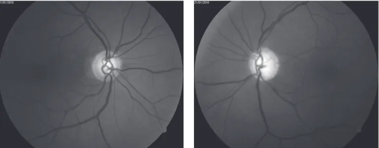
dis-Figure 1. Fundus images showing a normal optic disc with cupping of 0.6 x 0.7 in the right eye and a pale optic disc with cupping of 0.8 x 0.9 in the left eye
Figure 2. Automated perimetry (24:2, white-white) showing inferior arcuate defect in the right eye and diffuse reduction of sensitivity in the left eye. The findings in the left eye are compatible the severe cupping and optic disc pallor observed in retinography
ease, and smoking.
Upon examination, he had a corrected visual acuity of 20/ 20 in the right eye (RE) and counting fingers from a distance of 4 metres in the LE. A decreased pupillary photomotor reflex and a relative afferent pupillary defect (RAPD) 3+/4+ were observed in the LE. Biomicroscopy showed a clear cornea, a deep anterior chamber without reaction, a trophic iris, and in-cipient nuclear cataract in BE. Intraocular pressure (IOP) was 35 mmHg in the RE and 36 mmHg in the LE. The patient had an open anterior chamber angle at 360 degrees and a visible scleral spur in BE. Ophthalmoscopy showed an ON rim of normal colour and cupping of 0.6 x 0.7 in the RE and a pale ON rim with cup-ping of 0.8 x 0.9 in the LE (Figure 1).
24:2 white-white automated perimetry (Humphrey Systems, San Leandro, California) showed an inferior arcuate defect in the RE and diffuse reduction of sensitivity in the LE (Figure 2). These findings were confirmed in at least three visual field tests. The asymmetric progression with indolent visual loss, ON pallor, RAPD in the LE, and diffuse central visual field defect led to the hypothesis of a left pre-chiasmal compressive optic neuropathy. Thus, neuro-imaging tests (magnetic resonance im-aging [MRI] and magnetic resonance angiography) were done, showing a dolichoectatic left ICA compressing the ipsilateral ON and pushing the left part of the optic chiasm upward (Figure 3). Given the high IOP values, we opted for medical treat-ment with hypotensive eye drops (0.03% bimatoprost, 0.2% Atypical progression of visual loss in a patient with primary open-angle glaucoma
42
brimonidine tartrate, and 2% dorzolamide hydrochloride). The IOP was reduced (12/14mmHg) without progression of visual field defects in the RE. In the LE, no progression of the visual field defect was observed during a three-year follow-up period. As for the left optic pathway compression, after weighting the risks and benefits we opted for medical management.
D
ISCUSSIONDolichoectasia is a condition characterised by thinning of arterial walls, with substitution of elastic and reticular fibres by fibrous tissue. This results in increased length, tortuosity, and ir-regularity of the lumen. The most affected intracranial arteries are the internal carotid, vertebral, and basilar arteries(2).
Although the normal limits for the position and diameter of the basilar artery have been well defined, the definition of a dolichoectatic ICA remains subjective. It is worth noting that the normal anatomical variability in the relationship between the ICA and the ON may predispose some patients to symptom-atic compression(3).
Glaucoma involves a characteristic loss of the superior or inferior arcuate retinal nerve fibres, but the papillo-macular bundle tends to be spared during the early stages of the condi-tion, as seen in the RE of the case presented here. In compres-sive optic neuropathies, however, there is visual loss with an ab-normal pupillary reflex and central or cecocentral visual field defects, and dolichoectasia of the ICA can be shown to be one of the causes of the neuropathy(2).
Although POAG is a bilateral disease, it usually manifests asymmetrically, and there may be asymmetric cupping and vi-sual field defects between the two eyes(2). In the present case,
there were significant differences between the two eyes (worse on the left) along with other findings (e.g., relative afferent de-fect and rapid progression of asymmetric glaucomatous disease) that raised the suspicion of an associated neuro-ophthalmic dis-ease. For these reasons, neuro-imaging tests were done to clarify the diagnosis.
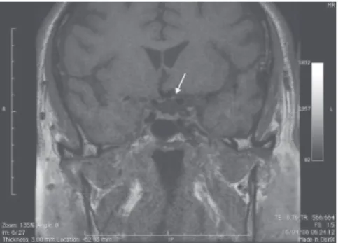
Figure 3. MRI showing contact between the internal carotid artery and the left optic nerve and elevation of the optic chiasm (white arrows). Angio-MRI showing asymmetric calibre of intracranial carotid arteries, larger on the left (white arrow tip).
Dolichoectasia of the ICA can mimic normal tension glau-coma (NTG), as ON compression or insufficient vascular supply can cause ipsilateral visual field loss and increased cupping. Ra-diological studies have shown manifestations such as calcifica-tion, dilacalcifica-tion, and ectasia of the intracavernous carotid arteries adjacent to the intracranial opening of the optic canal in patients with NTG. It is unclear whether there is a correlation between ON cupping asymmetry and the severity of carotid disease. The gradual widening of the ICA lumen (e.g., due to aging and high blood pressure) can result in compression of the adjacent ON in predisposed individuals(2).
Chronic compression of the ON can also compromise re-gional perfusion, producing ischemia in addition to the sive damage to the nerve fibres. Moreover, long-term compres-sion of the intracranial ON can produce a pattern of visual field loss in nerve fibre bundles and ON cupping, consistent with glau-coma(3). In the case presented here, in addition to the
glaucoma-tous damage in BE, the effects of ON compression by the left dolichoectatic ICA should be considered, resulting in asymmet-ric cupping and a pale ON rim.
A study using MRI showed that anatomical compression of the intracranial ON by the supra-clinoid ICA occurs relatively frequently in asymptomatic patients. Thus, identifying this rela-tionship, by itself, may not be clinically relevant. Still, there are reports of patients with occult or progressive optic neuropathy in which no other plausible mechanism of ON damage exists(3).
Some authors suggest that the most reliable specific signs of non-glaucomatous damage associated with compressive le-sions are age under 50 years, ON pallor with increased cupping, and visual field defects respecting the vertical meridian. In such cases, neuro-imaging tests would be indicated(4).
ON decompression often does not result in significant vi-sual recovery and is associated with a higher morbidity and mor-tality(3). Therefore, in the case presented here we opted for
medi-cal management.
After reviewing the literature on the subject, the authors found no similar cases in the Brazilian literature.
In conclusion, when atypical signs are found in glaucoma, Lavezzo MM, Battistella R, Oyamada MK
43
the ophthalmologist should be alert to the possibility of an asso-ciated compressive lesion. For an accurate diagnosis it is neces-sary to evaluate not only the appearance of the cupping, but also the residual neural rim, the pattern of retinal nerve fibre loss, and the visual field defect, as well as the pupillary reflex and colour vision. Once there is a suspicion of optic nerve or chiasm compression, neuro-imaging tests such as MRI and angio-MRI should be done.
R
EFERENCES1. Purvin V, Kawasaki A, Zeldes S. Dolichoectatic arterial compression of the anterior visual pathways: neuro-ophthalmic features and clini-cal course. J Neurol Neurosurg Psychiatry. 2004;75(1):27-32. 2. Ellis MF, Scott M, Erwin G. Carotid artery ectasia coexistent with
pri-mary open angle glaucoma. Clin Experiment Ophthalmol. 2001;29(1):44-6.
3. Jacobson DM. Symptomatic compression of the optic nerve by the carotid artery: clinical profile of 18 patients with 24 affected eyes identified by magnetic resonance imaging. Ophthalmology. 1999;106(10):1994-2004. Comment in Freeman JY, Newman NJ. Ca-rotid artery compression of the optic nerve. Ophthalmology. 2000;107(10):1798-9.
4. Greenfield DS, Siatkowski RM, Glaser JS, Schatz NJ, Parrish RK 2nd. The cupped disc. Who needs neuroimaging? Ophthalmology. 1998;105(10):1866-74. Comment in Lee AG. Differentiating glauco-matous from nonglaucoglauco-matous optic atrophy. Ophthalmology. 1999;106(5):855.
Corresponding author:
Marcelo Mendes Lavezzo
Rua Capote Valente, 136 – apto. 54
05409-000 Jardim América – São Paulo (SP), Brazil. Tel: +5511 3062 4968 / +5511 3069 6289
E-mail: [email protected]
Atypical progression of visual loss in a patient with primary open-angle glaucoma