EFFECTIVENESS OF SPEECH THERAPY IN EVOLUTION
OF ORAL INGESTION IN PATIENTS WITH POST STROKE
OROPHARYNGEAL DYSPHAGIA
Efetividade da intervenção fonoaudiológica na progressão da
alimentação via oral em pacientes com disfagia orofaríngea pós AVE
Clarissa Inaoka(1), Christiane Albuquerque(2)
(1) Centro de Especialização em Fonoaudiologia Clínica CEFAC – Saúde e Educação, Rio De Janeiro, RJ, Brasil. (2) Hospital Universitário Pedro Ernesto, Rio De Janeiro, RJ,
Brasil.
Conlict of interest: non-existent
pneumonia and other pulmonary problems that can be linked to a dysphagia without symptoms2.
Cerebrovascular diseases are considered the
leading cause of death worldwide and the second in Brazil, accounting for numerous sequels that
produce highly disability3,4. Among them there is the
oropharyngeal dysphagia, which has an incidence
that varies from 40% to 90%, becoming, therefore, a common manifestation of stroke2-5.
Changes may vary depending on the site and
extent of the lesion as well as on the age at which
the stroke occurs. The elderly population is most
commonly affected by stroke, and may have more dificulty in compensating changes in muscle tone
that reduce chewing and decrease tongue pressure3.
There is no doubt that the high incidence of dysphagia represents co-factor for mortality and morbidity. Thus, the diagnosis of dysphagia should not be restricted to the acute phase of stroke4.
The patient with dysphagia, while in hospital,
needs care of a multidisciplinary team of speech
INTRODUCTION
It is understood as swallowing, the passage of stomach contents into the mouth, and it may refer to the low of bolus or saliva. It is programmed in
successive phases: preparatory, oral, pharyngeal and esophagic1.
The disorder in the process of swallowing is called
dysphagia and can be caused by a mechanical
or neurological problem. Clinically it may manifest itself through symptoms like disorder in chewing, dificulty in initiating swallowing, nasal regurgitation,
decreased saliva control, coughing and/or choking
during meals. There may be further dehydration,
ABSTRACT
Purpose: to analyze the effectiveness of speech therapy in the evolution of oral ingestion of patients
with dysphagia symptoms, who have suffered previous or current stroke, admitted to a federal hospital
in Rio de Janeiro. Methods: a retrospective study was made from medical records of 20 patients,
for which was requested speech therapy for dysphagia. A functional scale was used to compare the oral ingestion level of each patient, before and after the therapy. Possible interference factors in the progression on the scale were studied: age, duration and incidence of previous stroke, clinical
complications. Results: over 20 patients, 15 showed improvement in the oral intake scale after
speech therapy. Clinical complications were considered statistically signiicant for the lack of evolution in oral feeding. Other analyzed factors were not statistically signiicant, and they did not interfere in the improvement or worsening of the patient. Conclusion: speech therapy is effective in improving
food intake by mouth in patients treated in hospitals with neurogenic dysphagia after stroke, except if
clinical complications appear during the process.
disorders and designed to describe the change in
functional communication of the individual and/or
ability to swallow over time. The audiologist marks the level at which the patient is on admission and
at discharge to describe the amount of change in communication and swallowing after intervention. By examining the admission and discharge of banknotes, one can evaluate the amount of change, and thus the beneits of treatment11.
The objective of this study was to analyze the effectiveness of speech therapy on progression of oral feeding of patients with symptoms of dysphagia
that underwent prior or current stroke, admitted to a
federal hospital in Rio de Janeiro, using the scale-ASHA NOMS.
METHODS
A retrospective study of medical records of patients with stroke admitted to a federal hospital in the city of Rio de Janeiro, with symptoms of neurogenic oropharyngeal dysphagia, and for which language intervention was requested by attending physicians. Data from January to August 2011 were
collected.
Of the 161 patients enrolled in the Speech
Therapy in the analyzed period, 20 adults and elderly were included in the survey, as meeting the criteria
for participation, namely: having suffered previous or current stroke, with symptoms of oropharyngeal
dysphagia; breathing ambiet air; present clinically stable and respond to simple verbal commands. Patients who showed abnormalities in phonatory structures, tracheostomy, ventilator dependency,
lowered level of consciousness and clinically very committed were excluded.
The sample comprised 20 patients, 14 females (70.0 %). The average age was 71.85 (+/-14.41
years). The minimum age was 38 years and
maximum 88 years. The median was 75 years. According to the need of each patient, a program of rehabilitation of swallowing consists of evalua
-tions, analysis of case severity and risk of dysphagia
in a managerial9 perspective was performed.
Speech therapy focused was also performed for the rehabilitation of swallowing during hospital
-ization, with techniques described in the literature
as thermal stimulation, swallowing maneuvers and
myofunctional exercises12,13.
ASHA NOMS scale was used to check the devel
-opment of oral intake in two stages, before and after speech therapy. To mark the irst level at which the patient was in the range, it was considered the food of this status before the initial speech evaluation. And to mark the level after speech therapy, dietary therapists, doctors of different specialties, physio
-therapist, nutritionist, nurse, occupational therapist
and psychologist. This team has a focus in minimizing the risks of early complications and preparing for the rehabilitation of sequels. The early speech-language intervention (twenty-four to forty-eight hours after
the event and with the patient clinically stable) in a
hospital environment aims an early identiication of dysphagia and prevention of clinical complications6
and may shorten the use of alternative feeding means, time of hospitalization, and contribute to the improvement of pulmonary condition. The development of safe and functional oral intake of the patient, associated with the maintenance of lung health and nutritional status is a signiicant evidence of the therapeutical effectiveness7. Therefore, there
is a necessity of checking which alternative feeding means and food consistencies the patient with oropharyngeal dysphagia will present before and after speech therapy.
Some factors can interfere in patient outcomes in relation to food intake by mouth, as the clinical worsening of the patient, the clinical complications and decreased level of consciousness. Other factors
analyzed in studies, such as age and underlying
disease, were not statistically signiicant, suggesting not interfere with improvement or worsening of
patient8.
It is of utmost importance the realization of a
speech-language service management by standard
indicators, facilitating the analysis of performance over time, also the inclusion of new processes and
technologies, and comparison with other services
judged as references, called benchmark. This management contributes to highlight the eficiency and effectiveness of rehabilitation programs9.
The effectiveness of rehabilitation in oropha -ryngeal dysphagia can be proven when the patients
can eat by mouth adequately. In order to measure this effectiveness, current research sought to establish scales of functional control of swallowing, with features such as: rehabilitation time compared to their functional effects, type of alternative feeding
means that the patient began rehabilitation and which changes occurred during the process,
increased volume, change in consistency of oral
intake and others10.
A measuring scale was developed by the
American Association of Speech-Language
Pathology (ASHA) in 1997, called the National
System of Measurement Results (NOMS) – which consists of a collection of data to illustrate the value of speech therapy in adults and children referring
to communication and swallowing. Those measures
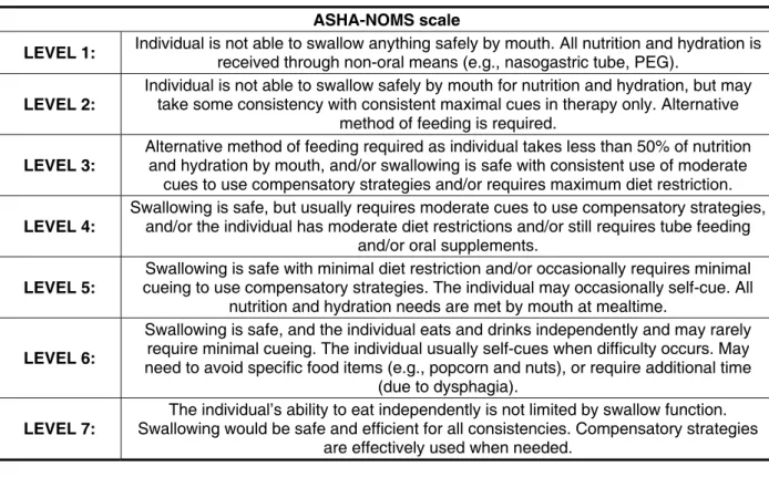
ASHA NOMS scale describes whether there was a change in functional status after the speech therapy of patients with dysphagia (Figure 1)
swallowing . The scale has not yet been validated
in Portuguese translation therefore will be used in
English.
status was veriied at speech therapy discharge. This discharge encompasses the following reasons: functional swallowing rehabilitation, hospital
discharge, gastrostomy, putting sector in disposal
due to no possibility of intervention at the time (eg, in the case of tracheal intubation).
ASHA-NOMS scale
LEVEL 1: Individual is not able to swallow anything safely by mouth. All nutrition and hydration is
received through non-oral means (e.g., nasogastric tube, PEG).
LEVEL 2:
Individual is not able to swallow safely by mouth for nutrition and hydration, but may take some consistency with consistent maximal cues in therapy only. Alternative
method of feeding is required.
LEVEL 3:
Alternative method of feeding required as individual takes less than 50% of nutrition and hydration by mouth, and/or swallowing is safe with consistent use of moderate
cues to use compensatory strategies and/or requires maximum diet restriction.
LEVEL 4:
Swallowing is safe, but usually requires moderate cues to use compensatory strategies, and/or the individual has moderate diet restrictions and/or still requires tube feeding
and/or oral supplements.
LEVEL 5:
Swallowing is safe with minimal diet restriction and/or occasionally requires minimal cueing to use compensatory strategies. The individual may occasionally self-cue. All
nutrition and hydration needs are met by mouth at mealtime.
LEVEL 6:
Swallowing is safe, and the individual eats and drinks independently and may rarely require minimal cueing. The individual usually self-cues when difficulty occurs. May need to avoid specific food items (e.g., popcorn and nuts), or require additional time
(due to dysphagia).
LEVEL 7:
The individual’s ability to eat independently is not limited by swallow function. Swallowing would be safe and efficient for all consistencies. Compensatory strategies
are effectively used when needed.
Figure 1 – National Measurement System Results – American Speech-Language-Hearing Association
– National Outcomes Measurement System – ASHA NOMS
Improvement was considered when an increase in the level scale in the post speech therapy occurred, and worsening considered when lowering the level in the post speech therapy.
It was also veriied if the following factors could interfere with the progression of oral feeding of
patients with speech therapy, such as:
• Age: age was analyzed in the group that did not
develop in ASHA NOMS scale and the group that evolved, the average was compared to verify
whether age has an impact on the improvement in scale.
• Time of stroke and incidence of previous strokes:
we veriied whether these factors inluence the level progression in the ASHA NOMS scale.
• Clinical deterioration and lowering the level of consciousness: these items were considered when the patient did not respond to simple
verbal commands for different reasons, namely infection, worsening of respiratory symptoms, worsening of neurological symptoms; compa -rison was made between patients who presented
these factors during language intervention with
patients who did not, checking which group most improved in the scale. There are reports in which
these events may affect the mechanisms of
airways protection8,14.
The effectiveness of swallowing rehabilitation
through outcome indicators was also analyzed: • Time to pull the alternative feeding way. • Time to return to oral feeding.
These indicators are expressed in relation to the number of days from the irst speech evaluation9.
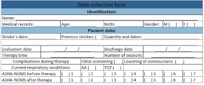
Data were collected from medical records with speciic form, based on a questionnaire created by
Furkim and Sacco8 (Figure 2).
of patients who have the alternative feeding way removed and return to oral feeding safely within a
time scale : 0 to 5 days, 6 to 10 days, 11 to 15 days and above 15 days15 .
Legend: AA: ambient air; TQT: tracheostomy
Figure 2 – Form for the data to be collected from medical records
First the acquired data from medical records using the form were addressed descriptively, as well as evolution or regression in ASHA NOMS scale. Subsequently, this variation was compared with the factors that could interfere with speech therapy.
This study was approved by the Ethics Committee
in Research of the institution, no. 02-2011.
The indings of the study were statistically analyzed with the following tests: Fischer’s exact test, which measures the degree of relationship
between the two traits in independent samples.
And the “Student t test”, which is to use data from a
sample to calculate the statistic and then compare
it with the distribution of T student, to identify the probability of having obtained the observed result if
the null hypothesis is true.
RESULTS
Table 1, based on a study done by Furkim and Sacco8, shows that 15 (75.0%) patients improved
after intervention, 4 (20.0%) patients did not develop in ASHA NOMS scale, and 1 (5.0%) patient had
worsening.
Table 2 shows the statistical analysis between
the average ages of the patients divided into two
groups, those who progressed and those who did
not develop the ASHA NOMS scale. It was found
that the group that did not evolved had higher average (77.20 years) compared to the group that
evolved (70.06 years). However, the difference is not statistically signiicant .
Table 3 shows the relationship between the
time of the stroke and changes in the ASHA NOMS
scale. The patients were divided into two groups
according to the time of stroke occurrence in the day of evaluation: acute (30 days) and not acute phase (after 31 days). Of the patients who developed the scale, the majority (66.7% – 10 individuals) had
had stroke 1 month ago, and the group that did not
evolved, 40.0% (2 subjects) also had had a recent stroke. There was no signiicant difference between the time of stroke and evolution in scale.
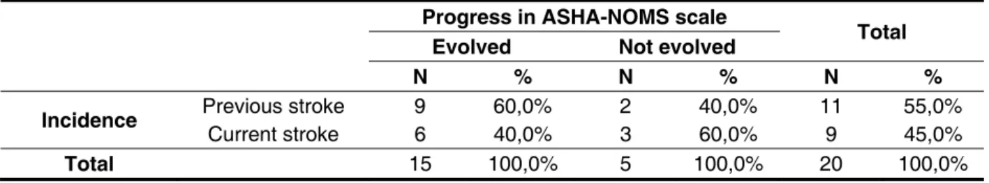
Table 4 shows the relationship between the
incidence of previous strokes and progression of feed consistency. It was veriied that in the group that progressed, 9 (60.0%) had already been affected by previous strokes, and in the group that did not evolved, 2 subjects (40.0 %). There was no signiicant statistical difference.
After
1 2 3 4 5 6 7 Total
Before
1 4 0 0 0 3 7 1 15
2 0 0 0 0 0 0 0 0
3 0 0 0 0 1 1 0 2
4 1 0 0 0 0 2 0 3
Total 5 0 0 0 4 10 1 20
Table 1 – Evolution of the patients according to levels of functional evaluation of oral intake
Legend: Dark gray: Number of patients who remained on the same level after speech therapy. Black: Number of patients who worsened after speech therapy.
Light gray: Number of patients who improved after voice therapy.
N Group Average age
15 Evolved 70,06
5 Not evolved 77,20
Table 2 – Average age-related developments in ASHA NOMS scale
Student’s t test: p = 0.074 (not signiicant, p <0.05)
Progress in ASHA-NOMS scale
Total Evolved Not evolved
N % N % N %
Stroke phases Acute phase 10 66,7% 2 40,0% 12 60,0%
Not acute phase 5 33,3% 3 60,0% 8 40,0%
Total 15 100,0% 5 100,0% 20 100,0%
Table 3 – Relationship between time of occurrence of stroke and evolution in ASHA NOMS scale
Fischer’s exact test: p = 0.347 (not signiicant, p <0.05)
Table 4 – Relationship between previous incidence of stroke and evolution in ASHA NOMS scale
Fischer’s exact test: p = 0.616 (not signiicant, p <0.05)
Progress in ASHA-NOMS scale
Total Evolved Not evolved
N % N % N %
Incidence Previous stroke 9 60,0% 2 40,0% 11 55,0%
Current stroke 6 40,0% 3 60,0% 9 45,0%
evolved, only 3 (21.4 %) patients demonstrated some problems during the process of swallowing rehabilitation. This shows that the onset of these complications contribute to the lack of progress in the ASHA NOMS scale, statistically proven.
Table 5 presents the clinical complications
during speech therapy, namely: onset of infection, worsening of respiratory symptoms and/or neuro
-logical symptoms. All 5 (100 %) patients in the group
that did not develop showed clinical worsening or
decreased level of consciousness. In the group that
Progress in ASHA-NOMS scale
Total Evolved Not evolved
N % N % N %
Complications
No complications 12 80,0% 0 0,0% 12 60,0%
Clinical worsening or lowering of
consciousness 3 20,0% 5 100,0% 8 40,0%
Total 15 100,0% 5 100,0% 20 100,0%
Table 5 – Clinical complications during therapy and its relation to progression in ASHA NOMS scale
Fischer’s exact test: p = 0.003 (signiicant, p <0.05)
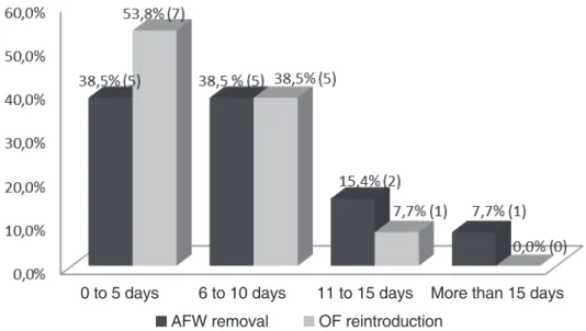
In the group of patients who developed the ASHA NOMS scale, two indicators of outcome, time to remove the alternative feeding way (AFW) and time to reintroduction of oral feeding (OF) were analyzed . Of the 15 patients who had improvement in the scale, 2 were already without AFW and released OF, and were presenting symptoms of dysphagia for some food consistencies. Therefore, 13 patients
were evaluated in these indicators. In Figure 3 it can
be seen that the vast majority of patients (76.9% – 10 subjects) could have the AFW removed before 10 days, and almost all patients (92.3 % – 12 individuals) obtained reintroduction of oral feeding before 10 days. The average for the two indicators was 3.25 days with a standard deviation of 2.06 for the AFW removal and 3.30 for the reintroduction of oral feeding.
Legend: AFW: alternative feeding means; OF: oral feeding.
Figure 3 – Proportion of patients who returned to oral feeding and had the alternative feeding way removed in a scale of time (in days)
AFW removal OF reintroduction
pharyngeal residue. It suggested that it is possible
that swallowing recovers functionally but remains
impaired at a more intricate level. This may also
explain the increased incidence of dysphagia after stroke for the second or third time25. In the present
study it was not possible to check this, since most
of the patients who had progressed on ASHA scale
repeat strokes, and in the group that did not evolved
half had a previous stroke history. This may be due
to compensatory mechanisms that patients develop
after some involvement. This compensation may
cause the patient who had recurrent stroke, to adapt
better to the different pattern of swallowing than a stroke patient who presents for the irst time. A more speciic comparative research would be important to verify the relationship between the incidence of
stroke and dysphagia, particularly in establishing
a relationship between the extent of injury and
swallowing disorders.
Aging can cause change in swallowing, called presbifagia. It can still be considered a risk factor
for dysphagia26. In this study the average age of the
patients in the group that developed in the ASHA
scale (70.06 years) showed no statistically signif
-icant difference in average age of the group who did not progress (77.20 years). This fact is due to the
age given by the participants, mostly elderly, which prevented a comparison with younger patients.
There are reports in the literature regarding
the level of consciousness, which can inluence the supply of safe diet by mouth8,14,27. It can be
considered insuficient for protecting airways28.
The results of this research were similar to these
reports. All patients in the group that did not progress in ASHA scale had clinical worsening or decreased
level of consciousness, veriied by not responding
to simple verbal commands. In these situations,
the oral feeding is suspended, which prevents food
progression and compromises speech therapy,
since there are no reports on the effectiveness of passive speech therapy for swallowing rehabilitation
literature. In the group that evolved on the scale,
only 18.2 % had some medical complications.
In the group that evolved on scale , it was
possible to withdraw the alternative feeding way in 77% of patients within 10 days. It was still possible to reintroduce oral feeding safely in most of these patients (92.3 %) , also in less than 10 days. This shows the importance of speech therapy services to minimize prolonged hospitalization of patients with
swallowing disorders.
Most patients (75.0%) at the end of the speech
rehabilitation, was located above the level in 5 ASHA
scale, which is not needed alternative feeding way because safe oral intake is viable, without risk of
complications. It is assumed that with the reduction Regarding the 5 patients who did not develop the
ASHA NOMS scale, 3 of them were submitted to gastrostomy. The other 2 patients had worsening of
respiratory symptoms and underwent endotracheal intubation and remained in this condition until the
end of the period analyzed in this study (September
2011) .
DISCUSSION
In this study the effects of speech therapy in the progression of food consistency in patients with stroke were analyzed. Most pacients presented
developments at ASHA scale during the period
studied, which demonstrates the effectiveness of voice therapy in the progression of oral feeding safely in hospital.
Despite the delineation of pathology studied, this presents a wide range of occurrence, linked to the type of stroke (ischemic or hemorrhagic) , site and extent of lesion15,17,18. The present study included
a heterogeneous sample about these factors. Regarding the type of stroke, only one patient
had hemorrhagic stroke, which prevented larger
comparisons. It is suggested that further research on speciic populations on the size and location of the lesion is needed, checking for relationship with the eficacy of speech therapy.
Another factor to be considered is the time of the stroke, which can inluence the rehabilitation of swallowing. The clinical impression is a sponta
-neous recovery of swallowing. This improvement
is relatively common and occurs over days or weeks15,19-21. In the study of Smithard et al, using
videoluoroscopic examination of swallowing, an incidence of aspiration of 22% by an average of two days after the stroke and 15% was found in a
month22. This can be explained by the distribution
of bilateral control of the swallowing muscles in the motor cortex. After hemispheric stroke, neuroplas -ticity promotes an adaptation and allows the control
of the muscles of swallowing, it is reorganized in the hemisphere not affected14. Other studies also
reported the recovery of dysphagia months or years after a stroke, but the recovery rate remained
low23,24.
In this research, the majority of participants were in the acute phase of stroke, in both groups:
that developed and did not develop in ASHA scale.
Therefore, it was not possible to verify statistically signiicant difference between patients with recent
and not recent stroke. Researches with larger samples are needed.
Logemann (1983) reported a delay in time after the action of swallowing recovery post
obtained a validated translation into Portuguese, to
facilitate and disseminate its use in Brazil.
Studies with larger and more speciic populations in these affections are needed, yet the effectiveness of speech therapy on progression of oral intake of hospitalized patients with dysphagia after stroke
was observed.
CONCLUSION
This work has been possible to verify the effectiveness of speech therapy in the evolution of food consistencies of patients with dysphagia who
underwent previous or current stroke, using the
ASHA NOMS scale as a marker.
It was also found that the onset of clinical deterio
-ration or loss of consciousness level directly affects
the speech swallowing rehabilitation, hindering
the progression of oral feeding, described by no evolution in the ASHA NOMS scale.
of hospital costs. However, this was not the purpose of this study, and it is suggested the realization of speciic research on the effects of speech rehabili
-tation of swallowing in reducing hospital costs. The American Speech-Language-Hearing
Association (ASHA) in their assignments and responsibilities includes the speech language
pathologist (SLP) in dysphagia management: identi
-fying and using appropriately measures of functional outcomes; knowing the quality improvement policies
established by accrediting bodies; knowing the
methods used to measure and monitor the quality of
important processes and results11.
It was observed that the ASHA scale is a practical
and easy to apply label to verify the oral intake of
hospitalized patients. Patients can be monitored
objectively the evolution of levels on the scale before and after speech therapy. It also showed the effects of the onset of clinical events in this progression. It can be considered an instrument for measuring effectiveness of speech therapy on progression of oral feeding safely. It is important that this scale
RESUMO
Objetivo: analisar a efetividade na progressão da alimentação via oral de pacientes com sintomas de
disfagia, que sofreram acidente vascular encefálico prévio ou atual, internados em um hospital federal
do Rio de Janeiro. Métodos: foi feito estudo retrospectivo do prontuário de 20 pacientes com acidente
vascular encefálico, para os quais foi solicitada fonoterapia para disfagia. Para comparação do nível de ingestão oral de cada paciente, antes e depois da terapia, foi utilizada uma escala funcional. Foram estudados os possíveis fatores de interferência na progressão na escala como: idade, intercorrências clínicas, tempo e incidência prévia do AVE. Os seguintes indicadores de resultado foram analisados:
tempo para retirada de via alternativa de alimentação e tempo para reintrodução de alimentação via oral. Resultados: dos 20 pacientes, 15 apresentaram melhora na escala de ingestão oral após a
fonoterapia. As intercorrências clínicas foram consideradas estatisticamente signiicantes para a não evolução da alimentação via oral. Os outros fatores analisados não demonstraram signiicância esta
-tística, sugerindo não interferir na melhora ou piora do paciente. Foi possível reintroduzir alimentação
via oral e retirar via alternativa de alimentação antes de 10 dias. Conclusão: a fonoterapia é efetiva
para melhorar a ingestão de alimentos por via oral nos pacientes com AVE e disfagia neurogênica, atendidos em ambiente hospitalar, salvo se apresentarem intercorrências clínicas e rebaixamento do nível de consciência durante o processo.
c1997-2012. [acesso em 2012 Mai 28]. Disponível
em: http://www.asha.org/members/research/noms/.
12. Cola PC, Gatto AR, Silva RG, Schelp AO, Henry MACA. Reabilitação em Disfagia Orofaríngea Neurogênica: sabor azedo e temperatura fria. Rev CEFAC [Internet]. 2008. [acesso em 2012 Mai 28];10(2):200-5. Disponível em: http://www.scielo. br/scielo.php?pid=S151618462008000200009&scri pt=sci_arttext.
13. Marchesan IQ, Furkim AM. Manobras utilizadas na reabilitação da deglutição. In: Costa M, Castro LP. Tópicos em deglutição e disfagia. Rio de Janeiro: Medsi; 2003. p. 375-84.
14. Bassi AER, Mitre EI, Silva MAOM, Arroyo MAS, Pereira MC. Associação entre disfagia e o topodiagnóstico da lesão encefálica pós-acidente vascular encefálico. Rev CEFAC [Internet]. 2004. [acesso em 2011 Set 27];6(2):135-42. Disponível em: http://www.cefac.br/revista/revista62/ Artigo%203.pdf.
15. Moraes DP. Estruturação de um Serviço de Fonoaudiologia Hospitalar [dissertação] São Paulo: Faculdade de Medicina da Universidade de São
Paulo; 2010.
16. Singh S, Hamdy S. Dysphagia in stroke
patients. Postgrad Med J [Internet]. 2006. [acesso em 2011 Sep 27];82(968):383-91. Disponível em: http://www.ncbi.nlm. nih.gov/pmc/articles/PMC256 3739/?tool=pubmed.
17. Assencio-Ferreira VJ, Neurologia e Fonoaudiologia. São José dos Campos: Pulso;
2003.
18. Barros AFF, Fabio SRC, Furkim AM.
Correlação entre os achados clínicos da deglutição
e os achados da tomograia computadorizada de crânio em pacientes com acidente vascular cerebral isquêmico na fase aguda da doença. Arq. Neuro-Psiquiatr. [Internet]. 2006. [acesso em 2011 Set 27];64(4):1009-14. Disponível em: http://www.scielo.br/scielo.php?pid=S0004-282X2006000600024&script=sci_ arttext.
19. Gordon C, Hewer RL, Wade DT. Dysphagia in acute stroke. BMJ (Clin Res Ed) [Internet]. 1987. [acesso em 2011 Set 27];295(6595):411-4.
Disponível em: http://www.ncbi.nlm.nih.gov/pmc/
articles/PMC1247273/
20. Mann G, Hankey GJ, Cameron D. Swallowing function after stroke: prognosis and prognostic factors at 6 months. Stroke [Internet]. 1999. [acesso em 2012 Set 27]; 30:744-8. Disponível em: http://
www.ncbi.nlm.nih.gov/pubmed/10187872.
21. Barer DH. The natural history and functional consequences of dysphagia after hemispheric stroke. J Neurol Neurosurg Psychiatry [Internet]. 1989. [acesso em 2011 Set 27];52(2):236-41.
REFERÊNCIAS
1. Douglas CR. Fisiologia aplicada à Fonoaudiologia. 2a ed. Rio de Janeiro: Guanabara Koogan; 2006.
2. Furkim AM, Santini CRQS. Disfagias Orofaríngeas. 2a ed. Barueri: Pró-fono; 2008. 3. Martino R, Foley N, Bhogal S, Diamant N, Speechley M, Teasell R. Dysphagia after stroke:
incidence, diagnosis, and pulmonary complications.
Stroke [Internet]. 2005. [acesso em 2010 Set 02];36(12):2756-63. Disponível em http://stroke.
ahajournals.org/content/36/12/2756.long.
4. Schelp AO, Cola PC, Gatto AR, Silva RG, Carvalho LR. Incidência de disfagia orofaríngea após acidente vascular encefálico em hospital público de referência. Arq Neuropsiquiatria [Internet]. 2004. [acesso em 2010 Set 05];62(2b):503-6. Disponível em: http://www.scielo.br/pdf/anp/v62n2b/ a23v622b. pdf.
5. Okubo PCMI. Detecção de disfagia na fase aguda do acidente vascular cerebral isquêmico: proposição de conduta baseada na caracterização dos fatores de risco [Tese]. Ribeirão Preto: Faculdade de Medicina da Universidade de São Paulo; 2008. 6. Abdulmassih EMS, Filho EDM, Santos RS, Jurkiewicz AL. Evolução de Pacientes com Disfagia Orofaríngea em Ambiente Hospitalar. Arq. Int. Otorrinolaringol [Internet]. 2009. [acesso em 2010 Set 08];13(1):55-62. Disponível em: http:// www.arquivosdeorl.org.br/conteudo/acervo_port. asp?id=589.
7. Silverio CC, Hernandez AM, Gonçalves MIR. Ingestão oral do paciente hospitalizado com disfagia orofaríngea neurogênica. Rev. CEFAC [Internet]. 2009. [acesso em 2010 Set 27];12(6):964-70. Disponível em: http://www. scielo.br/scielo.php?script=sci_arttext&pid=S1516-18462010005000090&lng=en.
8. Furkim AM, Sacco ABF. Eicácia da fonoterapia em disfagia neurogênica usando a escala funcional de ingestão por via oral (FOIS) como marcador.
Rev. CEFAC. 2008;10(4):503-12.
9. Moraes DP, Andrade CRF. Indicadores de qualidade para o gerenciamento da disfagia em
Unidades de Internação Hospitalar. J Soc Bras Fonoaudiol. 2011;23(1):89-94.
10. Silva RG. A eicácia da reabilitação em disfagia orofaríngea. Pró-Fono R. Atual. Cient [Internet]. 2007. [acesso em 2010 Set 21];19(1):123-30. Disponível em: http://www.scielo.br/pdf/pfono/ v19n1/13.pdf.
11. ASHA: American Speech and Hearing
Association. National Outcomes Measurement System (NOMS) [Internet]. [local desconhecido]:
25. Logemann JA. Evaluation and treatment of
swallowing disorders. 2ª ed. Austin (TX): Pro-Ed; 1983.
26. Russo, IP. Intervenção fonoaudiológica na
Terceira idade. Rio de Janeiro: Revinter; 2004.
27. Padovani AR, Moraes DP, Medeiros GC, Almeida TM, Andrade CRF. Intubação orotraqueal e disfagia: comparação entre pacientes com e sem dano cerebral. Einstein [Internet]. 2008. [acesso em 2011 Set 27];6(3):343-9. Disponível em: http:// apps.einstein.br/revista/arquivos/PDF/1000-v6n3aAO1000portp343-9.pdf.
28. Park M, et al. Traqueostomia percutânea no doente crítico: a experiência de uma unidade de terapia intensiva clínica. J. bras. pneumol. [Internet]. 2004. [acesso em 2011 Set 27];30(3):237-42. Disponível em: http://www.scielo.br/scielo. php?script=sci_artte xt&pid=S1806-37132004000300009.
Disponível em: http://www.ncbi.nlm.nih.gov/ pubmed/ 2564884.
22. Smithard DG, O’Neill PA, England RE, Park CL, Wyatt R, Martin DF, et al. The natural history of dysphagia following a stroke. Dysphagia [Internet]. 1997. [acesso em 2011 Set 27]. 12(4):188-93.
Disponível em: http://www.ncbi.nlm.nih.gov/ pubmed/9294937.
23. Wanklyn P, Cox N, Belield P. Outcome in patients who require a gastrostomy after stroke. Age Ageing [Internet]. 1995. [acesso em 2011 Set 27];24(6):510-4. Disponível em: http://www.ncbi.
nlm.nih.gov/pubmed/8588542.
24. Hull MA, Rawlings J, Murray E, et al. Audit of outcome of longterm enteral nutrition by percutaneous endoscopic gastrostomy. Lancet [Internet]. 1993. [acesso em 2011 Set 27];341(8849):869-72.
Disponível em: http://www.ncbi.nlm. nih.gov/ pubmed/8096573.
Received on: May 28, 2012
Accepted on: September 04, 2012
Endereço para correspondência:
Clarissa Inaoka Rua Polar, 280
São José dos Campos – SP
CEP: 12230-240