HLA-A*31 as a marker of genetic susceptibility
to sepsis
INTRODUCTION
Bacterial proliferation in the bloodstream triggers the innate immunity-mediated systemic inlammatory process that characterizes sepsis, a syndrome with a lethality rate between 20 to 50% in intensive care patients,(1) and whose most severe presentation, septic shock, has challenged medical practitioners for decades. Ironically, in the case of sepsis, medicine could be falling victim to its own success, whereby the signiicant increase in modern invasive procedures performed to save lives exposes the body to sepsis-inducing organisms. Additionally, the new and powerful antibiotics used to ight sepsis increase the drug resistance that sustains its lethality.
he HLA haplotypes are one of the genetic feature that most strongly associates with autoimmunity.(2) Since the irst description of the strong association of ankylosing spondylitis and HLA-B*27,(3) many other associations with autoimmune diseases have been described. Although these associations have been thoroughly studied and documented, little progress has been made in understanding how a particular HLA haplotype afects the corresponding disease. Multiple sclerosis,(4) psoriasis(5) and type 1 diabetes(6) are some classical
Fabiano Pinheiro da Silva1, Germano Preuhs
Filho2, Eduardo Finger3, Hermes Vieira Barbeiro1,
Fernando Godinho Zampieri1, Alessandra
Carvalho Goulart1, Francisco Torggler Filho1,
Nicolas Panajotopoulos2, Irineu Tadeu Velasco1,
Jorge Kalil2, Heraldo Possolo de Souza1, Luiz
Monteiro da Cruz Neto1, Hélcio Rodrigues2
1. Department of Emergency Medicine, Universidade de São Paulo - USP - São Paulo (SP), Brazil.
2. Immunology Department, Instituto do Coração, Universidade de São Paulo - USP - São Paulo (SP), Brazil.
3. Laboratórios Salomão Zoppi - São Paulo (SP), Brazil.
Objective: he HLA haplotype has been associated with many autoimmune diseases, but no associations have been described in sepsis. his study aims to investigate the HLA system as a possible marker of genetic sepsis susceptibility.
Methods: his is a prospective
cohort study including patients admitted to an intensive care unit and healthy controls from a list of renal transplant donors. Patients with less 18 years of age; pregnant or HIV positive patients; those with metastatic malignancies or receiving chemotherapy; or with advanced liver disease; or with end-of-life
Conflicts of interest: None.
Submitted on September 26, 2013 Accepted on October 31, 2013
Corresponding author:
Fabiano Pinheiro da Silva
Departamento de Emergências Clínicas da Universidade de São Paulo
Avenida Dr. Arnaldo, 455, sala 3.189 Zip code: 01246-000 - São Paulo (SP), Brazil E-mail: pinheirofabiano@hotmail.com
HLA-A*31 como marcador de suscetibilidade genética em sepse
ABSTRACT
Keywords: Sepsis; HLA-A antigens; Inlammation; Genetic markers
conditions were excluded. he DNA was extracted from the whole blood and HLA
haplotypes determined using MiliPlex®
technology.
Results: From October 2010 to
October 2012, 1,121 patients were included (1,078 kidney donors, 20 patients admitted with severe sepsis and 23 with septic shock). HLA-A*31 positive subjects had increased risk of developing sepsis (OR 2.36, 95%CI 1.26-5.35). Considering a p value <0.01, no other signiicant association was identiied.
Conclusion: HLA-A*31 expression is associated to risk of developing sepsis.
examples. Some exceptions exist though, such as the process whereby peptide deamination may lead to celiac disease,(7) or the scenario in which molecular mimicry between bacterial and endogenous molecules can produce Lyme disease.(8) Moreover, recent studies suggest that HLA contribution to autoimmunity may be polygenic.(9)
Furthermore, the relevance of HLAs is growing in areas such as immunization,(10,11) anthropology(12)
and pharmacogenomics.(13) Indeed, some adverse
drug reactions have shown strong HLA associations, as described for abacavir (HLA-B*5701), allopurinol (HLA-B*58) and carbamazepine (HLA-B*1502). Other conditions associated with the HLA system include, smoking behavior, schizophrenia, Parkinson's disease, narcolepsy and coronary heart disease.(14) his article investigated the HLA class I and II loci as possible genetic markers for sepsis susceptibility.
METHODS
he current study was a prospective cohort study conducted in the intensive care unit (ICU) of the Hospital das Clínicas da Universidade de São Paulo (USP), in São Paulo (Brazil), and conceived as part of the BRISK Project (Brazilian Initiative for Sepsis Knowledge), which was launched in 2009 to investigate diferent molecular aspects of sepsis. All patients admitted to the study unit, a 14 beds ICU, were consecutively included upon a signed informed consent form. Surgical, trauma and coronary syndrome patients are usually admitted to speciic ICUs in our hospital, making our ICU populations very homogeneous. Patients who were under 18 years of age, pregnant or HIV-positive; those who had metastatic malignancies or were undergoing chemotherapy; those who had advanced hepatic diseases; those with end-of-life conditions and those who refused to participate were excluded from this study. he control group consisted of a sample of kidney transplant donors received from the HLA service of the
Instituto do Coração, from the Universidade de São Paulo. Severe sepsis and septic shock were deined according to the American College of Chest Physicians/Society of Critical Care Medicine (ACCP/SCCM) Consensus Conference Committee proposed in 1992.(15) he control group was constituted by kidney donors evaluated at the Laboratory of Histocompatibility of the Immunology Department of the Instituto do Coração of USP during routine screening tests prior to donation. he protocol was approved by the Hospital das Clínicas Ethics Committee. Additionally, patients (or their close relatives) received
detailed explanations and were provided written consent for inclusion in the study protocol (protocol number 1,207/09).
DNA was extracted from whole blood, and HLA haplotyping was performed with MiliPlex®
technology. All statistical analyses were performed with the Statistical Package for the Social Sciences (SPSS) version 19.0 and R statistical software. A p value ≤0.01 was considered to be statistically signiicant. his signiicance level was chosen to reduce the risk of type I error, considering the small sample size.
RESULTS
his study included 1,121 patients (1,078 kidney donors, 20 severe sepsis patients and 23 patients admitted for septic shock) enrolled between October 2010 and October 2012. Sepsis, stroke, consciousness level changes, pulmonary edema and asthma/chronic obstructive pulmonary disease (COPD) accounted for more than 90% of the conditions afecting patients included in this study.
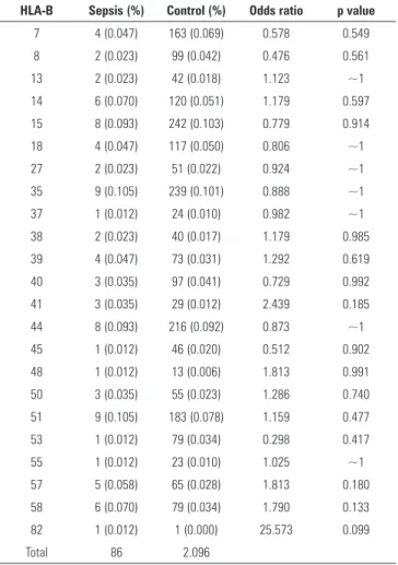
A signiicant association was found between HLA-A*31 and sepsis risk. HLA-A*31-positive individuals had a 2.36-fold higher risk of developing sepsis than HLA-A*31-negative patients. No other signiicant HLA association was identiied in our analysis using a 0.01 signiicance level. However, the comparison of the prevalence of HLA-A*30 between the control and septic groups showed a bordering value (Tables 1 to 5).
DISCUSSION
HLA classes I and II gene polymorphisms are the strongest and most consistent susceptibility alleles for autoimmune diseases. he HLA region is located at 6p21.3 and encompasses more than 400 genes. Single nucleotide polymorphism (SNP) genotyping technologies have identiied several SNPs that confer a very strong autoimmunity risk; however, strong linkage disequilibrium across the HLA region(16) complicates this interpretation. Indeed, the classes DR and DQ loci have shown strong association with a plethora of autoimmune diseases(17) and HLA-A and HLA-B are also commonly found to contribute to disease risk.
Table 1 - Prevalence of HLA*A in septic and control patients
HLA-A Sepsis (%) Control (%) Odds ratio p value
1 5 (0.058) 185 (0.078) 0.653 0.627
2 16 (0.186) 497 (0.211) 0.778 0.676 3 7 (0.081) 216 (0.092) 0.783 0.895
11 3 (0.034) 114 (0.048) 0.636 0.751
23 3 (0.035) 123 (0.052) 0.589 0.643 24 3 (0.035) 201 (0.085) 0.361 0.144
26 5 (0.058) 76 (0.032) 1.590 0.312 29 6 (0.070) 99 (0.042) 1.464 0.328
30 9 (0.105) 168 (0.071) 1.294 0.034
31 9 (0.105) 92 (0.039) 2.364* 0.006 32 3 (0.035) 57 (0.024) 1.272 0.783
33 3 (0.035) 94 (0.040) 0.771 ~1
34 2 (0.023) 22 (0.009) 2.196 0.466 66 2 (0.023) 25 (0.011) 1.933 0.564
68 8 (0.093) 128 (0.054) 1.510 0.194
74 2 (0.093) 45 (0.019) 1.074 ~1
Total 86 2.142
Results expressed as number (%). The percentage is in regard of the total number of alleles in the studied population.* CI95% (1.26-5.35).
Table 2 - Prevalence of HLA*B in septic and control patients
HLA-B Sepsis (%) Control (%) Odds ratio p value
7 4 (0.047) 163 (0.069) 0.578 0.549 8 2 (0.023) 99 (0.042) 0.476 0.561
13 2 (0.023) 42 (0.018) 1.123 ~1
14 6 (0.070) 120 (0.051) 1.179 0.597 15 8 (0.093) 242 (0.103) 0.779 0.914
18 4 (0.047) 117 (0.050) 0.806 ~1
27 2 (0.023) 51 (0.022) 0.924 ~1 35 9 (0.105) 239 (0.101) 0.888 ~1
37 1 (0.012) 24 (0.010) 0.982 ~1
38 2 (0.023) 40 (0.017) 1.179 0.985 39 4 (0.047) 73 (0.031) 1.292 0.619
40 3 (0.035) 97 (0.041) 0.729 0.992
41 3 (0.035) 29 (0.012) 2.439 0.185 44 8 (0.093) 216 (0.092) 0.873 ~1
45 1 (0.012) 46 (0.020) 0.512 0.902 48 1 (0.012) 13 (0.006) 1.813 0.991
50 3 (0.035) 55 (0.023) 1.286 0.740
51 9 (0.105) 183 (0.078) 1.159 0.477 53 1 (0.012) 79 (0.034) 0.298 0.417
55 1 (0.012) 23 (0.010) 1.025 ~1
57 5 (0.058) 65 (0.028) 1.813 0.180 58 6 (0.070) 79 (0.034) 1.790 0.133
82 1 (0.012) 1 (0.000) 25.573 0.099
Total 86 2.096
Table 3 - Prevalence of HLA*C in septic and control patients
HLA-C Sepsis (%) Control (%) Odds ratio p value
1 0 (0) 16 (0.025) 0 0.270
2 6 (0.070) 34 (0.054) 4.264 0.722 3 8 (0.093) 75 (0.119) 2.577 0.604
4 13 (0.151) 106 (0.168) 2.963 0.816
5 3 (0.035) 33 (0.052) 2.196 0.669 6 9 (0.105) 62 (0.098) 3.507 ~1
7 16 (0.186) 115 (0.182) 3.361 ~1 8 9 (0.105) 49 (0.078) 4.438 0.513
12 3 (0.035) 39 (0.062) 1.858 0.454
14 4 (0.047) 22 (0.035) 4.393 0.812 15 4 (0.047) 28 (0.044) 3.451 ~1
16 6 (0.070) 31 (0.049) 4.676 0.579
17 3 (0.035) 13 (0.021) 5.575 0.650 18 2 (0.023) 9 (0.014) 5.369 0.864
Total 86 632
Table 4 - Prevalence of HLA*DQ in septic and control patients
HLA-DQA Sepsis (%) Control (%) Odds ratio p value
1 28 (0.333) 265(0.430) 2.491 0.116 2 11 (0.131) 76 (0.123) 3.412 0.983
3 14 (0.167) 84 (0.136) 3.929 0.560
4 5 (0.060) 42 (0.068) 2.806 0.948 5 26 (0.310) 146(0.237) 4.198 0.189
6 0 (0.000) 3 (0.005) 0 ~1
Total 84 616
HLA-DQB Sepsis (%) Control (%) Odds ratio p value
2 17 (0.202) 135 (0.211) 3.113 0.969 3 32 (0.381) 180 (0.281) 4.395 0.078
4 7 (0.083) 50 (0.078) 3.461 ~1
5 12 (0.143) 134 (0.209) 2.214 0.199 6 16 (0.190) 141 (0.220) 2.805 0.629
Total 84 640
for HIV, HTLV-1, malaria, hepatitis C virus, tuberculosis and leprosy. Because it most likely drives haplotype selection, infection may ultimately be related to most HLA associated-diseases.
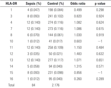
Table 5 - Prevalence of HLA*DR in septic and control patients
HLA-DR Sepsis (%) Control (%) Odds ratio p value
1 4 (0.047) 198 (0.084) 0.499 0.298
3 8 (0.093) 241 (0.102) 0.820 0.924 4 12 (0.140) 274 (0.116) 1.082 0.624
7 12 (0.140) 273 (0.116) 1.086 0.615
8 6 (0.070) 144 (0.061) 1.030 0.919 10 1 (0.012) 41 (0.017) 0.603 ~1
11 12 (0.140) 258 (0.109) 1.150 0.484 12 3 (0.035) 50 (0.021) 1.483 0.632
13 12 (0.140) 277 (0.117) 1.071 0.651
14 5 (0.058) 94 (0.040) 1.315 0.571 15 8 (0.093) 231 (0.098) 0.856 ~1
16 1 (0.012) 95 (0.040) 0.260 0.289
Total 84 2.176
publications implicate low HLA-DR membrane expression as a predictor of sepsis development(21) and poor septic outcome,(22,23) and it has become the most reliable ICU-acquired immunosuppression marker.(24,25) In our study, however, complete genetic screening of the HLA-DR locus was unable to ind any allele more prevalent in the septic population. hus, in our opinion, low HLA-DR membrane expression is a marker of cell exhaustion and not a locus related to genetic susceptibility.
HLA-A*31 is expressed in 5 to 10% of the world's population, with the highest expression being observed in the Brazilian Amerinds (65%) and the lowest in the Eskimo population (0%). HLA-A*31 has been strongly associated with carbamazepine-induced adverse drug reactions in a Japanese population and recognizes an antigen encoded by gastric signet ring cell carcinoma.(26,27) Indeed, HLA-A*31 is a target for vaccine development in cancer.(28) Interestingly, when we performed a BLAST search to investigate this cancer peptide, we found
a strong similarity with a Pseudomonas aeruginosa
hypothetical protein.
More than 1,000 HLA-A and HLA-B alleles have been characterized. Both the peptide-binding groove and the lanking regions are highly variable.(29) It would be interesting to decipher the peptides that are loaded into HLA-A*31 and are thereby eliciting severe sepsis and septic shock. hese peptides may be self or non-self proteins. he importance of bacterial infection cross priming has been studied in bacteria, including L. monocytogenes and Shigella, which can cause phagosome escape. In contrast, Mycobacterium tuberculosis and Salmonella typhimurium can survive within phagosomes and have to be cross-processed to
initiate MHC-I-dependent CD8 responses. Dendritic cells acquire antigen following pinocytosis of infected cell debris.(30) Patients in the present study, however, were not infected by these pathogens. Notably, Staphylococcus aureus, Acinetobacter baumanni and extended-spectrum beta-lactamase (ESBL) microorganisms account for more than 90% of the nosocomial infections in the present study.
Sequencing of the HLA ligands from a variety of antigen presenting cells (APC) revealed predominantly self-antigens found on the cell surface of these cells or inside their endosomes.(31,32) Additionally, cytoplasmic or nuclear antigens accounted for 10 to 30% of those peptides.(33,34)
Interestingly, a large fraction of proteins are immediately degraded following synthesis, as a result of factors such as defective transcription or translation, alternative reading frame usage, failed assembly into larger complexes or altered ubiquitin modiications.(35)
Many cells are linked by gap junctions that allow the transfer of small cytosolic molecules or ions into the cytosol of their neighboring cells. hese molecules and ions can then enter into the antigen presentation pathway of the neighboring cells.(36) Most HLA-B alleles are always loaded with peptides; however, only a proportion of HLA-A and HLA-C alleles are loaded. It has been suggested that HLA molecules that fail to present self-peptides are more available for peptides that arise during infection or stressful conditions.(37) Several HLA-I machinery proteins have been observed to be associated with phagosomes. It has been suggested that this is a result of endoplasmic reticulum membranes fusing with the phagosome during phagocytosis.(38,39)
Interestingly, recent indings show that besides cross-presentation, CD8+ T cells can be activated through the transfer of preformed peptide-HLA class I complexes from the surface of infected cells to uninfected APCs, a processed that has been named cross-dressing.(40) It seems that trogocytosis is the mechanism involved in this process. Whatever the origin, HLA-A*31 is an interesting marker of sepsis susceptibility and may help guide clinicians in the care of the critically ill.
More studies are necessary to investigate the mechanisms leading the HLA-A*31 expression to increase patients susceptibility to sepsis. In a near future this molecule may become an useful marker to identify patients more prone to develop severe infections.
CONCLUSION
he expression of HLA-A*31 is associated with the risk of developing sepsis.
Author’s contributions
F Pinheiro da Silva and H Rodrigues planned the experiments and wrote the irst draft. G Preuhs Filho performed the experiments. he remaining authors contributed to data collection and revised the manuscript.
ACKNOWLEDGEMENTS
he current study is part of an ongoing prospective cohort study that is designed to store clinical data and biological samples for multiple projects and was conceived by HP Souza and LM Cruz Neto (Brazilian Initiative for Sepsis Knowledge, BRISK Project).
F Pinheiro da Silva was supported by São Paulo Research Foundation (FAPESP), grant # 2009/17731-2.
We would like to express our gratitude to the patients and their families for consenting to participate in this study.
We are indebted to Paulo Starzynski for his great help with the statistical analyses.
Objetivo: Haplótipos do HLA têm sido associados a muitas doenças autoimunes, mas não foi descrita qualquer associação na sepse. O objetivo desse estudo é investigar o sistema HLA como um possível marcador de suscetibilidade genética à sepse.
Métodos: Estudo prospectivo de coorte, incluindo
pacientes admitidos em unidade de terapia intensiva e controles-saudáveis obtidos em lista de doadores de transplante renal. Foram excluídos pacientes abaixo dos 18 anos de idade, gestantes ou HIV positivos, pacientes com doença maligna metastática ou sob quimioterapia, pacientes com hepatopatia avançada, com condições de im de vida. O DNA foi extraído
de sangue total, e a haplotipagem de HLA foi realizada com a tecnologia MiliPlex®
.
Resultados: Foram incluídos 1.121 pacientes (1.078 doadores de rim, 20 pacientes com sepse grave e 23 pacientes admitidos por choque séptico) entre outubro de 2010 e outubro de 2012. Os participantes positivos para HLA-A*31 tiveram risco aumentado de desenvolver sepse (OR: 2,36 IC95%: 1,26-5,35). Não foi identiicada outra associação signiicativa, quando considerado como nível de signiicância o valor de p<0,01.
Conclusão: A expressão de HLA-A*31 está associada ao risco de desenvolvimento de sepse.
RESUMO
Descritores: Sepse; Antígenos HLA-A; Inlamação;
Marcadores genéticos
REFERENCES
1. Martin GS, Mannino DM, Eaton S, Moss M. The epidemiology of sepsis in the United States from 1979 through 2000. N Engl J Med. 2003;348(16):1546-54.
2. Cho JH, Gregersen PK. Genomics and the multifactorial nature of human autoimmune disease. N Engl J Med. 2011;365(17):1612-23.
3. Brewerton DA, Hart FD, Nicholls A, Caffrey M, James DC, Sturrock RD. Ankylosing spondylitis and HL-A 27. Lancet. 1973;1(7809):904-7. 4. Habek M, Brinar VV, Borovecki F. Genes associated with multiple sclerosis:
15 and counting. Expert Rev Mol Diagn. 2010;10(7):857-61.
5. Liu Y, Helms C, Liao W, Zaba LC, Duan S, Gardner J, et al. A genome-wide association study of psoriasis and psoriatic arthritis identifies new disease loci. PLoS Genet. 2008;4(3):e1000041.
6. Viken MK, Blomhoff A, Olsson M, Akselsen HE, Pociot F, Nerup J, et al. Reproducible association with type 1 diabetes in the extended class I region of the major histocompatibility complex. Genes Immun. 2009;10(4):323-33.
7. Fallang LE, Bergseng E, Hotta K, Berg-Larsen A, Kim CY, Sollid LM. Differences in the risk of celiac disease associated with HLA-DQ2.5 or HLA-DQ2.2 are related to sustained gluten antigen presentation. Nat Immunol. 2009;10(10):1096-101.
8. Gross D, Huber BT, Steere AC. Molecular mimicry and Lyme arthritis. Curr Dir Autoimmun. 2001;3:94-111.
9. International MHC and Autoimmunity Genetics Network, Rioux JD, Goyette P, Vyse TJ, Hammarström L, Fernando MM, Green T, et al. Mapping of multiple susceptibility variants within the MHC region for 7 immune-mediated diseases. Proc Natl Acad Sci U S A. 2009;106(44):18680-5.
10. Depla E, Van der Aa A, Livingston BD, Crimi C, Allosery K, De Brabandere V, et al. Rational design of a multiepitope vaccine encoding T-lymphocyte epitopes for treatment of chronic hepatitis B virus infections. J Virol. 2008;82(1):435-50.
12. Sanchez-Mazas A, Fernandez-Viña M, Middleton D, Hollenbach JA, Buhler S, Di D, et al. Immunogenetics as a tool in anthropological studies. Immunology. 2011;133(2):143-64.
13. Bharadwaj M, Illing P, Theodossis A, Purcell AW, Rossjohn J, McCluskey J. Drug hypersensitivity and human leukocyte antigens of the major histocompatibility complex. Annu Rev Pharmacol Toxicol. 2012;52:401-31. 14. Trowsdale J. The MHC, disease and selection. Immunol Lett.
2011;137(1-2):1-8.
15. Bone RC, Sibbald WJ, Sprung CL. The ACCP-SCCM consensus conference on sepsis and organ failure. Chest. 1992;101(6):1481-3.
16. Rai E, Wakeland EK. Genetic predisposition to autoimmunity--what have we learned? Semin Immunol. 2011;23(2):67-83.
17. Handunnetthi L, Ramagopalan SV, Ebers GC, Knight JC. Regulation of major histocompatibility complex class II gene expression, genetic variation and disease. Genes Immun. 2010;11(2):99-112.
18. Zhang WW, Li XF, Zhang X, Li JP, Li ZQ. A novel HLA-A31 allele, A*31:22, was identified by sequence-based typing. Tissue Antigens. 2012;80(4):380-1.
19. Ji Y, Sun Y, Liu Y, Xie J, Du K. Two novel HLA-A alleles: HLA-A*31:01:09 and HLA-A*33:30. Tissue Antigens. 2011;78(3):218-9.
20. Lim AH, Song SN, Lee JY, Cho NS, Kwon SY. A novel HLA-A*31 allele, A*31:57, identified by sequence-based typing. Tissue Antigens. 2012;79(5):386-7.
21. Gouel-Chéron A, Allaouchiche B, Guignant C, Davin F, Floccard B, Monneret G; AzuRea Group. Early interleukin-6 and slope of monocyte human leukocyte antigen-DR: a powerful association to predict the development of sepsis after major trauma. PloS One. 2012;7(3):e33095.
22. Genel F, Atlihan F, Ozsu E, Ozbek E. Monocyte HLA-DR expression as predictor of poor outcome in neonates with late onset neonatal sepsis. J Infect. 2010;60(3):224-8.
23. Wu JF, Ma J, Chen J, Ou-Yang B, Chen MY, Li LF, et al. Changes of monocyte human leukocyte antigen-DR expression as a reliable predictor of mortality in severe sepsis. Crit Care. 2011;15(5):R220.
24. Monneret G, Venet F, Pachot A, Lepape A. Monitoring immune dysfunctions in the septic patient: a new skin for the old ceremony. Mol Med. 2008;14(1-2):64-78.
25. Boomer JS, To K, Chang KC, Takasu O, Osborne DF, Walton AH, et al. Immunosuppression in patients who die of sepsis and multiple organ failure. JAMA. 2011;306(23):2594-605.
26. Yasoshima T, Sato N, Hirata K, Kikuchi K. The mechanism of human autologous gastric signet ring cell tumor rejection by cytotoxic T lymphocytes in the possible context of HLA-A31 molecule. Cancer. 1995;75(6 Suppl):1484-9.
27. Suzuki K, Sahara H, Okada Y, Yasoshima T, Hirohashi Y, Nabeta Y, et al. Identification of natural antigenic peptides of a human gastric signet ring cell carcinoma recognized by HLA-A31-restricted cytotoxic T lymphocytes. J Immunol. 1999;163(5):2783-91.
28. Takedatsu H, Shichijo S, Azuma K, Takedatsu H, Sata M, Itoh K. Detection of a set of peptide vaccine candidates for use in HLA-A31+ epithelial cancer patients. Int J Oncol. 2004;24(2):337-47.
29. Eagle RA, Traherne JA, Ashiru O, Wills MR, Trowsdale J. Regulation of NKG2D ligand gene expression. Hum Immunol. 2006;67(3):159-69. 30. Kurts C, Robinson BW, Knolle PA. Cross-priming in health and disease. Nat
Rev Immunol. 2010;10(6):403-14.
31. Hunt DF, Michel H, Dickinson TA, Shabanowitz J, Cox AL, Sakaguchi K, et al. Peptides presented to the immune system by the murine class II major histocompatibility complex molecule I-Ad. Science. 1992;256(5065):1817-20.
32. Zhou D, Blum JS. Presentation of cytosolic antigens via MHC class II molecules. Immunol Res. 2004;30(3):279-90.
33. Dongre AR, Kovats S, deRoos P, McCormack AL, Nakagawa T, Paharkova-Vatchkova V, et al. In vivo MHC class II presentation of cytosolic proteins revealed by rapid automated tandem mass spectrometry and functional analyses. Eur J Immunol. 2001;31(5):1485-94.
34. Dengjel J, Schoor O, Fischer R, Reich M, Kraus M, Müller M, et al. Autophagy promotes MHC class II presentation of peptides from intracellular source proteins. Proc Natl Acad Sci U S A. 2005;102(22):7922-7.
35. Schubert U, Antón LC, Gibbs J, Norbury CC, Yewdell JW, Bennink JR. Rapid degradation of a large fraction of newly synthesized proteins by proteasomes. Nature. 2000;404(6779):770-4.
36. Neijssen J, Herberts C, Drijfhout JW, Reits E, Janssen L, Neefjes J. Cross-presentation by intercellular peptide transfer through gap junctions. Nature. 2005;434(7029):83-8.
37. Neefjes J, Jongsma ML, Paul P, Bakke O. Towards a systems understanding of MHC class I and MHC class II antigen presentation. Nat Rev Immunol. 2011;11(12):823-36.
38. Guermonprez P, Saveanu L, Kleijmeer M, Davoust J, Van Endert P, Amigorena S. ER-phagosome fusion defines an MHC class I cross-presentation compartment in dendritic cells. Nature. 2003;425(6956):397-402. 39. Ackerman AL, Kyritsis C, Tampé R, Cresswell P. Early phagosomes in
dendritic cells form a cellular compartment sufficient for cross presentation of exogenous antigens. Proc Natl Acad Sci U S A. 2003;100(22):12889-94. 40. Wakim LM, Bevan MJ. Cross-dressed dendritic cells drive memory CD8+