2 artigo 456
REVIEw ARtICLE
1 – Instructing Professor and Head of the Knee Surgery Group, Department of Orthopedics and Traumatology, School of Medical Sciences, Santa Casa de Misericórdia de São Paulo (SCMSP), São Paulo, SP, Brazil.
2 – Professor in the Physiotherapy Course, São Camilo University Center and São Judas Tadeu University; Researcher in the Biomechanics Laboratory, São Judas Tadeu University, São Paulo, SP, Brazil.
3 – Researcher in the Biomechanics Laboratory, São Judas Tadeu University, São Paulo, SP, Brazil.
4 – Professor in the Musculoskeletal Physiotherapy Specialization Course and Physiotherapist in the Knee and Sports Traumatology Groups, Santa Casa de Misericórdia de São Paulo, São Paulo, SP, Brazil.
5 – Instructing Professor in the Knee Surgery Group, Department of Orthopedics and Traumatology, School of Medical Sciences, Santa Casa de Misericórdia de São Paulo (SCMSP), São Paulo, SP, Brazil.
6 – Adjunct Professor in the Knee Surgery Group, Department of Orthopedics and Traumatology, School of Medical Sciences, Santa Casa de Misericórdia de São Paulo (SCMSP), São Paulo, SP, Brazil.
Work performed in the Knee Surgery Group, Department of Orthopedics and Traumatology, School of Medical Sciences, Santa Casa de Misericórdia de São Paulo (SCMSP), São Paulo, SP, Brazil.
Correspondence: Rua Barata Ribeiro 380/64, Bela Vista, 01308-000 São Paulo, SP, Brazil. E-mail: [email protected] Work received for publication: December 22, 2010; accepted for publication: October 4, 2011.
REHABILITATION PROTOCOL AFTER ISOLATED POSTERIOR
CRuCIATE LIgAmENT RECONSTRuCTION
Ricardo de Paula Leite Cury1, Henry Dan Kiyomoto2, Gustavo Fogolin Rosal3, Flávio Fernandes Bryk4, Victor Marques de Oliveira5, Osmar Pedro Arbix de Camargo6
ABstRACt
To create a rehabilitation protocol following reconstruction of the posterior cruciate ligament (PCL), through a literature review. The literature review was conducted in the Medline and Embase databases, to search for data on biomechanical concepts and analyses relating to the posterior cruciate liga-ment of the knee. The search strategy was set up using the following rules: problem or injury in association with ana-tomical location terms; or surgical intervention procedure in association with rehabilitation terms. We began the process in this manner and subsequently introduced restrictions on certain terms to improve the search specificity. To design the protocol, a table was created for better data assessment, based
on the time that elapsed between surgery and the start of phy-siotherapy. A rehabilitation protocol was created to improve weight-bearing control in the initial weeks after surgery, with the aid of a knee brace. Our aim was to achieve gains in total range of motion of the knee, which should be attained by the third month, thereby avoiding contractures resulting from the tissue healing process. Strengthening exercises and sensory-motor training were guided accordingly, thus avoi-ding overload on the graft and respecting the healing phases. The protocol proposed through this review was based on the current evidence relating to this subject.
Keywords –Posterior Cruciate Ligament; Knee; Rehabilitation
The authors declare that there was no conflict of interest in conducting this work
this article is available online in Portuguese and English at the websites: www.rbo.org.br and www.scielo.br/rbort
INtRODUCtION
There is a lack of biomechanical, histological and clinical studies on knee rehabilitation following pos-terior cruciate ligament (PCL) injury, in relation both to cases treated conservatively and to cases that un-derwent reconstruction. The existing studies are often based on aspects of the integration and rehabilitation of the anterior cruciate ligament (ACL), transposed to the PCL. The aim of the present study was to review the points presented in the current literature and, to-gether with tacit knowledge of the last few years at our clinic, to put forward a rehabilitation protocol.
MEtHODs
A search in the literature was conducted using the Medline database through the PubMed website and using the Embase database through the Patient, Inter-vention, Comparison and Outcome (PICO) strategy. The investigation was divided into search strategies that emphasized range of motion and therapeutic exer-cises, as described below.
replacement were used to clean up the search for related articles. In addition, the Mesh terms Rehabilitation and Range of motion were also combined in an attempt to only retrieve articles relating to ROM. In this manner, 33 articles were identified. Of these 11 reported the ROM and/or showed programs for ROM gain.
Regarding exercise programs: Posterior cruciate ligament [Mesh] was combined with Physical therapy modalities [Mesh], Rehabilitation [Mesh], Exercise [Mesh], Exercise therapy [Mesh] and Exercise test [Mesh] as a strategy, and 19 articles were identified. Of these, six had the objective of analyzing the reha-bilitation protocol.
In addition, because few in vivo studies were avai-lable, we also used a strategy with greater sensitivity, through analyzing in vitro biomechanical studies and mathematical modeling studies on knee-related exercises.
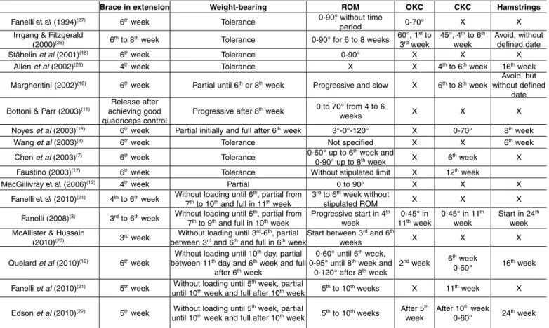
From using a filter for meta-analyses or rando-mized controlled clinical trials, only one study was identified, and this did not cover all aspects of reha-bilitation. Thus, the present review (Table 1) was con-ducted mainly on basic science studies and on cadaver models, because of the few randomized controlled clinical trials found. The protocol was constructed in a spreadsheet with a format that accompanied the
variable of postoperative time. Thus, the protocol was made to be easy to view and to consult (Annex 1).
REsULts
The protocol presented shows the period of early release for weight-bearing over the first weeks, done partially through use of two crutches and a long immobilizer locked into extension.
Passive mobilization for improving ROM should be done early on; for this, we recommend that pro-gressive gain should be envisaged, with the parame-ters of 70° of flexion in the fourth week and 90° in the sixth week. Following this, full ROM needs to be achieved by the third month in order to avoid contrac-tures resulting from the tissue healing process. Note that active flexion movement of the knee should be delayed for two months.
The post-surgical reconstruction period for the PCL may be accompanied by pain. In this case, analgesia provided through electrotherapeutic means is benefi-cial for the rehabilitation process, with regard to the patient’s comfort. Cryotherapy should be used whe-never the knee presents conditions of pain or edema. The greatest limitation of physiotherapy in the
table 1 – Review with systematized search of the literature.
Brace in extension Weight-bearing ROM OKC CKC Hamstrings
Fanelli et al (1994)(27) 6th week Tolerance 0-90° without time
period 0-70° X X
Irrgang & Fitzgerald
(2000)(25) 6th to 8th week Tolerance 0-90° for 6 to 8 weeks 60°, 1
st to 3rd week 45°, 4
th to 6th
week
Avoid, without defined date
Stähelin et al (2001)(15) 6th week Tolerance 0-90° X X X
Allen et al (2002)(28) 4th week Tolerance X X 4th to 6th week 16th week
Margheritini (2002)(18) 6th week Partial until 6th or 8th week Progressive and slow X 6th to 8th week without defined Avoid, but date
Bottoni & Parr (2003)(11) achieving good Release after quadriceps control
Progressive after 8th week 0 to 70° from 4 to 6
weeks X X X
Noyes et al (2003)(16) 6th week Partial initially and full after 6th week 3°-0°-120° X 0-70° 8th week
Wang et al (2003)(8) 6th week Tolerance Not specified X X 6th week
Chen et al (2003)(7) 6th week Tolerance 0-60° up to 6th week and
0-90° up to 8th week X 6th week X Faustino (2003)(17) 6th week Tolerance Without stipulated limit X 12th week
MacGillivray et al (2006)(12) 4th week Partial 0 to 90° X X X
Fanelli et al (2010)(21) 4th to 6th week Without loading until 6th, partial from
7th to 10th and full in 11th week
3rd to 6th week without
stipulated ROM X X X
Fanelli (2008)(3) 3rd to 6th week Without loading until 6th, partial from
7th to 9th and full in 10th week Progressive start in 4 th
week
0-45° in
11th week 0-45° in 11 th
week
Start in 24th week McAllister & Hussain
(2010)(20) 3rd week Without loading until 3
rd-6th, partial
between 3rd and 6th and full in 6th weekStart between 3 rd and 6th
weeks X X X
Quelard et al (2010)(19) 6th week Without loading until 10
th day, partial between 11th day and 6th week and full
after 6th week
0-60° until 6th week, 0-95° until 8th week and
0-120° after 8th week
2nd week 6th week
0-60° 16th week
Fanelli et al (2010)(21) 5th week Without loading until 5th week, partial
until 10th week and full after 10th week 5th to 10th weeks X 11th week X
Edson et al (2010)(22) 5th week Without loading until 5th week, partial
until 10th week and full after 10th week 5th to 10th weeks After 5 th
week
After 10th week
patient rehabilitation process relates to strengthening exercises. In our protocol, we delay open kinetic chain (OKC) exercises for the knee flexors until the eighth week after the operation, while closed kinetic chain (CKC) and OKC exercises for the extensors remain in the second week.
Sensory-motor work should start together with the release to perform CKC exercises for the extensors, and the progression from stable ground surfaces to unstable surfaces should be done by around the fourth month, along with stressing for anteroposterior, side--to-side and rotational displacement, respectively. Over this period, we begin the process of plyometric trai-ning, which is reserved for the population of athletes. The time taken for non-athletic individuals to be released for general activities is around six months, with a further two months for sports activities at com-petitive level.
DIsCUssION
The rehabilitation process for PCL injuries is assessed as a complementary but essential point within functional recovery of the knee(1). Rehabilitation protocols prioritize protection of the reconstructed ligament, so as to avoid excessive stress on the graft during the rehabilitation until the graft has become integrated(2). However, it is not known with any certainty what the safe tensions would be and how much provocation can be allowed during rehabilitation exercises(3).
Little is known on the structural modifications of grafts after ligament reconstruction. Bosch and Kas-perczyk(4) studied the histochemical and biochemical characteristics of grafts from the central third of the patellar tendon for ACL reconstruction, in sheep, with the aim of understanding the integration process. They found a necrotic phase with diminution of the resis-tance to stress particularly in the eighth week after reconstruction. It is noteworthy that graft necrosis continued to be seen until the 104th week, i.e. two years after the reconstruction.
Moreover, it is a difficult task to determine the stress that the ligaments are subjected to during pas-sive movement of the knee in weight-bearing and muscle force activities and whether these are preju-dicial to the graft. Direct measurement methods such as placement of load cells (measurement devices) in the ligament are difficult to do in vivo. Thus, studies on cadavers and indirect biomechanical methods such as inverse dynamics are the methods most used.
Points relating to rom
To avoid loss of ROM, Irrgang and Harner(5) di-vided the care relating to reconstructed knees into three phases: before the surgery, the focus should be on elimination of edema and pain and restoration of ROM; during the operation, ROM seems to be clo-sely related to the positioning of the bone tunnels and to the surgical technique; after the surgery, early mobilization and gains in mobility are recommended, with extension restored after two to three weeks and flexion achieved by the third month(6).
Restrictions relating to the limits on knee flexion gains are discussed in the literature, with divergences between the rehabilitation protocols presented. Some authors have prioritized limiting the range of angles to between 0 and 60º(7-10), 0 and 70º(11), 0 and 90º(12-15) or 0 and 120º(16), or without any stimulated limit(17) or according to the patient’s tolerance(18). Quelard et al(19) recommended a gradual protocol for gaining passive mobility of the knee, such that a range of 0-60° would be achieved in the first six weeks, 0-90° from the sixth to the eighth week and 0-120° from the eighth week onwards.
Some studies have used a slower protocol and have not included passive mobilization of the knee in the first weeks. McAllister and Hussain(20) started the protocol between the third and sixth weeks, Fanelli et al(21) between the fifth and tenth weeks, Fanelli(3) in the fourth week and Edson et al(22) in the fifth week.
The criteria of ROM progression are not discussed in the protocols that we found, and there is no biome-chanical explanation to explain why passive gain of movement is limited. The protocols used in the literatu-re seem to be based on personal clinical experience(22). In situ studies on PCL tensions(23) have demons-trated that with increasing degree of passive flexion of the knee, there is also an increase in the tension in the PCL. Moreover, the varus stress and posterior shear stress in the tibia may also generate increased force on the PCL(24).
Because of this evidence, caution is needed in rela-tion to gains in passive knee ROM. On the other hand, delayed gain in movement may have consequences such as restriction of joint ROM and functional loss.
studies on cadavers(24) and was advocated by Irrgang and Fitzgerald(25) in their rehabilitation protocol.
Our protocol restricts the gain in passive ROM to 70º for four weeks and progresses to 90º for ano-ther two weeks. After the sixth week, gains in passive ROM are progressive, according to the patient’s to-lerance, but we maintain the passive anteriorization force applied to the tibia until the tenth week.
release for weight-bearing (walking)
Early release for weight-bearing in isolated recons-truction of the PCL is a common practice among the rehabilitation protocols cited in the literature(11,12,16), but there is no consensus regarding how much this could be done without causing deleterious effects to the graft undergoing healing. Many protocols(7,15,17) favor early weight-bearing according to the patient’s toleran-ce. In other words, the introduction of weight-bearing may be completed in the first weeks of reconstruction.
Through a study with a mathematical model, Shel-burne and Pandy(26) demonstrated that because of the forces exerted on the knee during weight-bearing, the tibia presents a tendency towards anterior shearing in relation to the femur, which theoretically would not overload the PCL.
Bosch and Kasperczyk(4) conducted an experi-ment on sheep and found that moveexperi-ment and early weight-bearing did not cause ruptures and did not increase the length of the graft. Corroborating this concept, Toutoungi et al(2) found that the effect of axial compression tended to diminish the femoroti-bial shearing and consequently the stresses generated on the central ligaments.
In the study by Noyes and Barber-Westin(6), which involved PCL reconstruction, weight-bearing was introduced progressively, with a protective orthosis locked in extension for four weeks, until full weight-bearing was reached around the fifth week. However, other studies are divergent. Some authors have recommended that weight-bearing should be introduced according to the patient’s tolerance and should be started in the first week(7,8,15,17,20,27,28), while one study restricted weight-bearing until the sixth week(16) and others until the eighth week(11,18).
In some protocols, weight-bearing is not recommen-ded during the first days after reconstruction. Quelard et al(19) used a protocol without weight-bearing over the first 10 days, progressing to partial weight-bearing on the 11th day, which continued until the fifth week, with full weight-bearing from the sixth week onwards.
McAllister and Hussain(20) did not used weight-bearing for three weeks and progressed to partial weight-bearing in the fourth and fifth weeks and full weight-bearing in the sixth week.
Edson et al(22) did not use weight-bearing for five weeks and progressed to partial weight-bearing in the sixth week and full weight-bearing in the 10th week. Other authors have used different protocols (Table 1); however, all of them used a protective orthosis locked in extension, in association with weight-bearing.
Based on the studies cited above, our group feels increasingly secure in recommending partial bearing, with evolution to full weight-bearing according to the patient’s tolerance, for isolated PCL injuries.
muscle strengthening
There have been divergences of opinion regarding the use of OKC or CKC exercises as rehabilitation op-tions for the process of muscle strengthening, in re-lation to efficacy of strength gains, control over knee muscles and stress on ligaments. There is a tendency towards using CKC exercises at the start of protocols, with complementation using OKC exercises at the more advanced phases(29-34). CKC exercises generate axial compression forces on the joint, which diminishes the shearing forces on the knee, as well as leading to simul-taneous contraction of the quadriceps and hamstrings, which are desirable in the initial phase of rehabilitation.
In the rehabilitation protocols cited in previous studies(7,11,17), OKC and CKC exercises were introdu-ced in an arbitrary manner, without backing from any studies quantifying the tensions in the PCL or their consequences in relation to ligament laxity during the rehabilitation process. Quelard et al(19) recommended that OKC exercises for strengthening the quadriceps should be started from the second week. Some studies have suggested starting these exercises in the first three weeks(7-9,11), while others have introduced them only between the fourth and sixth weeks(10,13,14,22). Fanelli(3) only started quadriceps strengthening with OKC exercises in the 11th week, at angles of 0-45°.
Certain protective angle ranges have been re-commended for OKC quadriceps strengthening exercises. Ranges of 0 to 60º have been used(9,13,14), while other authors have recommended that this strengthening should be done from 0 to 70º(27).
diminish the stress on the PCL, especially at the end of the knee extension. Thus, these were the preferred exercises at the start of the rehabilitation process.
One proviso needs to be made in relation to OKC exercises for the quadriceps and their implications in post-reconstruction rehabilitation of the PLC. By con-sidering only the protection angles in relation to PCL grafts, excessive stress may be placed on the femoro-patellar joint and may consequently cause lesions in the cartilage coating this joint(36). Therefore, the safe angle for the neoligament is between 0 and 70º(25), and protection for the femoropatellar joint involves angles from 45 to 90º(36). Thus, with the aim of protecting the graft and the femoropatellar joint, our group uses angles from 45 to 70º to stimulate the quadriceps. Since this only leaves a small range of motion, we undertake the OKC exercises in isometric form at multiple angles within this safety range. The aim of OKC exercises in the initial phase is not related to gains in strength, re-sistance or muscle power, but to recruitment of the ma-ximum number of muscle fibers. Thus, we undertake OKC exercises by associating the patient’s maximum voluntary contraction with neuromuscular electrosti-mulation, with the aim of combating the arthrogenic inhibition that is present in diseased knees(37).
The rehabilitation protocols that we found introdu-ced CKC exercises for quadriceps strengthening at di-fferent times during the rehabilitation protocol. These times included the fourth(9,10), sixth(19,27), eighth(11,14), tenth(22), eleventh (3,21) and twelfth weeks(17).
Regarding the protection angles, three variants were found in our investigation. Some authors started with mini-squats from 0 to 45º(3,25), others introduced CKC exercised at angles from 0 to 70º(16) and yet others(19,22) started with 0 to 60°.
In vivo studies(38-40) analyzing the length of the na-tive PCL through measurements made using magne-tic resonance imaging have demonstrated that CKC exercises increased the length of the two bands of the PCL at greater flexion angles. However, this type of measurement is unable to define the amount of tension generated in the PCL during active rehabilitation exer-cises. Therefore, only direct measurements by means of load cells would be capable of defining these ten-sions, but the methodology of this procedure makes it very difficult to assess the PCL.
CKC exercises are safe in relation to anterior shea-ring forces on the tibia(26) and should be performed ca-refully in the initial phase of the rehabilitation process
following PCL surgery. The factors that may have an influence on the stresses in the cruciate ligaments are the forces generated by muscle contractions, such as co-contractions and ground reaction forces(32).
Shelburne and Pandy(26) demonstrated that from 10º of flexion onwards, in CKC exercises, the PCL presents increased tension, even though the peak stress occurs at around 80º of flexion.
In our protocol, CKC exercises are started in the second week and are initially performed in situations of controlled overload. We use exercises on stable surfaces, such as leg press exercises, mini-squats and functional activities such as getting up from and sit-ting down on high chairs.
The ROM should respect the angles of 0 to 45º, since shearing of the tibia occurs anteriorly, which spares the PCL from excessive tension, as well as protecting the femoropatellar joint(25). Beyond 70 to 80º, the tensions increase considerably, thus causing excessive stress on the PCL(26).
When the hamstring muscles are contracted in iso-lation, i.e. in OKC exercises, the tension on the PCL is increased because of the traction force of these muscles on the tibia(2,26,34,35). Shelburne and Pandy(26) demons-trated that the hamstrings are responsible for constant posterior tension and that as the knee flexion increases, the forces favoring anteriorization of the tibia diminish.
The protocols generally postpone the introduction of exercises directed towards the hamstrings with the aim of not excessively tensioning the graft during the initial postoperative phase. There is divergence be-tween studies regarding when to start to work on the posterior muscles of the thigh, such that the suggested start is in the sixth(8), eighth(16), ninth(13), 16th(28) or 24th week(3,19,22). In our protocol, hamstring exercises are postponed until the eighth week with the aim of sparing the posteriorization forces during the initial phase of the rehabilitation protocol.
Andersen et al(41) found that beyond 10 to 12 weeks after PCL surgery, for patients with functional ROM, normalized gait and little or no significant clinical complaint, there was less concern regarding the type of exercise, speed at which the exercise was perfor-med and the muscles to be emphasized for normali-zing these patients’ muscle strength and restoring their remaining functional deficits.
sensory-motor training
quantity of mechanoreceptors found in this ligament. The proprioceptive effect of the PCL has mainly been studied and discussed in relation to preservation or not of this ligament in total knee prosthesis surgery. There have been divergent results regarding compari-sons between knees with and without the ligament, in assessing functional outcomes for the knee(43).
Because of the PCL injury process and the role of the PCL in proprioception, sensory-motor training should always be performed. There should be progression from stable ground surfaces with static exercises to unstable
surfaces with dynamic exercises that are increasingly specific to the functional objective(43).
FINAL REMARKs
The majority of the protocol proposed fits within the current evidence on this subject. The protocol has been used in our clinic with good tolerance among the patients. The present state of evidence has allowed us to analyze each phase of the rehabilitation pro-cess, but further studies of clinical nature with greater strength of evidence need to be conducted.
REFERENCEs
1. Wilk KE. Rehabilitation of isolated and combined posterior cruciate ligament injuries. Clin Sports Med. 1994;13(3):649-77.
2. Toutoungi DE, Lu TW, Leardini A, Catani F, O’Connor JJ. Cruciate ligament forces in the human knee during rehabilitation exercises. Clin Biomech (Bristol,Avon). 2000;15(3):176-87.
3. Fanelli GC. Posterior cruciate ligament rehabilitation: how slow should we go? Arthroscopy. 2008;24(2):234-5.
4. Bosch U, Kasperczyk WJ. Healing of the patellar tendon autograft after pos-terior cruciate ligament reconstruction--a process of ligamentization? An ex-perimental study in a sheep model. Am J Sports Med. 1992;20(5):558-66. 5. Irrgang JJ, Harner CD. Loss of motion following knee ligament reconstruction.
Sports Med. 1995;19(2):150-9.
6. Noyes FR, Barber-Westin SD. Reconstruction of the anterior and posterior cruciate ligaments after knee dislocation. Use of early protected postoperative motion to decrease arthrofibrosis. Am J Sports Med. 1997;25(6):769-78. 7. Chen CH, Chen WJ, Shih CH. Double-bundle posterior cruciate ligament reconstruction
with quadriceps and semitendinosus tendon grafts. Arthroscopy. 2003;19(9):1023-6. 8. Wang CJ, Chen HS, Huang TW. Outcome of arthroscopic single bundle
recons-truction for complete posterior cruciate ligament tear. Injury. 2003;34(10):747-51. 9. Leighton MM, Bach BR. Reabilitação das lesões dos ligamentos do joelho. In: Tria AJ. Lesões ligamentares do joelho. Rio de Janeiro: Revinter; 2002. p. 289-319. 10. Weber MD, Woodall WR. Reabilitação do joelho. In: Andrews JR, Harrelson GL,
Wilk KE. Reabilitação física do atleta. Rio de Janeiro: Elsevier; 2005. p. 399-456. 11. Bottoni CR, Parr RR. Double bundle arthroscopic posterior cruciate ligament
reconstruction using a new medial femoral cortical bridge technique. Techni-ques in Knee Surgery. 2003;2(4):239-49. Available from: www.en.aspetar.com/ mritems/pdf/pcl%20op%20techs.pdf. Accessed in 2010 (Ago 11).
12. MacGillivray JD, Stein BE, Park M, Allen AA, Wickiewicz TL, Warren RF. Compa-rison of tibial inlay versus transtibial techniques for isolated posterior cruciate liga-ment reconstruction: minimum 2-year follow-up. Arthroscopy. 2006;22(3):320-8. 13. Kisner C, Colby LA. O Joelho. In: Kisner C, Colby LA. Exercícios terapêuticos.
São Paulo: Manole; 1998. p. 407-57.
14. Monteiro CG, Forgas CB. Assistência fisioterapêutica nas lesões do ligamento cruzado posterior. In: Cohen M, Abdalla RJ. Lesões nos esportes. Rio de Janeiro: Revinter; 2003. p. 558-59.
15. Stähelin AC, Südkamp NP, Weiler A. Anatomic double-bundle posterior cruciate ligament reconstruction using hamstring tendons. Arthroscopy. 2001;17(1):88-97. 16. Noyes FR, Medvecky MJ, Bhargava M. Arthroscopically assisted quadriceps double--bundle tibial inlay posterior cruciate ligament reconstruction: An analysis of techniques and a safe operative approach to the popliteal fossa. Arthroscopy. 2003;19(8):894-905. 17. Faustino CAC. Reconstrução do ligamento cruzado posterior com os enxertos dos
tendões dos músculos flexores do joelho. Acta Ortop Bras. 2003;11(2):95-101. 18. Margheritini F, Rihn J, Musahl V, Mariani PP, Harner C. Posterior cruciate
liga-ment injuries in the athlete: an anatomical, biomechanical and clinical review. Sports Med. 2002;32(6):393-408.
19. Quelard B, Sonnery-Cottet B, Zayni R, Badet R, Fournier Y, Hager JP, et al. Isolated posterior cruciate ligament reconstruction: is non-aggressive reha-bilitation the right protocol? Orthop Traumatol Surg Res. 2010;96(3):256-62. 20. McAllister DR, Hussain SM. Tibial inlay posterior cruciate ligament reconstruc-tion: surgical technique and results. Sports Med Arthrosc. 2010;18(4):249-53. 21. Fanelli GC, Beck JD, Edson CJ. Double bundle posterior cruciate ligament
recons-truction: surgical technique and results. Sports Med Arthrosc. 2010;18(4):242-8. 22. Edson CJ, Fanelli GC, Beck JD. Postoperative rehabilitation of the posterior
cruciate ligament. Sports Med Arthrosc. 2010;18(4):275-9.
23. Dürselen L, Claes L, Kiefer H. The influence of muscle forces and external loads on cruciate ligament strain. Am J Sports Med. 1995;23(1):129-36. 24. Markolf KL, O’Neill G, Jackson SR, McAllister DR. Effects of applied quadriceps
and hamstrings muscle loads on forces in the anterior and posterior cruciate ligaments. Am J Sports Med. 2004;32(5):1144-9.
25. Irrgang JJ, Fitzgerald GK. Rehabilitation of the multiple-ligament-injured knee. Clin Sports Med. 2000;19(3):545-71.
26. Shelburne KB, Pandy MG. Determinants of cruciate-ligament loading during rehabilitation exercise. Clin Biomech (Bristol, Avon). 1998;13(6):403-413. 27. Fanelli GC, Giannotti BF, Edson CJ. The posterior cruciate ligament
arthrosco-pic evaluation and treatment. Arthroscopy. 1994;10(6):673-88.
28. Allen CR, Kaplan LD, Fluhme DJ, Harner CD. Posterior cruciate ligament injuries. Curr Opin Rheumatol. 2002;14(2):142-9.
29. Draganich LF, Vahey JW. An in vitro study of anterior cruciate ligament strain induced by quadriceps and hamstrings forces. J Orthop Res. 1990;8(1):57-63. 30. Escamilla RF, Fleisig GS, Zheng N, Barrentine SW, Wilk KE, Andrews JR. Biomechanics of the knee during closed kinetic chain and open kinetic chain exercises. Med Sci Sports Exerc. 1998;30(4):556-69.
31. Escamilla RF. Knee biomechanics of the dynamic squat exercise. Med Sci Sports Exerc. 2001;33(1):127-41.
32. Palmitier RA, An KN, Scott SG, Chao EY. Kinetic chain exercise in knee reha-bilitation. Sports Med. 1991;11(6):402-13.
33. Lutz GE, Palmitier RA, An KN, Chao EY. Comparison of tibiofemoral joint forces during open-kinetic-chain and closed-kinetic-chain exercises. J Bone Joint Surg Am. 1993;75(5):732-9.
34. Mesfar W, Shirazi-Adl A. Biomechanics of changes in ACL and PCL material properties or prestrains in flexion under muscle force-implications in ligament reconstruction. Comput Methods Biomech Biomed Engin. 2006;9(4):201-9. 35. Zavatsky AB, Beard DJ, O’Connor JJ. Cruciate ligament loading during
iso-metric muscle contractions. A theoretical basis for rehabilitation. Am J Sports Med. 1994;22(3):418-23.
36. Steinkamp LA, Dillingham MF, Markel MD, Hill JA, Kaufman KR. Biomecha-nical considerations in patellofemoral joint rehabilitation. Am J Sports Med. 1993;21(3):438-44.
37. Fischer-Rasmussen T, Krogsgaard M, Jensen DB, Dyhre-Poulsen P. Inhibition of dynamic thigh muscle contraction by electrical stimulation of the posterior cruciate ligament in humans. Muscle Nerve. 2001;24(11):1482-8.
38. Komatsu T, Kadoya Y, Nakagawa S, Yoshida G, Takaoka K. Movement of the posterior cruciate ligament during knee flexion--MRI analysis. J Orthop Res. 2005;23(2):334-9.
39. Li G, DeFrate LE, Sun H, Gill TJ. In vivo elongation of the anterior cruciate ligament and posterior cruciate ligament during knee flexion. Am J Sports Med. 2004;32(6):1415-20. 40. DeFrate LE, Gill TJ, Li G. In vivo function of the posterior cruciate ligament
during weightbearing knee flexion. Am J Sports Med. 2004;32(8):1923-8. 41. Andersen LL, Magnusson SP, Nielsen M, Haleem J, Poulsen K, Aagaard P.
Neuromuscular activation in conventional therapeutic exercises and heavy re-sistance exercises: implications for rehabilitation. Phys Ther. 2006;86(5):683-97. 42. Pagnano MW, Cushner FD, Scott WN. Role of the posterior cruciate ligament
in total knee arthroplasty. J Am Acad Orthop Surg. 1998;6(3):176-87. 43. Lephart SM, Fu FH. Proprioception and neuromuscular control in joint stability.
Annex 1 – Rehabilitation protocol for posterior cruciate ligament.
Date of surgery
ACtIVItIEs OF DAILy LIVINg
FULL LOADING PARTIAL LOADING
DRIVING
GOING UP AND DOWN STAIRS GOING UP AND DOWN STAIRS RUNNING
ROM (extension-flexion)
PASSivE
FREE ACTIVE
PATELLAR MOBILIZATION
ANALgEsIA
ELECTROANALGESIA (MINIMUM 30 MINUTES) CRYOTHERAPY (20 – 30 MINUTES)
MOBILIZATION
KINEsIOtHERAPy
STRETCHING (HAMSTRINGS AND TS) STRETCHING (QUADRICEPS) OKC (HIP FLExION) OKC (HIP ExTENSION) OKC (HIP ADDUCTION) OKC (HIP ABDUCTION) OKC (KNEE FLExION) OKC (KNEE ExTENSION) OKC (ANKLE)
CKC (stable ground surface)
CKC (unstable ground surface, without support) ERGOMETRIC BICYCLE
EENM
sensory-motor
STABLE GROUND SURFACE (two feet)
STABLE GROUND SURFACE (one foot)
UNSTABLE GROUND SURFACE (two feet)
UNSTABLE GROUND SURFACE (one foot)
DISPLACEMENT (anteroposterior)
DISPLACEMENT (side-to-side) DISPLACEMENT (pivot)
Plyometry TWO FEET
vERTiCAL JUmP
HORIZONTAL jUMP ONE FOOT
vERTiCAL JUmP
HORIZONTAL jUMP
REtURN tO sPORts ACtIVIty
* 1 CRUTCH; ** 2 CRUTCHES; *** WALKING FRAME
# RELEASE FOR WEIGHT-BEARING, ACCORDING TO PATIENT’S TOLERANCE ^ WITH ExTENSION BRACE
0REMOVAL OF BRACE
NOTE: MINIMUM ExPECTED GAIN * EMPHASIS ON ExTENSION (0°) ^ TIBIA STABILIZED POSTERIORLY
NOTE: CAN BE DONE EVERY TWO HOURS
# RELEASE ACCORDING TO PATIENT’S TOLERANCE ^ WITH jOINTED BRACE
# RELEASE FOR WEIGHT-BEARING, ACCORDING TO PATIENT’S TOLERANCE
# RELEASE FOR WEIGHT-BEARING, ACCORDING TO PATIENT’S TOLERANCE
week Month
week Month
week Month
week Month
week Month
week Month
week Month
Progressivegain
Progressive gain
Until muscle inhibition disappears