© 2005 Science Publications
Corresponding Author: Hidekatsu Yanai, MD, PhD and Hiroshi, Yoshida, MD, PhD, Assistant Professor, Technical Editor of AJBB, Division of General Medicine, Department of Internal Medicine, Kashiwa Hospital,
Oxidized Low-density Lipoprotein Upregulates GM2 Activator Protein Gene Expression
1,2Hidekatsu Yanai, 2Hiroshi Yoshida, 1Hironobu Fujiwara, 1Shigeru Yoshida, 1Hirotoshi Fuda
3Shu-Ping Hui, 4Hironori Nagasaka, 2Norio Tada and 1Hitoshi Chiba
1Department of Laboratory Medicine, Hokkaido University School of Medicine, Sapporo, Japan
2 Division of General Medicine, Department of Internal Medicine, Kashiwa Hospital
The Jikei University School of Medicine, Kashiwa, Japan
3Faculty of Pharmaceutical Sciences, The Health Sciences University of Hokkaido
Ishikari-Tobetsu, Japan
4Division of Metabolism, Chiba Children's Hospital, Chiba, Japan
Abstract: Oxidized low-density lipoprotein (LDL) has been shown to be a powerful regulator of gene expression in monocyte-derived macrophage. To determine the effects of oxidized LDL on macrophage gene expression, macrophages incubated with native or oxidized LDL were analyzed by differential display technique. The differentially expressed cDNA (387 bp) fragment by oxidized LDL showed 100% homology to the part of 3’-untranslated region of the gene of human GM2 activator protein. Quantitative RT-PCR data using specific TaqMan probe showed the increased GM2 activator protein mRNA expression in macrophages incubated with oxidized LDL by about twice (215.4 31.6 %) of that in macrophages incubated with native LDL. Our results shows that ox-LDL upregulates GM2 activator protein gene expression, indicating that GM2 activator protein may be associated with the pathogenesis of atherosclerosis.
Key words: Oxidized LDL, macrophage, GM2 activator protein, differential display
INTRODUCTION
Human monocyte-derived macrophages are thought to be involved in the foam cell formation of by taking up oxidized low-density lipoprotein (LDL)[1-4]. In the early stages of atherosclerosis, monocytes are thought to attach to the arterial wall and to differentiate into macrophages. They take up oxidized LDL, which is present in atherosclerotic lesion in vivo and then become foam cells.
Oxidized LDL has been demonstrated to activate peroxisome proliferator- activated receptor (PPAR) gamma-dependent transcription through a signaling pathway involving scavenger receptor-mediated particle uptake, suggesting that ox-LDL may be a powerful regulator of macrophage gene expression[5]. Recently, severel macrophage gene expressions have been reported to be regulated by oxidized LDL[6-11].
The differential display is one of outstanding technique for such comparative studies by allowing a systematic and non-biased screening for molecular differences at the level of mRNA expression between or among different cells or tissues[12]. To determine the effects of oxidized LDL on macrophage gene expression, we identified altered gene expression in macrophages incubated with oxidized LDL compared
with those incubated with native-LDL, using the differential display technique.
MATERIALS AND METHODS
Isolation and modification of plasma lipoproteins:
Serum samples were obtained from normolipidemic and normotensive healthy subjects and lipoproteins were isolated by ultracentrifugation and gel filtration[11,13]. In the serum samples, ethylenediaminetetraacetic acid (EDTA)-2Na (pH 7.4; 0.27 mmol L¯1) was added and present throughout the LDL separation procedures to avoid oxidation. LDL fractions were concentrated to 2 mL by ultrafiltration using Amicon XM-50 (Amicon, Palo Alto, CA) and dialyzed at 4°C for 36 h twice against 2 liters of 2 mmol L¯1 sodium phosphate buffer (PBS, pH 7.4) containing 0.15 mol L¯1 NaCl in order to remove EDTA. After dialysis, LDL was stored under nitrogen at 4°C in the dark to avoid oxidation. Protein contents were determined by a modified Lowry method[14]. Oxidized LDL was prepared by incubating LDL with CuSO4 (0.8 g L¯1 lipoprotein in 20 mmol L¯1
CuSO4 ) at 37°C for 24 h.
0 10 20 30 40 50 60 70 80 90 100 %
* density gradient centrifugation of peripheral blood
using Ficoll-paque (Pharmacia Biotech, uppsala, Sweden). Twenty mL of blood, treated with EDTA-2Na to inhibit coagulation, was layered over 10 mL of Ficoll-Paque and centrifuged at 1,000 g for 30 min. Mononuclear cells were plated at a density of 2 x 10⁶ cells in a 25 cm2 well (Becton Dickinson Labware, NJ) in RPMI-1640 (GIBCO BRL, N.Y., WI) containing 10% (vol/vol) human lipoprotein deficient serum. After 2h of incubation at 37°C in 5% CO2, nonadherent cells
were removed. Monocyte-derived macrophages were cultured for 12 days before incubation with native LDL or oxidized LDL (20 mg mL¯1 lipoprotein in 5 mL RPMI1640) for 24 h. Then, macrophages were stained by Giemsa method and we counted foam cells among 500 macrophages.
Differential display and determination of differentially expressed bands: Total RNA was isolated from macrophages incubated with native LDL or oxidized LDL and then treated with DNase to remove genomic DNA. The differential display was carried out by using three 3’-anchor primers, oligodeoxy thymidylic acid primers, used to reverse transcribe total RNA into first-strand cDNA, which was amplified subsequently by polymerase chain reaction (PCR) using 25 arbitrary upstream primers (Nippongene Co. Ltd., Tokyo, Japan). PCR products were electrophoresed on 12.5% native polyacryl-amide gels and stained by SYBER Green I (Molecular Probes, Leiden, Netherlands). The differentially expressed bands were cut out from gel, eluted and re-amplified with the same primers by PCR. The re-amplified cDNA bands were cloned by using TA cloning kit (Promega, N.Y., WI). The isolated fragment was sequenced using a 310 Genetic Analyzer (Perkin-Elmer, Branchburg, NJ).
Quantitative measurement of mRNA with quantitative reverse transcription-PCR: Transcript levels of differentially expressed gene (GM2 activator protein gene) in 100 ng total RNA from macrophages were measured by real time reverse transcription (RT)-PCR. A 105-bp fragment starting at position nt1463 in the GM2 activator protein mRNA sequence (GenBank) was amplified with a forward primer sense 5’-AGCCTCCCAAGTAGCTTGGA-C-3’ and a reverse primer 5’-CAAGGCCAAGGTGGACAGAT-3’. A TaqMan probe (5’-(6-carboxy- fluorescein)- CATGTTGGCCAGGATGGTCTCGATCT-3’-(6’-carboxy-tetramethyl- rhodamine) was included in the reaction. PCR products were analyzed by Gene Amp 5700 Sequence Detection System (Perkin-Elmer, Branchburg, NJ). A standard curve was plotted by using
β a a a and the significant correlation (r=-0.984) was observed in this measurement technique.
RESULTS
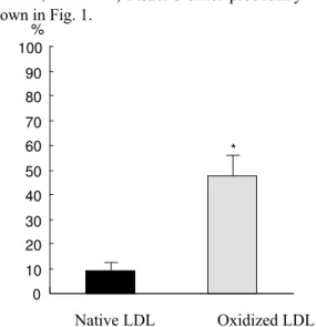
Foam cell formation: More foam cells were observed in macrophages incubated with ox-LDL (n=5,
± %) than those incubated with n-LDL (n=5, ± P< , Fisher’s exact probability test) as shown in Fig. 1.
Native LDL Oxidized LDL
Fig. 1: Foam cells formation by native LDL and oxidized LDL. Back and shaded boxes indicate the rate of foam cell in macrophages incubated with native LDL and oxidized LDL, respectively. *means P < 0.0001 by Fisher’s exact probability test
Fig. 2: Differentially displayed bands on 12.5% native polyacryl-amide gel electrophoresis. Band A was differentially displayed band. bp and numbers indicates base pair. Ox and N means transcripts using mRNA obtained from macrophages incubated with oxidized LDL and native LDL, respectively
bp
A
Ox N
154--0 2 4 6 8 10 12
14 *
Fig. 3: Sub-cloning of differentially displayed band A. The differentially displayed cDNA fragments was 387 bp length and showed 100% homology to the part (nt2036-2422) of 3’-untranalated region of Homo Sapiens GM2 activator protein gene (GENEBANK-NONST/ BLASTN). nt means nucleotide number. Nucleotides underlined mean the primers we used for differential display technique
mg mL¯1
Native LDL Oxidized LDL
Fig. 4: Quantification of GM2 activator protein mRNA. Black and shaded boxes indicate GM2AP mRNA levels (ng mL¯1) on macrophage incubated with native LDL and oxidized LDL, respectively. * means P < 0.01 by Mann-Whitney U test
Detection and identification of differentially displayed band:An approximately 400bp cDNA band was present in reverse transcripts using RNA derived from macrophages incubated with oxidized LDL, but was absent in those incubated with n-LDL (Fig. 2). The isolated and differentially expressed cDNA fragments was 387 bp length and showed 100% homology to the part (nt2036-2422) of 3’-untranalated region of Homo
Sapiens GM2 activator protein (GM2-AP) gene (Fig. 3) (GENEBANK-NONST/ BLASTN)
Expression of GM2 activator protein mRNA: GM2-activator protein mRNA expression in macrophages incubated with oxidized LDL (n=5, 11.81±2.15 ng mL¯1) was significantly higher than that incubated with n-LDL (n=5, 5.36±0.23, P < 0.01, Man-Whitney U test) as shown in Fig. 4.
DISCUSSION
A characterization of regulated gene expression in eukaryotic cells is essential for studying cell growth and differentiation as well as for understanding the molecular mechanisms of diseases. The differential display was developed for such comparative studies by allowing a systematic and non-biased screening for molecular differences at the level of mRNA expression between or among different cells or tissues[12].
Macrophage oxidized LDL uptake is enhanced by oxidized LDL itself, resulting in the direct contribution to foam cell formation and it may also adversely affect many other aspects of arterial wall and thus contribute further to the atherogenic process[15,16]. Oxidized LDL has been reported to regulate various gene expression in macrophages[6-11]. As a result of the study with the differential display, we found the increased expression of the activator protein of GM2 ganglioside, in macrophages and confirmed the increased level of mRNA quantified by using real-time TaqMan RT-PCR assay.
2031 CCTCT GATCT CAGAC CGTGC ATGCCTTGTC CTCTTAAGACA ACTCCTGTGG
2081 CACCGTTTCT CCCTCCACAG GGCCAAAGCC ATAGTGTCCGG TCCCAAGGAC
2131 AAGGCTCTTC CAGTGCTAGG AGAGGTATGA GCAGCCTCTCA CCTGTGAGCT
2181 GTGGGGATCA CAAGGCTGCC TGCCTCAGTC TTGGAGTCCTG TTGGGTGAAT
2231 GAGGCAGATG GGAAAGAGCC TCACCAGCAG CTGCTTTTGGA GCAGGGGTCC
2281 AAGGAAGAGA GGGTGGCCTC GACATCAAAC TGCCAGGATTT TTCTACCACC
2331 CTGTTACATC ATAACAACTT CTGAAACACA CACCAGCCCTG AGTTCTGGGC
The GM2 activator protein is a low-molecular-mass (22 kDa) soluble monomeric glycoprotein that acts as a substrate specific cofactor for the hydrolysis of GM2 ganglioside by lysosomal β x a a A[17]. A Deficiency of the GM2 activator protein results in the storage of GM2 ganglioside and severe neurological disease, Sandhoff disease[17].
Recently, considerably higher ganglioside levels in atherosclerotic lesions of human aorta than those in unaffected areas of aorta and increased concentrations of serum gangliosides in atherosclerotic patients have been observed, suggesting that high ganglioside levels in the aorta and serum may be an additional risk factor for atherosclerosis[18]. The intimal tissue containing fatty streaks and atherosclerotic plaques has been reported to accumulate glycosphingolipids, predominantly glucosylceramide, lactosylceramide and ganglioside GM3. Glycosphingolipid levels in plaques were highest: glucosylceramide was 18- and 8-fold, lactosylceramide was 8- and 7-fold and GM3 was 2.5- and 12-fold higher than in musculoelastic and elastic- hyperplastic intimal layers of normal regions, respectively[19].
Furthermore, markedly increased
glycosphingolipids have been also observed in the aorta of the Watanabe hereditable hyperlipidemic (WHHL) rabbit, an animal model for human familial hypercholesterolemia, as compared with the normal rabbit. Glucosylceramide, lactosylceramide and GM3 ganglioside increased to over 10 times the normal level in the aorta of WHHL rabbits[20]. Neutral glycosphingolipid and ganglioside levels were increased in the serum and aorta of the apolipoprotein E gene knockout mice which shares many features of human atherosclerosis[21]. Therefore, accumulated glycosphingolipids in the aortic wall has been indicated to be another feature of human atherosclerosis as well as oxidized adducts of lipid and proteins.
By this means, accumulating data suggest a significant association between glycosphingolipids and atherosclerosis, however, the underlying mechanism for glycosphingolipid metabolism including uptake, synthesis, transportation and degradation, in atherosclerotic lesion remains to be elucidated. GM2 activator protein is a secretory and lysosomal protein and its relevant cells possess a carbohydrate- independent mechanism to re-capture the activator, with or without bound lipid, from the extracellular fluid. In addition, GM2 activator protein has been shown to bind, solubilize and transport a broad spectrum of lipid molecules, such as glycolipids, gangliosides and phosphoacylglycerol, suggesting that GM2 activator protein may serve as a general intra- and/or inter-cellular lipid transport protein in vivo[22]. Although several experiments in terms of expression mechanisms including GM2 activator protein itself are further required, the present results presumably suggest that oxidized LDL may upregulate
GM2 activator protein, a general lipid transporter in macrophages and consequently enhance the glycosphingolipid accumulation, related to the atherogenesis. Increased levels of GM2 activator protein in the arterial wall or serum may be another feature of human atherosclerosis, as well as increased glycosphingolipid levels.
ACKNOWLEDGEMENTS
We wish thank members in the Department of Laboratory Medicine, Hokkaido University Medical Hospital, for technical help. This work was primarily performed at the Department of Laboratory Medicine, Hokkaido University School of Medicine, Sapporo, Japan.
REFERENCES
1. Gerrity, R.G., 1981. The role of the monocytes in atherogenesis, I:transition of blood-borne monocytes into foam cells in fatty lesions. Am. J. Pathol., 103: 181-190.
2. Takahashi, K., M. Naito, T. Kodama, H. Suzuki, T. Mori and A. Matsumoto, 1992. Expression of macrophage scavenger receptors in various human tissues and atherosclerotic lesions. Clin. Biochem., 25: 365-368.
3. Goldstein, J.L., Y.K. Ho, S.K. Basu and M.S. Brown, 1979. Binding site on macrophages that mediates uptake and degradation of acetylated low density lipoprotein, producing massive cholesterol deposition. Proc. Natl. Acad. Sci. USA., 76: 333-337.
4. Yla-Herttuala, S., W. Palinski, M.E. Rosenfeld, S. Parthasarathy, T.E. Carew, S. Butler, J.L. Witztum and D. Steinberg, 1989. Evidence for the presence of oxidatively modified low-density lipoproteins in atherosclerotic lesions of rabbit and man. J. Clin. Invest., 84: 1086-1095.
5. Nagy, L., P. Tontonoz, J.G. Alvarez, H. Chen and R.M. Evans RM, 1989. Oxidized LDL regulates macrophage gene expression through ligand activation of (PPAR) gamma. Cell, 93: 229-240. 6. Tang, C.K., G.H. Yi, J.H. Yang, L.S. Liu, Z.
Wang, C.G. Ruan and Y.Z., 2004. Oxidized LDL upregulated ATP binding cassette transporter-1 in THP-1 macrophages. Acta. Pharmacol. Sin., 25:581-586.
7. Mikita, T., G. Porter, R.M. Lawn and D. Shiffman, 2001. Oxidized low density lipoprotein exposure alters the transcriptional response of macrophages to inflammatory stimulus. J. Biol. Chem., 276: 45729-45739.
9. Nakazato, K., T. Ishibashi, K. Nagata, Y. Seino, Y. Wada, T. Sakamoto, R. Matsuoka, T. Teramoto, M. Sekimata, Y. Homma and Y. Maruyama, 2001. Expression of very low density lipoprotein receptor mRNA in circulating human monocytes: its up-regulation by hypoxia. Atherosclerosis, 155: 439-444.
10. Fu, Y., N. Luo and M.F. Lopes-Virella, 2000. Oxidized LDL induces the expression of ALBP/aP2 mRNA and protein in human THP-1 macrophages. J Lipid. Res., 41: 2017-2023.
11. Yoshida, H., O. Quehenberger, N. Kondratenko, S. Green and D. Steinberg, 1998. Minimally oxidized low-density lipoprotein increases expression of scavenger receptor A, CD36 and macrosialin in resident mouse peritoneal macrophages. Arterioscler. Thromb. Vasc. Biol., 18: 794-802. 12. Liang, P. and A.B. Pardee, 1989. Differential
display. A general protocol. Mol. Biotechnol. 10: 261-267.
13. Yoshida, H., K. Sasaki, Y. Namiki, N. Sato and N. Tada, 2005. Edaravone, a novel radical scavenger, inhibits oxidative modification of low-density lipoprotein (LDL) and reverses oxidized LDL-mediated reduction in the expression of endothelial nitric oxide synthase. Atherosclerosis, 179: 97-102. 14. Lowry, O.H., N.J. Rosebrough, A.J. Farr and R.J.
Randall, 1951. Protein measurement with the folin phenol reagent. J. Biol. Chem., 193: 265-275 15. Steinberg, D., 1997. Low density lipoprotein
oxidation and its pathological significance. J Biol. Chem. 272: 20963-20967.
16. Witztum, J.L. and D. Steinberg, 2001. The oxidative modification hypothesis of atherosclerosis: does it hold for humans? Trends. Cardiovasc. Med., 11:93-102.
17. Meier, E.M., G. Schwarzmann, W. Furst and K. Sandhoff, 1991. The human GM2 activator protein. A substrate specific cofactor of beta-hexosaminidase A. J. Biol. Chem., 266: 1879-1887.
18. Prokazova, N.V. and L.D. Bergelson, 1994. Gangliosides and atherosclerosis. Lipids, 29: 1-5. 19. Mukhin, D.N., F.F. Chao and H.S. Kruth, 1995.
Glycosphingolipid accumulation in the aortic wall is another feature of human atherosclerosis. Arterioscler. Thromb. Vasc. Biol., 15: 1607-1615. 20. Hara, A. and T. Taketomi, 1991. Characterization
and changes of glycosphingolipids in the aorta of the Watanabe hereditable hyperlipidemic rabbit. J. Biochem. (Tokyo), 109: 904-908.
21. Garner, B., D.A. Priestman, R. Stocker, D.J. Harvey, T.D, Butters and F.M. Platt, 2002. Increased glycosphingolipid levels in serum and aortae of apolipoprotein E gene knockout mice. J. Lipid. Res., 43: 205-214.