Regulate Lifespan and Stress Response Together with
the Anaphase-Promoting Complex
Spike D. L. Postnikoff, Mackenzie E. Malo, Berchman Wong, Troy A. A. Harkness*
Department of Anatomy and Cell Biology, College of Medicine, University of Saskatchewan, Saskatoon, Canada
Abstract
Forkhead box O (FOXO) transcription factors have a conserved function in regulating metazoan lifespan. A key function in this process involves the regulation of the cell cycle and stress responses including free radical scavenging. We employed yeast chronological and replicative lifespan assays, as well as oxidative stress assays, to explore the potential evolutionary conservation of function between the FOXOs and the yeast forkhead box transcription factorsFKH1andFKH2. We report that the deletion of bothFKHgenes impedes normal lifespan and stress resistance, particularly in stationary phase cells, which are non-responsive to caloric restriction. Conversely, increased expression of theFKHsleads to extended lifespan and improved stress response. Here we show the Anaphase-Promoting Complex (APC) genetically interacts with the Fkh pathway, likely working in a linear pathway under normal conditions, asfkh1Dfkh2Dpost-mitotic survival is epistatic to that observed inapc5CAmutants. However, under stress conditions, post-mitotic survival is dramatically impaired inapc5CAfkh1D
fkh2D, while increased expression of eitherFKHrescues APC mutant growth defects. This study establishes theFKHs role as evolutionarily conserved regulators of lifespan in yeast and identifies the APC as a novel component of this mechanism under certain conditions, likely through combined regulation of stress response, genomic stability, and cell cycle regulation.
Citation:Postnikoff SDL, Malo ME, Wong B, Harkness TAA (2012) The Yeast Forkhead Transcription Factors Fkh1 and Fkh2 Regulate Lifespan and Stress Response Together with the Anaphase-Promoting Complex. PLoS Genet 8(3): e1002583. doi:10.1371/journal.pgen.1002583
Editor:Gregory P. Copenhaver, The University of North Carolina at Chapel Hill, United States of America ReceivedSeptember 16, 2011;AcceptedJanuary 20, 2012;PublishedMarch 15, 2012
Copyright:ß2012 Postnikoff et al. This is an open-access article distributed under the terms of the Creative Commons Attribution License, which permits unrestricted use, distribution, and reproduction in any medium, provided the original author and source are credited.
Funding:This work was supported by an operating grant from the Canadian Institutes for Health Research (CIHR) and an infrastructural grant from the Canadian Foundation for Innovation (CFI) New Investigators award to TAAH. SDLP is supported by a Natural Sciences and Engineering Research Council (NSERC) Doctorate award. The funders had no role in study design, data collection and analysis, decision to publish, or preparation of the manuscript.
Competing Interests:The authors have declared that no competing interests exist.
* E-mail: [email protected]
Introduction
The evolutionarily conserved insulin-signaling pathway plays a critical role in multiple cellular processes [1–4]. Perhaps most important is the decisive role it plays in cellular, and organismal, survival. This pathway must be tightly regulated, as overactive insulin-signaling leads to increased survival of cells that would otherwise be shunted down the apoptotic pathway. This occurs by increased repression of stress response, pro-apoptotic, and DNA repair genes, thereby increasing the proliferative capacity, and oncogenic potential of these cells. Although the lifespan of damaged cells is increased under these conditions, this situation increases the probability that the organism will die prematurely due to disease onset. On the other hand, reduced insulin-signaling relieves repression of the stress response, cell cycle arrest and DNA repair pathways, increasing cell maintenance capacity and survival. It is now clear that there is a link between diabetes and cancer [5–7], diseases associated with the insulin-signaling pathways, highlighting the importance of understanding the precise activity of this pathway.
The insulin-signaling phosphorylation cascade activates AKT, which targets cellular factors that switch metabolism from catabolic to anabolic reactions, favoring growth and reproduction over maintenance and repair [8]. Major AKT targets include the forkhead box O family (FOXO) transcription factors (reviewed in [9–14]). The FOXOs are believed to serve diverse rolls in
longevity determination and tumor suppression in metazoans from nematodes to humans. The FOXOs integrate signals from energy, growth factor and stress signaling cascades to regulate cell
differentiation, cell-cycle progression, apoptosis, autophagy,
DNA-damage repair, and scavenging reactive oxygen species. FOXO proteins have been shown to interact with multiple cofactors that mediate their activity through posttranslational modifications. Phosphorylation, ubiquitination, methylation, and acetylation regulate transcription factor efficiency and nuclear shuttling (reviewed in [9,10]). Specifically, nutrient (insulin) and growth factor signals lead to cytosolic sequestering and ubiquitina-tion of the FOXOs, targeting them for degradaubiquitina-tion; conversely, internal reactive oxygen species (ROS), DNA damage sensing and starvation signals can cause nuclear shuttling, and transcription factor activity. Thus, a dynamic and complex molecular network controls FOXO protein function, yet specific downstream targets remains speculative.
Saccharomyces cerevisiaeis often utilized to elucidate regulation of fundamental eukaryotic mechanisms. However, the individual deletion of any of the four Forkhead box orthologs does not affect lifespan [15], suggesting a lack of functional conservation.
However, two of the orthologs, FKH1 and FKH2, show genetic
redundancy, as deletion of both genes is necessary to alter growth, cell morphology and gene transcription phenotypes [16–20].
Further evolutionary conservation for FKH1 and FKH2 is
[18], and cell cycle regulation through the regulation of both G1 and G2/M gene clusters [17], hallmarks of the human FOXO genes.
The Fkh1/2 regulated CLB2 gene cluster [17] encodes genes required for Anaphase-Promoting Complex (APC) activity (APC1,
CDC5,CLB2, andCDC20), as well as APC targets (CLB2,CDC5,
CDC20andIQG1) [21–23]. The APC is a highly conserved multi-subunit ubiquitin-protein ligase (E3) that promotes mitotic progression and G1 maintenance by targeting cell cycle regulators, such as the securin Pds1 and the cyclin Clb2, for proteasome-dependent degradation [21,23,24]. The APC has been demon-strated to be critical for regulating genomic stability, and longevity in yeast and higher eukaryotic organisms [25–30]. In yeast, mutation to multiple APC subunits decreases replicative lifespan (RLS; measures mitotic longevity) and chronological lifespan (CLS; measures post-mitotic survival), while over-expression of
APC10 increases RLS [29]. In mice, mutations to the APC regulator BubR1, a component of the spindle checkpoint, lead to premature aging defects [27,31]. Consistent with this, we and others have provided evidence that the yeast APC plays a role in stress response by possibly targeting proteins that block proper stress response for degradation [28,32,33].
Our data supports a model whereFKH1andFKH2function is
evolutionarily conserved with higher eukaryotic FOXO proteins with regards to lifespan and oxidative stress resistance. We show that the FKHs are required for increased stress resistance and survival in response to severe caloric restriction (cultures maintained in water). Importantly, we identify the APC as a
potential target of the FKHs under normal conditions, while
functioning cooperatively under stress conditions.
Results
FKH1andFKH2 encode redundant longevity determinants
FOXO transcription factors regulate processes involved in the homeostasis of metazoan cells and tissues with the net outcome
being lifespan extension and tumor suppression, yet many of the downstream targets remain unknown [9–14]. The budding yeast
Saccharomyces cerevisiaeis a powerful tool used to elucidate genetic and molecular mechanisms mediating many cellular processes, but independent deletion of the four yeast forkhead box protein encoding genes (FKH1,FKH2,HCM1,FHL1) does not alter CLS [15]. However, Fkh1 and Fkh2 have been shown to be phenotypically redundant, as they are required for M/G1 progression and cell cycle arrest in response to hydrogen peroxide [18]. These characteristics lead us to examine the role of both Fkh1 and Fkh2 in the regulation of yeast lifespan. We investigated theFKHsusing the RLS assay, a measure of the mitotic lifespan of individual cells, finding that deletion of either individualFKHgene had no effect on RLS, as reported for CLS [15]. Double mutant cells could not be assayed using RLS due to their flocculent phenotype (data not shown). Therefore, we investigated the potential of theFKHsin regulating CLS, a measure of metabolic activity in post-mitotic stationary phase cells [34]. In cultures maintained in depleted complete media (DM), we also observed that single deletion of theFKHgenes did not impair CLS. Deletion
of both FKH1 and FKH2, on the other hand, reduced CLS
(Figure 1A), with mean (50%) survival reached by day 8.5 for WT cultures, day 11 forfkh2Dcultures, day 8 forfkh1Dcultures, and only day 4 forfkh1Dfkh2Dcultures.
Controversy exists as to whether higher eukaryotic FOXOs, downstream targets of nutrient/insulin signaling, are contributing factors to caloric restriction-induced lifespan extension [35–39].
To examine whether the yeast FKHs play a role in caloric
restriction, we examined the CLS of the FKH mutants by
maintaining the post-mitotic cultures in distilled H2O. Water is believed to act as a form of severe caloric restriction (SCR) that simulates the low-nutrient environment that yeast in the wild would most likely encounter [40,41]. Maintenance in H2O extended the mean survival of WT,fkh1D andfkh2Dcultures to
19–21 days, while little change was observed in fkh1D fkh2D
cultures with a mean survival of 5 days (Figure 1B). The lack of response infkh1Dfkh2Dcultures maintained in H2O suggests that Fkh1 and Fkh2 play a redundant role in SCR-induced lifespan extension.
Although Fkh1 and Fkh2 have not previously been associated with longevity in yeast, they have been linked with stress response in mitotically active cells [18], which is associated with an evolutionarily conserved role in long lifespan [42–44]. To address whether the Fkhs’ role in longevity is a manifestation of their involvement in stress resistance in post-mitotic cells, we treated WT andfkh1D fkh2D day 5 stationary phase cells maintained in either H2O or DM with 100 mM hydrogen peroxide (H2O2) for 1 hour (Figure 1C). WT day 5 stationary phase cultures exhibited
increased resistance to H2O2when maintained in water compared
to DM. However, this effect was nullified infkh1Dfkh2Dcultures, indicating that the Fkh proteins are required for stress resistance during stationary phase. A plate assay confirmed that stationary phase cells exhibit increased stress response compared to
mitotically active cells, and that deletion of FKH1 and FKH2
diminishes this effect (Figure 1D).
The Fkh proteins are present during stationary phase and have a higher nuclear content in H2O
To further assess the role of theFKHs in normal and stressed post-mitotic lifespan, the endogenous genes encoding Fkh1 and Fkh2 were C-terminally TAP (tandem affinity purification)-tagged, with protein levels analyzed as cells aged in DM media and H2O (Figure 2A). Fkh1-TAP was indeed expressed as cells aged during stationary phase in DM and H2O. Fkh2-TAP was also observed in Author Summary
aging stationary phase cells, but at much lower levels (data not shown). Fkh1-TAP levels appear slightly lower in day 5 stationary phase cells compared to day 1, with very little difference between H2O and DM. Nonetheless, Fkh1 and Fkh2 proteins are expressed in post-mitotic cells.
Next, endogenous C-terminal GFP-tagged Fkh1 and Fkh2 were analyzed in aging cells to determine cellular localization. In day 5
maintained in H2O (Figure 2C). The percentage of cells harboring nuclear Fkh1-GFP or nuclear Fkh2-GPF was consistently observed to be greater when the cells were maintained in H2O compared to DM (Figure 2D). This suggests the presence and nuclear localization of the Fkhs may be necessary for normal CLS and stress resistance, especially in an SCR environment.
Increased expression of theFKHsenhances stress resistance, CLS, and RLS
In higher eukaryotic systems increased expression of FOXO orthologues is associated with increased longevity and stress resistance [38,45]. Thus, we predicted that an increase in survival
would be expected with the overexpression of FKH1 and/or
Figure 2. The Fkh proteins are present in the nucleus of stationary phase cells.(A) Cells expressing endogenously TAP-taggedFKH1were grown to stationary phase and either left in DM or transferred to H2O for the remainder of the experiment. Samples were removed on days 1 and 5 for Western analysis using antibodies against the TAP epitope, or GAPDH as a load control. Fkh2-TAP was also observed in stationary phase cells (data not shown). (B) Cells expressing endogenously taggedFKH1- orFKH2-GFP were grown to day 5 stationary phase while maintained in DM. Cells were observed to harbor both Fkh1 and Fkh2 nuclear staining. (C) Day 13 stationary phase cells expressing Fkh2-GFP were imaged, showing reduced nuclear staining in cells maintained in DM. (D) The percentage of nuclear localized Fkh1-GFP or Fkh2-GFP was determined as cells aged in either DM or H2O.
FKH2. IncreasedFKHexpression was accomplished by integrating
the GAL1/10 inducible promoter immediately upstream of the
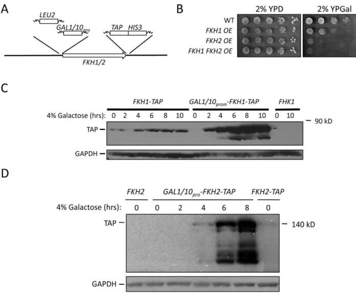
FKH1-TAP and FKH2-TAP start sites (Figure 3A). Cells
overex-pressing both FKH1 and FKH2 were created by crossing the
appropriate strains. Growth of these cells on 2% glucose was
comparable to WT, but growth was diminished when FKH
overexpressing (OE) cells were grown on 2% galactose-supple-mented media (Figure 3B). Fkh1-TAP and Fkh2-TAP were weakly expressed in 2% glucose, but massively expressed after 6 hrs in 2% galactose (Figure 3C, 3D). Lower concentrations of galactose (0.05–0.1%) did not influence vegetative growth (data not shown), but did improve stress resistance and longevity (Figure 4). First, we measured the ability of 5 day stationary phase cultures maintained in DM to survive a 1 hour treatment of 100 mM H2O2. For this experiment, once cells reached stationary phase the cells were split with one sample receiving a supplement of 0.05% galactose. After
5 days, samples were removed, treated with H2O2, and then diluted onto YPD plates to determine colony forming units.
Controls were cells that did not receive H2O2. The FKH OE
cultures exhibited increased survival in the absence of galactose, with improved resistance when supplemented with galactose (Figure 4A). These observations are consistent with a role for the Fkh proteins in stress resistance during stationary phase.
If the Fkh proteins do enhance stress resistance during stationary phase, then it is likely that increasedFKH expression may prolong metabolic activity in these cells. CLS of WT andFKH
OE cells was measured in DM in the presence and absence of 0.05% galactose (Figure 4B). Cells were grown in 2% glucose to stationary phase, then split, with one sample receiving a supplement of 0.05% galactose. Unaltered WT cells were used as a control. We observed that the addition of 0.05% galactose increased the CLS mean lifespan (50% survival) of the unaltered
Figure 3. IncreasedFKH1orFKH2expression improves stress resistance and extends CLS and RLS.(A) Schematic representation of scheme used to integrate theGAL1/10promoter upstream ofFKH1andFKH2. TheLEU2PCR product, containing 300 basepairs ofLEU2promoter (incorporated for selection purposes), was flanked by 60 basepairs of homology to theFKHpromoter and to theGAL1/10promoter. TheGAL1/10
promoter PCR fragment was flanked by 60 basepairs of homology to the 39end ofLEU2and to the 59end of theFKHgene. Cells were cotransformed with both products and selected on leu2plates. Cells harboringFKH1andFKH2under the control of theGAL1/10promoter were generated by crossing the single integrated strains. (B) Cells overexpressing (OE)FKH1and/orFKH2from theGAL1/10promoter were grown overnight in 2% YPD, then spot diluted onto the plates shown. The plates were incubated at 30uC for 2 to 5 days. (C)FKH1-TAPOE cells were grown overnight in 2% glucose. The next day, the cells were washed and resuspended in media containing 2% galactose. Samples were taken every two hours for 10 hours for Western analysis using antibodies against TAP and GAPDH. Cells expressingFKH1-TAPandFKH1under their own promoter were used as controls. (D)FKH2-TAPOE cells were analyzed as described above forFKH1-TAPOE cells.
WT control from approximately 8.5 to 12 days. In the absence of galactose the OE strains exhibited mean lifespans of 9.5 to 12 days. However, in the presence of 0.05% galactose, theFKH1OE strain
experienced a 15 day mean lifespan, while FKH2 OE strains
enjoyed mean lifespans of approximately 27 days. Yeast cell lifespan can be measured in post-mitotic stationary phase (CLS), or in rapidly dividing mitotic cells (RLS). Stress resistance plays a major role in determining both CLS and RLS, however not all genes that influence CLS also influence RLS. Sir2 in yeast is a good example of this [44]. To determine whether the Fkh proteins also influence RLS, we measured RLS in the cells employed in Figure 4B using 2% sucrose as a base carbon source in the presence and absence of 0.1% galactose (Figure 4C). In the absence of galactose, RLS of all strains was relatively unchanged.
Upon 0.1% galactose supplementation, FKH2 OE cells had a
markedly longer RLS. The mean lifespan ofFKH1OE cells was
also increased, but not to the same extent as theFKH2OE cells.
We also observed increased RLS inFKH2OE, but notFKH1OE
cells using 0.05% galactose (data not shown). The enhanced stress resistance and CLS observed in OE strains in the absence of galactose are not surprising as we previously documented the basal activity of the galactose promoter [33]. Our results are consistent with the Fkh proteins playing a role in responding to stress, which may indirectly lead to increased CLS and RLS. The effect appears to be greater during post-mitotic stationary phase cells, with Fkh2 perhaps playing a more pivotal role compared to Fkh1.
The Fkh proteins and the Anaphase-Promoting Complex (APC) work together to mediate post-mitotic survival
The advantage of using yeast for genetic studies is the relative ease of identifying interacting partners for proteins and genes of interest. Thus, we sought possible downstream Fkh targets that may be involved in stress response and longevity. One possible target of the Fkh transcription factors is the Anaphase-Promoting Complex (APC). The APC is an evolutionarily conserved ubiquitin-protein ligase (E3) that targets proteins that inhibit mitotic progression and exit, as well as G1 maintenance, for ubiquitin- and proteasome-dependent degradation [21,23]. We previously observed APC mutants to exhibit reduced CLS and
RLS, while increasedAPC10expression extended RLS [29,46].
Furthermore, APC mutants are sensitive to DNA damaging agents, and exhibit both chromatin assembly and histone modification defects [28,33,46–49]. Consistent with the APC’s involvement in histone biogenesis and lifespan, we recently demonstrated that yeast cells harboring histone modification defects are subject to reduced RLS [50]. The APC appears to play an evolutionarily conserved role in lifespan, as mutations to mouse BubR1, a component of the spindle checkpoint that inhibits APC function, resulted in inappropriate APC activity and premature aging phenotypes [27]. A possible link between the APC and the Fkhs was revealed by a previous microarray analysis offkh1Dfkh2D
cells, whereAPC1 (APC subunit),CDC5 (APC activator/target),
CLB2(APC activator/target),CDC20(APC activator/target), and
IQG1 (APC target), were identified as responsive to the Fkh
transcription factors [17,21,22]. A subsequent analysis of CLB2
mRNA expression during the cell cycle (CLB2mRNA synthesis is
cell cycle regulated) showed that it was defective infkh1Dfkh2D
mutants [16]. Clb2, a mitotic cyclin that is an important activator of the APC, later becomes targeted by the APC for degradation to allow exit from mitosis [21]. To test our hypothesis that APC activity may be targeted and activated by the Fkh proteins, we createdfkh1Dfkh2Dcells harboring a mutation in the APC subunit
Apc5 to enable genetic analyses.APC5encodes an essential APC
subunit (the apc5CA allele used in our studies contains a two
basepair deletion at the 59end of the coding region, most likely creating an N-terminally truncated protein [28]; unpublished data). Cells with theapc5CAmutation grow slowly at temperatures
above 36uC, which can be recovered or exacerbated by genetic
alteration of negative or positive regulators, making this an
excellent allele to identify APC5 interacting partners
[29,33,47,49,51].
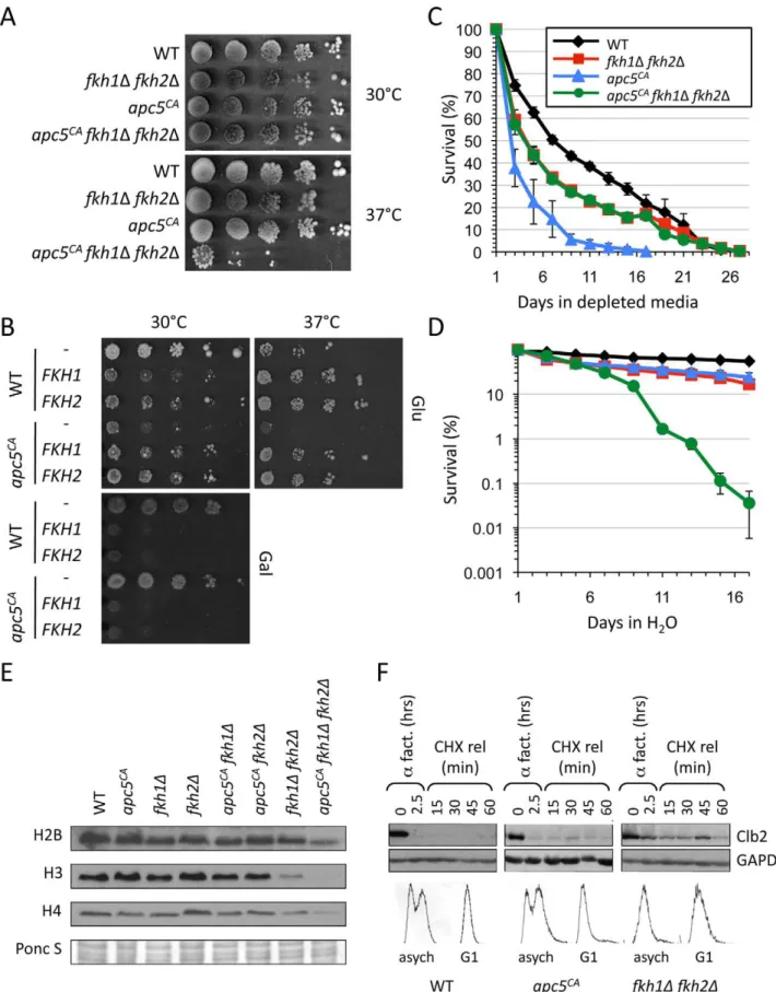
First, we examined the growth characteristics of apc5CAfkh1D
fkh2D mutants. Deletion of both FKH genes severely impaired
apc5CA growth at the restrictive temperature, but not at the
permissive temperature (Figures 5A). Deletion of bothFKHgenes also impaired the growth ofapc10D cells and was lethal in the
apc11-13 background (unpublished data). This preliminary Figure 4. Increased expression of the FKH genes increases
lifespan and stress response.(A) TheFKHOE cells were grown to stationary phase, then either maintained in DM, or 0.05% galactose was added. The cells were incubated for an additional 5 days, then split, with one half treated with 100 mM H2O2for 1 hour. The other half served as the untreated control. Following the 1 hour incubation, the cells were diluted and plated onto YPD until colony forming units formed. Survival was determined by dividing the treated cells by the untreated cells. Standard error is shown for at least 3 replicates. (B) CLS was determined for the OE strains when maintained solely in DM (left panel) or in DM supplemented with 0.05% galactose (right panel). Standard error is shown for at least 3 replicates. (C) RLS was determined for the OE strains on 2% sucrose plates or sucrose plates supplemented with 0.1% galactose. Typical results are shown.
investigation provides evidence that the APC and the Fkhs may share a common function.
If the Fkhs and the APC do share a common function, then
increased expression of the FKHs may restore the apc5CA
temperature sensitive (ts) growth phenotype. Thus, we expressed
plasmid borneFKH1andFKH2under the control of theGAL1/10
promoter in WT and apc5CA cell. The cells were grown in 2%
glucose supplemented media, then spot diluted onto plates containing either 2% galactose or 2% glucose (Figure 5B). At 30uC, expression of theFKHson glucose-supplemented media was slightly detrimental to WT cells, but beneficial to apc5CA
cells. Galactose-driven genes do have basal activity in the presence of glucose [33]. On galactose plates, overexpression of either FKH
was toxic, as observed above (Figure 3B). At 37uC, expression of
eitherFKHgene on glucose plates improved growth of both WT
and apc5CA cells. We have also observed that increased FKH1
expression suppressed the ts defect in additional APC mutants,
including apc10D, apc11-13, cdc16-1, cdc23-1, apc5CA apc10Dand
apc5CAcdc26Dcells (data not shown). These observations suggest that under conditions of stress, whether temperature or impaired
APC activity, moderately increasedFKHexpression improves the
capacity of the cell to cope.
Next, we asked whether deletion of bothFKHsinfluencedapc5CA
CLS. Cells expressing the different combinations of mutations were grown to stationary phase, then split, with one half resuspended in H2O, and the other half left in DM. Equal volumes were then plated on the days shown to generate survival curves (Figure 5C, 5D). In DM,apc5CAcells rapidly lost viability compared to WT andfkh1Dfkh2Dcells (Figure 5C). Interestingly, the triple mutant survived as long as fkh1D fkh2D cells. This suggests that deletion of bothFKH1andFKH2is epistatic to the
apc5CA allele under the conditions tested. In other words, under normal media conditions using DM, the Fkhs appear to act upstream of the APC. When the experiment was conducted by maintaining the cells in H2O for the duration of the experiment, a different survival profile was observed (Figure 5D). WT (20 days vs 7 days) andapc5CA
(5 days vs 2.5 days) cells both responded to
H2O conditions by exhibiting a longer mean CLS. However,
fkh1Dfkh2Dcells had the same CLS in H2O as in DM, which was similar to apc5CA cells in H2O, whereas the triple mutant had a greatly reduced CLS in H2O. As stated above, the failure offkh1D fkh2Dcells to survive longer under SCR conditions, such as H2O, suggests that the Fkhs are required for long life under SCR conditions. The similar H2O CLS observed infkh1D fkh2Dand
apc5CAcells, and the dramatically reduced H2O CLS in the triple mutant indicates that the Fkhs and the APC may have redundant functions under stress conditions. This contrasts with the Fkh/ APC epistatic interaction that appears to occur under DM conditions. This could reflect dual roles for the Fkh proteins; as cell cycle regulators under normal conditions, and as stress response proteins under stress conditions [17,18,52,53]. Lastly, these observations clearly identify another non-mitotic function for the APC. The APC has been shown to function in other non-mitotic
activities, such as meiosis, quiescence, differentiation, metabolism, maintenance of post-mitotic neurons, and interestingly, memory in mice [30,54–58].
In addition to controlling CLS, the APC and the Fkhs are also involved in histone metabolism, but likely through very different mechanisms. The Fkhs are redundant activators of cell cycle dependent histone expression [17]. On the other hand, histones and histone modifications are reduced when genes encoding
different APC subunits, such as APC5, APC9, APC10, APC11,
CDC16, CDC23and CDC26, are mutated [33]. The mechanism involved remains elusive, but it is likely post-transcriptional, as histone mRNAs are unaltered in APC mutants [33]. Our analysis of total histone levels in the different mutant combinations indicates that histone control is indeed through different redundant mechanisms, as histone levels are greatly reduced in the triple mutant compared to the single and double mutants (Figure 5E). However, it remains possible that the Fkhs drive histone synthesis through direct transcriptional control and indirectly via the APC.
As a direct assessment of whether the Fkhs control an APC function, we measured Clb2 stability inapc5CAand fkh1Dfkh2D
mutants. Clb2 is a B-type cyclin that is targeted by the APC for degradation in order to exit mitosis [21].CLB2transcripts are also controlled by the Fkhs [16,17]. WT,apc5CAandfkh1Dfkh2Dcells were grown to early log phase at 30uC, then arrested in G1 usinga
factor. The cells were determined to be arrested in G1 using microscopy (data not shown) and FACS analysis (Figure 5F). The cells were then released into cycloheximide, with samples removed every 15 minutes for protein analysis using antibodies against
endogenous Clb2. In both WT andapc5CAcells Clb2 levels were
decreased in G1 arrested cells when compared to asynchronous cells (Figure 5F). Infkh1Dfkh2Dcells however, the degradation of Clb2 was reduced in G1 arrested cells. However, it should be noted thatfkh1Dfkh2Dcells accumulated in G1 in asynchronously grown cells, as indicated by FACS, perhaps reflecting the inability to degrade mitotic cyclins. These observations are consistent with a model where the Fkh transcription factors act in a positive manner upstream of the APC, perhaps through transcriptional activation of APC subunits, activators and targets.
The APC and the Fkhs function in the stress response pathway
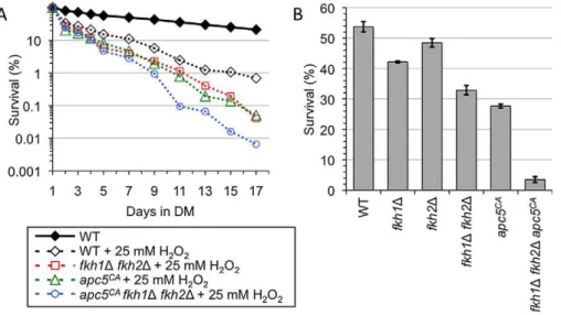
Since the APC is associated with maintaining lifespan in multiple systems [27,29,30,31], we tested whether the APC may also be involved in oxidative stress resistance. To test this hypothesis, we conducted CLS in the presence of oxidative stress in WT,apc5CA,fkh1Dfkh2Dandapc5CAfkh1Dfkh2Dcells. The cells were grown to stationary phase followed by the addition of 25 mM
H2O2(Figure 6A), with cell counts determined every other day.
The results show WT cells had a reduced lifespan in response to
25 mM H2O2, while the mutants were further impaired. The
lifespan of the triple mutant was dramatically reduced compared
to apc5CA and fkh1D fkh2D cells in H2O2, as it was in water
maintained in depleted media (DM) for the remainder of the experiment. Colony forming units were determined every other day and a survival curve was plotted. Standard error is shown for at least 3 repeats. (D) CLS was determined for the strains used above. Rather than maintenance in DM, the cells were washed once they reached stationary phase and maintained in H2O for the remainder of the experiment. Standard error is shown for at least 3 repeats. The experiments in (C) and (D) were started from the same cultures. (E) The panel of strains shown were grown overnight at 30uC to early log phase growth. Proteins were extracted and analyzed by Westerns to assess total histone H2B, H3 and H4 protein levels. A Ponceau S stained gel is included to shown equivalence of protein load. (F) WT,apc5CAandfkh1
Dfkh2Dcells encodingBAR1were grown to early log phase, then arrested with 2mg/mlafactor for 1.5 hours in pH 3.5 YPD media. Another 2mg/mlafactor was added followed by another hour of incubation. Cycloheximide was then added with continued incubation. Samples were removed at the times shown and Clb2 protein levels were determined by Western blotting. Antibodies against GAPDH were included as a load control. The same blot was divided and used for the Clb2 and GAPDH Westerns. Samples were taken before and after G1 arrest for FACS analysis.
(Figure 5D). These observations provide additional evidence that the triple mutant is extremely sensitive to stress and likely perceives water as a severe stress, rather than a form of caloric restriction. To our knowledge, few mutations have been described that act in a negative manner to SCR.
To investigate the roles of the APC and the Fkh proteins in stress resistance further, stationary phase cultures were treated
with 100 mM H2O2for 30 minutes at 30uC, and then plated to
determine cell viability. While 53.2% of WT cells survived
100 mM H2O2, only 21.3% of apc5CA cells and 31.3% of the
fkh1D fkh2D survived the treatment (Figure 6B). However, the
apc5CAfkh1Dfkh2Dmutant was dramatically impaired, with only 3.5% surviving this treatment. Taken together, our data indicates that the APC and the Fkh proteins have overlapping functions in response to oxidative stress in post-mitotic cells, opposed to the epistatic interaction observed in unstressed cells (Figure 5C, Figure 7). Consistent with an APC involvement in post-mitotic CLS, the APC is also required for stress response in post-mitotic cells.
Discussion
This report provides evidence to support an evolutionarily conserved role for the yeast forkhead transcription factors Fkh1 and Fkh2 in lifespan determination and stress response.FKH1and
FKH2act redundantly in controlling CLS and post-mitotic stress response, as deletion of both genes is required to observe CLS and stress response defects (Figure 1), as previously described for other phenotypes [16,17]. Importantly, we show that a modest increase
inFKH1orFKH2expression results in increased CLS, RLS and
stress resistance (Figure 4). The Fkhs may have a dual function in cell cycle progression and in stress response. A microarray analysis initially identified such a role, as arrest offkh1Dfkh2Dcells in G1 identified a series of genes involved in cell cycle progression, whereas the transcript profile identified in asynchronous fkh1D
fkh2D cells was composed of stress response genes [17].
Importantly, we identify an evolutionarily conserved cell cycle
regulator, the Anaphase-Promoting Complex (APC), as a potential downstream target of the Fkh transcription factors (Figure 5, Figure 6). Genetic interaction studies between APC and Fkh mutants revealed a possible division of Fkh labor. For example, the
CLS of apc5CA and fkh1D fkh2D cells showed an epistatic
interaction under normal conditions (Figure 5C), suggesting the Fkhs may be upstream of the APC. However, under stress conditions, such as maintenance in H2O (Figure 5D) and in the
presence of 25 mM H2O2 (Figure 6A), the CLS of the triple
mutant was dramatically impaired beyond any of the single and double mutants, defining a synergistic interaction. This most likely Figure 6. The APC and the Fkh proteins provide overlapping function to respond to oxidative stress.(A) CLS was performed using the strains shown in the presence of 25 mM H2O2. The cells were grown to stationary phase, and then H2O2was added to the cultures. Colony forming units were determined every other day for the remainder of the experiment. Survival curves are shown. (B) The strains described above were used to determine resistance to oxidative stress. Day 5 stationary phase cells were exposed to 100 mM H2O2for 1 hour at 30uC. Controls were not treated with H2O2. Diluted cells were then plated onto YPD plates until colonies formed. The percent survival was determined, with standard error shown for at least 3 replicates.
doi:10.1371/journal.pgen.1002583.g006
Figure 7. Model depicting how the APC and the Fkh proteins may interact under normal and stress conditions.Under normal conditions, the Fkh proteins play a role in activating the APC. This in turn mediates progression through mitosis and maintenance of G1. This interaction is depicted by bold arrows. Under stress conditions, both the APC and the Fkh proteins respond via different mechanisms. The Fkh proteins likely promote the expression of stress response proteins. The APC acts according to a mechanism that remains uncharacterized, but likely requires ubiquitin-dependent processes.
reflects a role for the Fkhs in activating the transcription of genes involved in stress response (Figure 7). Thus, under non-stress conditions, the Fkhs may play a role in driving APC activity that leads to controlled progression through mitosis and into G1. Since the APC is required for genomic stability, activating the APC would be predicted to increase the fidelity of chromosome segregation, reducing nondysjunction events, thereby increasing the potential for a healthier and longer cellular lifespan. The APC also plays a role in stress response (Figure 6B). Previously, it was shown cells lacking the Cdh1 APC activator were sensitive to multiple stresses, such as ethanol, caffeine and salt [32], and we have shown thatapc5CAandapc10Dcells are sensitive to UV and methylmethanesulfonate (MMS) [28,33]. Stabilization of the APC targets Clb2 and Hsl1 also increased stress sensitivity [32], indicating the APC may activate the stress response by alleviating inhibitory signals. The Fkhs’ contribution to longevity is most likely an indirect reaction to stress, occurring via at least two pathways, one involving transcription of stress genes [17,18], and the other through driving APC activity (Figure 7). It has long been established that cells better equipped to repair damage and respond to stress stand a better chance to live a healthier and potentially longer lifespan [34,42,43,59]. These observations demonstrate the evolutionary conservation of the FOXO transcription factors in yeast, which respond to stress and extend cellular lifespan.
Fkh1, Fkh2, and severe caloric restriction (SCR)
The initial focus of this work was to determine whether the conserved yeast Fkh proteins were involved in longevity, as shown with metazoan FOXOs. Our work clearly demonstrates a need for the Fkh proteins for extended CLS and RLS. However, our work also demonstrates that the Fkh proteins are necessary for extended lifespan in response to SCR. Recently, the Rim15 stress responsive transcription factor was identified as a major mediator of SCR
lifespan extension [15]. Deletion of RIM15 blocked extended
lifespan in ras2D, sch9D and tor1D strains, indicating that the phenomenon of SCR funnels through Rim15. Interestingly,
although deletion of RIM15 in the extremely long-lived ras2D
sch9Dmutant reduced lifespan under normal conditions, this strain could still respond to SCR, suggesting other factors can respond to SCR in the absence of Rim15. Our data provides the possibility that Fkh1/Fkh2 may fulfill this role. Future work will require an analysis of strains lackingFKH1,FKH2andRIM15.
The APC is likely a downstream target of the stress responsive Fkh proteins
We tested the hypothesis that the Fkh proteins play a role in lifespan by contributing to APC activation. Our results support
this hypothesis, as (i) low-level expression of FKH1 or FKH2
suppressed APC mutant growth phenotypes; (ii) deletion of both
FKH1 and FKH2 exacerbated APC mutant histone and growth
phenotypes in mitotically active cells; and (iii) deletion of FKH1
andFKH2stabilized the APC substrate Clb2. Furthermore, stress resistance and lifespan in the presence of stress were markedly worse in the triple mutant compared to the single and double mutants. These results demonstrate that the APC and the Fkhs function together in both mitotic and post-mitotic cells. While
fkh1D fkh2D cells do not respond to SCR, apc5CA fkh1D fkh2D
exhibit decreased CLS under these conditions. This suggests that H2O is perceived as much more than a low level stress in the triple mutant, which is consistent with the Hormesis hypothesis of aging, a theory that postulates low level stresses turn on stress defense mechanisms, leading to potenially longer life [60]. Nonetheless,
these observations implicate the APC as a player in caloric restriction and stress response.
We observed that APC andFKHmutants interacted differently
depending on the growth conditions. Figure 7 presents a model describing how this may occur. Under non-stress conditions, such as growth on YPD or maintenance of stationary phase cells in the depleted media (DM), there was little interaction (growth on YPD at 30u) or an epistatic interaction (CLS in DM). The interaction could be interpreted to define a pathway where the Fkhs act upstream of the APC. It could be as simple as the Fkhs driving the transcription of the APC subunit Apc1, and the APC activators Clb2, Cdc5 and Cdc20 [16]. This interaction is likely more complicated than direct classical epistasis, perhaps involving stoichiometric alteration of APC activators, subunits and/or substrates. The deletion of theFKHs in APC mutant strains may bring about a homeostatic balance between APC activity and substrate levels. On the other hand, under stress conditions, such as high temperature or the addition of H2O2,apc5CAfkh1Dfkh2D
cells exhibited a synergistic interaction. This type of interaction occurs when multiple proteins drive a similar activity in a redundant manner, thus requiring that more than one mutation must occur to expose a phenotype. Both the Fkhs and the APC are required for response to stress, but likely act in very different manners. The Fkhs drive expression of many stress response genes [16], whereas the molecular mechanisms employed by the APC to
combat stress may involve APCCdh1, which has been found to
regulate different stress responses through the degradation of substrates such as Clb2 and Hsl1 as a part of the APC’s role in G1 maintenance [32]. An additional role the APC likely plays in stress revolves around histone metabolism. We have shown that APC mutants exhibit defects in histone maintenance, chromatin assembly and histone modifications [28,33,47–49,51]. It is well established that histone modifications, such as phosphorylation of H2A/H2AX, methylation of H3 Lys79, and acetylation of H3 Lys56 and H4 Lys16, are required for recruitment of DNA repair enzymes to sites of DNA damage [61–65]. Therefore, under stress conditions, the Fkhs and the APC likely play separate, yet overlapping roles in ensuring the cell responds to stressful damaging agents in a positive manner.
Our studies provide a potential link between the Fkh transcription factors and the APC that ties the APC together with stress response. This provides insight into a molecular mechanism whereby the APC facilitates longevity. The evolutionarily conserved nature of our results, and the established role the APC plays in tumor development and progression, suggests that the APC may be a potential downstream target of the insulin signaling pathway. A recent report supports this scenario, as the insulin driven AKT1 kinase phosphorylates the APCCdh1substrate Skp2, an SCF component, which inhibits Skp2 degradation [66]. This enables SCF activity and promotes cell cycle progression. These studies offer the basis for further studies in understanding APC-dependent longevity.
Conclusions
This study identifies the yeast forkhead box containing proteins Fkh1 and Fkh2 as regulators of lifespan, allowing for the characterization of upstream and downstream regulation by these factors. This may provide insight into how the highly related FOXO proteins in mammals regulate both lifespan extension and
tumor suppression. Our data supports a model whereFKH1and
FKH2are functionally orthologous with the metazoan FOXOs,
Materials and Methods
Yeast strains and plasmids
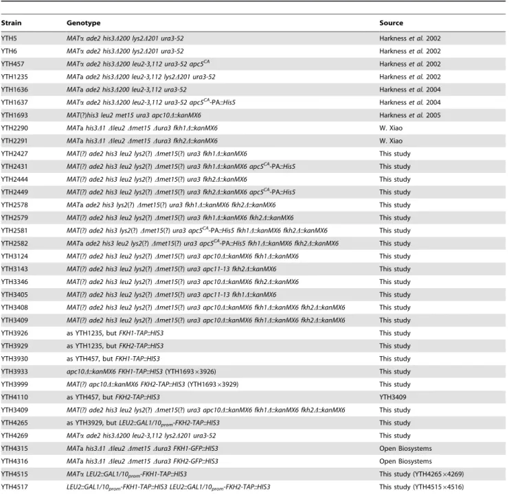
The yeast strains used in this study are shown in Table 1. The
fkh1D::kanMX6 and fkh2D::kanMX6 strains, obtained from the ResGen library (provided by W. Xiao, U. of Saskatchewan), were repeatedly backcrossed to our S288c background strains to generate apc5CA, apc10D and apc11-13 congenic partners. apc11-13 cells were a kind gift from T. Hunter (Salk Institute). Cells
harboringAPC9,APC10andCDC26deletions were acquired from
W. Xiao and backcrossed repeatedly to our S288c background.
cdc16-1, cdc23-1 and the isogenic wild type were generously
provided by D. Stuart (U. of Alberta). The C-terminalFKH1-TAP
andFKH2-TAPstrains were generously provided by A. Ghavidel
(U. of Toronto). PCR based methods were used to TAP-tagFKH1
and FKH2 in various mutants. Cells expressing endogenously
GFP-tagged FKH1 and FKH2 were obtained from Open
BioSytems. The galactose inducible FKH1-HA and FKH2-HA
encoding plasmids were obtained from the Research Genetics library of tandem affinity tagged plasmids purchased by W. Xiao
(U. of Saskatchewan). The GAL1/10 promoter was integrated
upstream of the FKH1 and FKH2 genes by PCR-based
homologous integration approach. Two sets of primers were designed for this approach. First, primers were designed to amplify theLEU2gene (plus 300 basepairs of the promoter) flanked on the
59 side by 60 nucleotides of sequence homologous to the
immediate promoter regions ofFKH1orFKH2, and the 39 side
by 60 nucleotides homologous to the GAL1/10 promoter. The
Table 1.Saccharomyces cerevisiaestrains used in this study.
Strain Genotype Source
YTH5 MATaade2 his3D200 lys2D201 ura3-52 Harknesset al.2002
YTH6 MATaade2 his3D200 lys2D201 ura3-52 Harknesset al.2002
YTH457 MATaade2 his3D200 leu2-3,112 ura3-52 apc5CA Harknesset al.2002
YTH1235 MATaade2 his3D200 leu2-3,112 lys2D201 ura3-52 Harknesset al.2002
YTH1636 MATaade2 his3D200 leu2-3,112 ura3-52 Harknesset al.2004
YTH1637 MATaade2 his3D200 leu2-3,112 ura3-52 apc5CA-PA::His5 Harknesset al.2004
YTH1693 MAT(?)his3 leu2 met15 ura3 apc10D::kanMX6 Harknesset al.2005
YTH2290 MATahis3D1Dleu2Dmet15Dura3 fkh1D::kanMX6 W. Xiao
YTH2291 MATahis3D1Dleu2Dmet15Dura3 fkh2D::kanMX6 W. Xiao
YTH2427 MAT(?) ade2 his3 leu2 lys2(?)Dmet15(?)ura3 fkh1D::kanMX6 This study
YTH2431 MAT(?) ade2 his3 leu2 lys2(?)Dmet15(?)ura3 fkh1D::kanMX6 apc5CA-PA::His5 This study
YTH2444 MAT(?) ade2 his3 leu2 lys2(?)Dmet15(?)ura3 fkh2D::kanMX6 This study
YTH2449 MAT(?) ade2 his3 leu2 lys2(?)Dmet15(?)ura3 fkh2D::kanMX6 apc5CA-PA::His5 This study
YTH2578 MATaade2 his3 lys2(?)Dmet15(?)ura3 fkh1D::kanMX6 fkh2D::kanMX6 This study
YTH2579 MAT(?) ade2 his3 leu2 lys2(?)Dmet15(?)ura3 fkh1D::kanMX6 fkh2D::kanMX6 This study
YTH2581 MAT(?) ade2 his3 lys2(?)Dmet15(?)ura3 apc5CA-PA::His5 fkh1D::kanMX6 fkh2D::kanMX6 This study
YTH2582 MATaade2 his3 leu2 lys2(?)Dmet15(?)ura3 apc5CA-PA::His5 fkh1
D::kanMX6 fkh2D::kanMX6 This study YTH3124 MAT(?) ade2 his3 leu2 lys2(?)Dmet15(?)ura3 apc10D::kanMX6 fkh1D::kanMX6 This study
YTH3143 MAT(?) ade2 his3 leu2 lys2(?)Dmet15(?)ura3 apc11-13 fkh2D::kanMX6 This study
YTH3346 MAT(?) ade2 his3 leu2 lys2(?)Dmet15(?)ura3 apc10D::kanMX6 fkh2D::kanMX6 This study
YTH3405 MAT(?) ade2 his3 leu2 lys2(?)Dmet15(?)ura3 apc11-13 fkh1D::kanMX6 This study
YTH3408 MAT(?) ade2 his3 leu2 lys2(?)Dmet15(?)ura3 apc10D::kanMX6 fkh1D::kanMX6 fkh2D::kanMX6 This study
YTH3409 MAT(?) ade2 his3 leu2 lys2(?)Dmet15(?)ura3 apc10D::kanMX6 fkh1D::kanMX6 fkh2D::kanMX6 This study
YTH3926 as YTH1235, butFKH1-TAP::HIS3 This study
YTH3929 as YTH1235, butFKH2-TAP::HIS3 This study
YTH3930 as YTH457, butFKH1-TAP::HIS3 This study
YTH3933 apc10D::kanMX6 FKH1-TAP::HIS3(YTH169363926) This study
YTH3999 MAT(?) apc10D::kanMX6 FKH2-TAP::HIS3(YTH169363929) This study
YTH4110 as YTH457, butFKH2-TAP::HIS3 YTH3409
YTH3409 MAT(?) ade2 his3 leu2 lys2(?)Dmet15(?)ura3 apc10D::kanMX6 fkh1D::kanMX6 fkh2D::kanMX6 This study
YTH4265 as YTH3929, butLEU2::GAL1/10prom-FKH2-TAP::HIS3 This study
YTH4269 MATaade2 his3D200 leu2-3,112 lys2D201 ura3-52 This study
YTH4315 MATahis3D1Dleu2Dmet15Dura3 FKH1-GFP::HIS3 Open Biosystems
YTH4316 MATahis3D1Dleu2Dmet15Dura3 FKH2-GFP::HIS3 Open Biosystems
YTH4515 MATaLEU2::GAL1/10prom-FKH1-TAP::HIS3 This study (YTH426564269)
YTH4517 LEU2::GAL1/10prom-FKH1-TAP::HIS3 LEU2::GAL1/10prom-FKH2-TAP::HIS3 This study (YTH451564516)
second primer set was designed to amplify GAL1/10 promoter flanked on the 59side by 60 nucleotides homologous to the 39end of theLEU2gene and on the 39end by 60 nucleotides homologous to the first 60 nucleotide ofFKH1orFKH2. Primer sequences are available upon request. The two PCR fragments were transformed
together into WT cells. Leu+ transformants were selected for
further analysis.
Media and methods
Media were prepared as previously described [28,33].
Segrega-tion of thekanMX6 cassette was determined by patching spores
onto 0.2 mg/ml geneticin-supplemented YPD plates. Mutants
containing two or morekanMX6marked alleles were generated by
selecting tetrads in which G418 resistance segregated in a 2:2
fashion. Escherichia coli strains JM109 and DH10B were used to
propagate DNA plasmids. DNA manipulations such as DNA minipreps, and yeast andE. colitransformations were carried out according to standard protocols [67]. Spot dilution assays were conducted by pipeting 3ml of cells from samples generated from a
10-fold dilution series onto the various media shown and grown at the temperatures indicated. The starting spot generally contained 56104cells. To assess resistance to oxidative stress, cultures were
grown in 2% YPD at 30uC for 5 days and then diluted to an
OD600 of 1 in depleted media (DM). Each of these cultures was
divided into two samples and 100 mM H2O2(EMD Chemicals)
was added to one sample. Both samples were then incubated at 30uC for 1 hour. Viability was determined by plating diluted cells
onto 2% YPD and comparing the growth of the H2O2 treated
culture to that of the non-treated control culture. Clb2 stability was determined by growing the indicated cells to early log phase at 30uC, then adding 2mg/mlafactor, when usingBAR1strains, in
media at pH 3.5 to arrest cells in G1. After 1.5 hours, another
2mg/ml was added, with continued incubation for another
1 hour. After this treatment, cells were arrested in G1. Cell cycle arrest was confirmed by microscopic visualization of the cells and
by flow cytometry. Theafactor was then washed away and fresh
media containing 10mg/ml cycloheximide was added. The
incubation was continued at 30uC, with samples removed every
15 minutes for Western analysis using antibodies against endog-enous Clb2 and GAPDH. FACS was performed as described previously [28]. To characterize fkh1D fkh2DFACS profiles the following changes were made to overcome the persistent cell/cell contacts inherent to this mutant. 1 ml of culture (OD6000.4) was resuspended in 50ml followed by the addition 10ml of 12.5 U/ml lyticase. This solution was incubated for 15 minutes at room temperature, followed by the addition of 500ml 50 mM Tris. The cells were then sonicated for 6 seconds at output 4. The cells were centrifuged, resuspended in 1 ml 70% EtOH and processed as usual past this step.
Lifespan assays
Chronological lifespan was performed as previously described [29,40,41]. Briefly, overnight CM (2% glucose) cultures were diluted to OD6000.5 in fresh CM with a flask to culture volume ratio of 5:1. The incubation continued (200 RPM) at 30uC. Each day the same volume of culture was diluted and plated to evaluate colony forming units (CFU) as a measure of viability. When the CFU counts peaked, this was deemed stationary phase and
denoted as Day 1. Every two days CFU were determined and compared to Day 1. For severe caloric restriction (SCR) experiments, once stationary phase was reached in CM (Day1), cultures were washed and resuspended in sterile distilled H2O, with washes of equal volume of water every two to four days to remove metabolites produced by the cells. Galactose and hydrogen peroxide were added to appropriate cultures upon reaching Day 1 to final concentrations of 0.05% and 25 mM respectively.
For fluorescent localization, samples were obtained from CLS cultures, washed and mounted in PBS or Ultracruz mounting medium (Santa Cruz Biotechnology sc-24941) and imaged using
1006 oil immersion with an Olympus BX51 fluorescent
microscope. Images were captured using an INFINITY 3-1UM camera, and analyzed with Infinity Analyse software version 5.0.3 (Lumenera).
Replicative lifespan experiments were performed as previously described [29]. The plates were stored at 4uC each night. The experiments were performed blind; the genotypes of the strains and conditions used were not revealed to the experimenter until the final mother stopped producing daughters.
Western analysis
Yeast whole cell protein extraction was performed as previously described [28]. Samples were resolved on a 15% acrylamide SDS-PAGE gel, which was stained with Coomassie brilliant blue R-250 (OmniPUR) or transferred directly to nitrocellulose membrane (PALL) at 400 mAmps for 1 hour. Protein loading was analyzed using ImageJ 1.37v software (NIH). Equalized samples were then transferred to nitrocellulose membrane. For Western analysis, the membranes were blocked in 5% non-fat milk (Biorad) and PBST
overnight at 4uC. The membranes were then incubated with
primary antibody in 5% non-fat milk and PBST for 1.5 hours at room temperature or overnight at 4uC. Rabbit polyclonal H2B (Abcam), polyclonal H3 (Abcam), and polyclonal anti-H4 (Abcam) were used at a 1:4000 dilution. Rabbit polyclonal anti-Clb2 (Santa Cruz; Y-180) was used at 1:2000. The TAP antibody (Open Biosystems) was used at a dilution of 1:1000. Mouse monoclonal anti-GAPDH (Sigma) was used at a dilution of 1:20,000. The membranes were then washed 3 times in PBST for 15 minutes, and incubated in 1:10,000 dilution of secondary antibody in 5% non-fat milk and PBST for 1 hour at room temperature. After another 3 washes in PBST for 15 minutes, the membranes were processed with Enhanced Chemiluminescence reagent (PerkinElmer) and exposed to Kodak film.
Acknowledgments
We kindly thank Ata Ghavidel, Tony Hunter, Dave Stuart, and Wei Xiao for strains and plasmids. Emma Turner is acknowledged and thanked for providing comments on the manuscript. We thank Donna Lindsay for her role in troubleshooting the over-expression replicative lifespan assay.
Author Contributions
Conceived and designed the experiments: SDLP TAAH. Performed the experiments: SDLP MEM BW TAAH. Analyzed the data: SDLP MEM TAAH. Contributed reagents/materials/analysis tools: SDLP MEM TAAH. Wrote the paper: SDLP TAAH.
References
1. Haigis MC, Yankner BA (2010) The aging stress response. Mol Cell 40: 333–344.
2. Maki RG (2010) Small is beautiful: insulin-like growth factors and their role in growth, development, and cancer. J Clin Oncol 28: 4985–4995.
3. Bartke A (2011) Single-gene mutations and healthy ageing in mammals. Philos Trans R Soc Lond B Biol Sci 366: 28–34.
5. Ben Sahra I, Le Marchand-Brustel Y, Tanti JF, Bost F (2010) Metformin in cancer therapy: a new perspective for an old antidiabetic drug? Mol Cancer Ther 9: 1092–1099.
6. Slawson C, Copeland RJ, Hart GW (2010) O-GlcNAc signaling: a metabolic link between diabetes and cancer? Trends Biochem Sci 35: 547–555. 7. Wysocki PJ, Wierusz-Wysocka B (2010) Obesity, hyperinsulinemia and breast
cancer: novel targets and a novel role for metformin. Expert Rev Mol Diagn 10: 509–519.
8. Salminen A, Kaarniranta K (2010) Insulin/IGF-1 paradox of aging: regulation via AKT/IKK/NF-kappaB signaling. Cell Signal 22: 573–577.
9. Berdichevsky A, Guarente L (2006) A stress response pathway involving sirtuins, forkheads and 14-3-3 proteins. Cell Cycle 5: 2588–2591.
10. Calnan DR, Brunet A (2008) The FoxO code. Oncogene 27: 2276–2288. 11. Burgering BM (2008) A brief introduction to FOXOlogy. Oncogene 27:
2258–2262.
12. Greer EL, Brunet A (2008) FOXO transcription factors in ageing and cancer. Acta Physiol 192: 19–28.
13. Zhao Y, Wang Y, Zhu WG (2011) Applications of post-translational modifications of FoxO family proteins in biological functions. J Mol Cell Biol 3: 276–282.
14. Monsalve M, Olmos Y (2011) The Complex biology of FOXO. Curr Drug Targets 12: 1322–1350.
15. Wei M, Fabrizio P, Hu J, Ge H, Cheng C, et al. (2008) Life span extension by calorie restriction depends on Rim15 and transcription factors downstream of Ras/PKA, Tor, and Sch9. PLoS Genet 4: e13. doi:10.1371/journal. pgen.0040013.
16. Hollenhorst PC, Bose ME, Mielke MR, Muller U, Fox CA (2000) Forkhead genes in transcriptional silencing, cell morphology and the cell cycle: overlapping and distinct functions forFKH1andFKH2inSaccharomyces cerevisiae. Genetics 154: 1533–1548.
17. Zhu G, Spellman PT, Volpe T, Brown PO, Botstein D, et al. (2000) Two yeast forkhead genes regulate the cell cycle and pseudohyphal growth. Nature 406: 90–94.
18. Shapira M, Segal E, Botstein D (2004) Disruption of yeast forkhead-associated cell cycle transcription by oxidative stress. Mol Biol Cell 15: 5659–5669. 19. Voth WP, Yu Y, Takahata S, Kretschmann KL, Lieb JD, et al. (2007) Forkhead
proteins control the outcome of transcription factor binding by antiactivation. EMBO J 26: 4324–4334.
20. Sherriff JA, Kent NA, Mellor J (2007) The Isw2 chromatin-remodeling ATPase cooperates with the Fkh2 transcription factor to repress transcription of the B-type cyclin gene CLB2. Mol Cell Biol 27: 2848–2860.
21. Harper JW, Burton JL, Solomon MJ (2002) The anaphase-promoting complex: it’s not just for mitosis any more. Genes Dev 16: 2179–2206.
22. Ko N, Nishihama R, Tully GH, Ostapenko D, Solomon MJ, et al. (2007) Identification of yeast IQGAP (Iqg1p) as an anaphase-promoting-complex substrate and its role in actomyosin-ring-independent cytokinesis. Mol Biol Cell 18: 5139–5153.
23. Barford D (2011) Structure, function and mechanism of the anaphase promoting complex (APC/C). Q Rev Biophys 44: 153–190.
24. Passmore LA (2004) The anaphase-promoting complex (APC): the sum of its parts? Biochem. Soc Trans 32: 724–727.
25. Hartwell LH, Smith D (1985) Altered fidelity of mitotic chromosome transmission in cell cycle mutants ofS. cerevisiae. Genetics 110: 381–395. 26. Palmer RE, Hogan E, Koshland D (1990) Mitotic transmission of artificial
chromosomes in cdc mutants of the yeast,Saccharomyces cerevisiae. Genetics 125: 763–774.
27. Baker DJ, Jeganathan KB, Cameron JD, Thompson M, Juneja S, et al. (2004) BubR1 insufficiency causes early onset of aging-associated phenotypes and infertility in mice. Nat Genet 36: 744–749.
28. Harkness TAA, Davies GF, Ramaswamy V, Arnason TG (2002) The ubiquitin-dependent targeting pathway inSaccharomyces cerevisiaeplays a critical role in multiple chromatin assembly regulatory steps. Genetics 162: 615–632. 29. Harkness TAA, Shea KA, Legrand C, Brahmania M, Davies GF (2004) A
functional analysis reveals dependence on the anaphase-promoting complex for prolonged life span in yeast. Genetics 168: 759–774.
30. Li M, Shin YH, Hou L, Huang X, Wei Z, et al. (2008) The adaptor protein of the anaphase promoting complex Cdh1 is essential in maintaining replicative lifespan and in learning and memory. Nat Cell Biol 10: 1083–1089. 31. Baker DJ, Chen J, van Deursen JM (2005) The mitotic checkpoint in cancer and
aging: what have mice taught us? Curr Opin Cell Biol 17: 583–589. 32. Simpson-Lavy KJ, Sajman J, Zenvirth D, Brandeis M (2009) APC/CCdh1
specific degradation of Hsl1 and Clb2 is required for proper stress responses ofS. cerevisiae. Cell Cycle 8: 3003–3009.
33. Turner EL, Malo ME, Pisclevich MG, Dash MD, Davies GF, et al. (2010) The
Saccharomyces cerevisiae Anaphase Promoting Complex interacts with multiple histone-modifying enzymes to regulate cell cycle progression. Eukaryot Cell 9: 1418–1431.
34. Zuin A, Castellano-Esteve D, Ayte´ J, Hidalgo E (2010) Living on the edge: stress and activation of stress responses promote lifespan extension. Aging 2: 231–237. 35. Wang F, Nguyen M, Qin FX, Tong Q (2007) SIRT2 deacetylates FOXO3a in
response to oxidative stress and caloric restriction. Aging Cell 6: 505–514. 36. Greer EL, Dowlatshahi D, Banko MR, Villen J, Hoang K, et al. (2007) An
AMPK-FOXO pathway mediates longevity induced by a novel method of dietary restriction inC. elegans. Curr Biol 17: 1646–1656.
37. Min KJ, Yamamoto R, Buch S, Pankratz M, Tatar M (2008) Drosophila lifespan control by dietary restriction independent of insulin-like signaling. Aging Cell 7: 199–206.
38. Giannakou ME, Goss M, Partridge L (2008) Role of dFOXO in lifespan extension by dietary restriction in Drosophila melanogaster: not required, but its activity modulates the response. Aging Cell 7: 187–198.
39. Greer EL, Brunet A (2009) Different dietary restriction regimens extend lifespan by both independent and overlapping genetic pathways inC. elegans. Aging Cell 8: 113–127.
40. Fabrizio P, Longo VD (2003) The chronological life span ofSaccharomyces cerevisiae. Aging Cell 2: 73–81.
41. Fabrizio P, Longo VD (2007) The chronological life span ofSaccharomyces cerevisiae. Methods Mol Biol 371: 89–95.
42. Pijl H (2011) Longevity. The allostatic load of dietary restriction. Physiol Behav Jun 2.
43. Tang BL (2011) Sirt1’s systemic protective roles and its promise as a target in antiaging medicine. Transl Res 157: 276–284.
44. Fabrizio P, Gattazzo C, Battistella L, Wei M, Cheng C, et al. (2005) Sir2 blocks extreme life-span extension. Cell 123: 655–667.
45. Giannakou ME, Goss M, Jacobson J, Vinti G, Leevers SJ, Partridge L (2007) Dynamics of the action of dFOXO on adult mortality in Drosophila. Aging Cell 6: 429–438.
46. Harkness TAA (2006) The Anaphase Promoting Complex and aging: the APCs of longevity. Curr Genomics 7: 263–272.
47. Harkness TAA, Arnason TG, Legrand C, Pisclevich MG, Davies GF, Turner EL (2005) Contribution of CAF-I to anaphase-promoting-complex-mediated mitotic chromatin assembly inSaccharomyces cerevisiae. Eukaryot Cell 4: 673–684. 48. Harkness TAA (2005) Chromatin assembly from yeast to man: Conserved
factors and conserved molecular mechanisms. Current Genomics 6: 227–240. 49. Islam A, Turner EL, Menzel J, Malo ME, Harkness TA (2011) Antagonistic
Gcn5-Hda1 interactions revealed by mutations to the Anaphase Promoting Complex in yeast. Cell Div 6: 13.
50. Feser J, Truong D, Das C, Carson JJ, Kieft J, et al. (2010) Elevated histone expression promotes life span extension. Mol Cell 39: 724–735.
51. Arnason TG, Pisclevich MG, Dash MD, Davies GF, Harkness TAA (2005) Novel interaction between Apc5p and Rsp5p in an intracellular signaling pathway inSaccharomyces cerevisiae. Eukaryot Cell 4: 134–146.
52. Kumar R, Reynolds DM, Shevchenko A, Shevchenko A, Goldstone SD, Dalton S (2000) Forkhead transcription factors, Fkh1p and Fkh2p, collaborate with Mcm1p to control transcription required for M-phase. Curr Biol 10: 896–906.
53. Pic A, Lim FL, Ross SJ, Veal EA, Johnson AL, et al. (2000) The forkhead protein Fkh2 is a component of the yeast cell cycle transcription factor SFF. EMBO J 19: 3750–3761.
54. Pesin JA, Orr-Weaver TL (2008) Regulation of APC/C activators in mitosis and meiosis. Annu Rev Cell Dev Biol 24: 475–499.
55. Manchado E, Eguren M, Malumbres M (2010) The anaphase-promoting complex/cyclosome (APC/C): cell-cycle-dependent and -independent functions. Biochem Soc Trans 38: 65–71.
56. Kuczera T, Stilling RM, Hsia HE, Bahari-Javan S, Irniger S, et al. (2010) The anaphase promoting complex is required for memory function in mice. Learn Mem 18: 49–57.
57. Homer H (2011) New insights into the genetic regulation of homologue disjunction in mammalian oocytes. Cytogenet Genome Res 133: 209–222. 58. Eguren M, Manchado E, Malumbres M (2011) Non-mitotic functions of the
Anaphase-Promoting Complex. Semin Cell Dev Biol 22: 572–578.
59. Fabrizio P, Pozza F, Pletcher SD, Gendron CM, Longo VD (2001) Regulation of longevity and stress resistance by Sch9 in yeast. Science 292: 288–290. 60. Masoro EJ (1998) Hormesis and the antiaging action of dietary restriction. Exp
Gerontol 33: 61–66.
61. Chambers AL, Downs JA (2007) The contribution of the budding yeast histone H2A C-terminal tail to DNA-damage responses. Biochem Soc Trans 35: 1519–1524.
62. Conde F, Refolio E, Cordo´n-Preciado V, Corte´s-Ledesma F, Arago´n L, et al. (2009) The Dot1 histone methyltransferase and the Rad9 checkpoint adaptor contribute to cohesin-dependent double-strand break repair by sister chromatid recombination inSaccharomyces cerevisiae. Genetics 182: 437–446.
63. Burgess RJ, Zhang Z (2010) Roles for Gcn5 in promoting nucleosome assembly and maintaining genome integrity. Cell Cycle 9: 2979–2985.
64. Le´vesque N, Leung GP, Fok AK, Schmidt TI, Kobor MS (2010) Loss of H3 K79 trimethylation leads to suppression of Rtt107-dependent DNA damage sensitivity through the translesion synthesis pathway. J Biol Chem 285: 35113–35122.
65. Vempati RK (2011) DNA damage in the presence of chemical genotoxic agents induce acetylation of H3K56 and H4K16 but not H3K9 in mammalian cells. Mol Biol Rep 39: 303–308.
66. Gao D, Inuzuka H, Tseng A, Chin RY, Toker A, Wei W (2009) Phosphorylation by Akt1 promotes cytoplasmic localization of Skp2 and impairs APCCdh1-mediated Skp2 destruction. Nat Cell Biol 11: 397–408.