Inhibitory Effect of Serotonin Antagonist on
Leukocyte-Endothelial Interactions
In Vivo
and
In Vitro
Hiroshi Kataoka1, Yuno Ariyama2, Michiyo Deushi2, Mizuko Osaka2, Kosaku Nitta1, Masayuki Yoshida2*
1Department of Medicine, Kidney Center, Tokyo Women’s Medical University, Tokyo, Japan,2Department of Life Sciences and Bioethics, Graduate School of Medical and Dental Sciences, Tokyo Medical and Dental University, Tokyo, Japan
Abstract
Background
Although 5-HT2Aserotonergic antagonists have been used to treat vascular disease in
patients with diabetes mellitus or obesity, their effects on leukocyte-endothelial interactions have not been fully investigated. In this study, we assessed the effects of sarpogrelate hydrochloride (SRPO), a 5-HT2Areceptor inverse agonist, on leukocyte-endothelial cell
interactions in obesity bothin vivoandin vitro.
Methods and Findings
In thein vivoexperiment, C57BL/6 mice were fed a high-fat high-fructose diet (HFFD), com-prising 20% fat and 30% fructose, with or without intraperitoneal injection of 5 mg/kg/day SRPO for 4 weeks. The body weight, visceral fat weight, and serum monocyte chemoattrac-tant protein-1 levels in the mice increased significantly with the HFFD, but these effects were prevented by chronic injections of SRPO. Intravital microscopy of the femoral artery detected significant leukocyte-endothelial interactions after treatment with HFFD, but these leukocyte-endothelial interactions were reduced in the mice injected with SRPO. In thein
vitroexperiment, pre-incubation of activated human umbilical vein endothelial cells
(HUVECs) with platelet-rich plasma (PRP) induced THP-1 cell adhesion under physiologi-cal flow conditions, but the adhesion was reduced by pretreatment of PRP with SRPO. A fluorescent immunobinding assay showed that PRP induced significant upregulation of E-selectin in HUVECs, but this upregulation was reduced by pretreatment of PRP with SRPO. In otherin vitroconditions, pre-incubation of THP-1 cells with phorbol 12-myristate 13-ace-tate increased the adhesion of THP-1 cells to activated HUVECs under rotational condi-tions, but this adhesion was reduced by pretreatment with SRPO. Western blotting analysis showed that protein kinase Cαactivation in THP-1 cells was inhibited by SRPO.
a11111
OPEN ACCESS
Citation:Kataoka H, Ariyama Y, Deushi M, Osaka M, Nitta K, Yoshida M (2016) Inhibitory Effect of Serotonin Antagonist on Leukocyte-Endothelial InteractionsIn VivoandIn Vitro. PLoS ONE 11(1): e0147929. doi:10.1371/journal.pone.0147929
Editor:Gianfranco Pintus, University of Sassari, ITALY
Received:August 9, 2015
Accepted:January 11, 2016
Published:January 29, 2016
Copyright:© 2016 Kataoka et al. This is an open access article distributed under the terms of the
Creative Commons Attribution License, which permits unrestricted use, distribution, and reproduction in any medium, provided the original author and source are credited.
Data Availability Statement:All relevant data are within the paper.
Funding:The authors have no support or funding to report.
Conclusion
Our findings indicated that SRPO inhibits vascular inflammation in obesity via inactivation of platelets and leukocytes, and improvement of obese.
Introduction
Serotonin/5-hydroxytryptamine (5-HT) and its receptor 5-HT2AR, a member of the G protein
—coupled receptor (GPCR) family, are known to have effects on atherosclerosis-associated
conditions, including body weight [1], abdominal fat weight [2], and glucose and lipid metabo-lism [3]. The 5-HT2AR signaling pathway includes diacylglycerol, protein kinase C (PKC),
mitogen-activated protein kinase (MAPK), AP-1, and NF-κB [4–7], which are associated with chronic inflammation in adipose tissue and they are thought to play pivotal roles in the devel-opment of atherosclerosis [8]. The 5-HT-dependent pathway plays a role in inflammatory cytokine production [5,9], leukocyte activation [10], and platelet activation [11]. In particular, activated platelets, which are often found in obese individuals, induce the recruitment of leuko-cytes to endothelial cells [12]. Activated platelets amplify inflammatory processes through their interactions with vascular cells, blood cells, and cytokines [13].
Sarpogrelate hydrochloride (SRPO), an anti-platelet drug that has been used to prevent thrombosis in atherosclerotic diseases, is a 5-HT2Areceptor antagonist. A previous study
showed that SRPO decreases the plasma plasminogen activator inhibitor activity [14], as well as reduces the plasma levels of monocyte chemoattractant protein-1 (MCP-1) [15] and soluble E-selectin [16]. In the present study, we assessed the anti-inflammatory effects of SRPO on leu-kocyte-endothelial cell interactions bothin vitroandin vivo.
Materials and Methods
Reagents
The reagents and antibodies used in this study comprised phorbol 12-myristate 13-acetate (PMA) from Wako Pure Chemicals (Tokyo, Japan), RPMI 1640 medium and Dulbecco's
phos-phate-buffered saline (PBS) from Sigma-Aldrich (St Louis, MO), and anti-PKCαfrom Santa
Cruz Biotechnology, Inc. (Santa Cruz, CA). Western blotting was performed using the stan-dard protocol and ECL reagents (Amersham Biosciences). SRPO was kindly provided by Mit-subishi Tanabe Pharma Corp, Yokohama, Japan.
Animals
Medical and Dental University (Permit Number: 0150026A). All surgery was performed under sodium pentobarbital anesthesia, and all efforts were made to minimize suffering.
Intravital microscopy
IVM examination of femoral arteries was performed after 4 weeks of feeding and intraperito-neal injection of SRPO, as described previously [18]. The mice were anesthetized and ventilated after tracheotomy, where the rectal temperature was kept at 36.0–37.0°C with a heating pad and an infrared heat lamp in order to maintain the body temperature and blood pH. The inter-action between rhodamine 6G-labeled leukocytes and the femoral artery was monitored with an epifluorescent microscope (BX51WI, Olympus, Tokyo) and a CCD camera (CoolSnap HQ, Olympus, Tokyo, Japan).
Quantification of rolling and adherent leukocytes in femoral artery of
mice
Leukocyte-endothelial interactions were clearly visualized on the anterior half of the vessel por-tion facing the objective. All movies were recorded with 5 frames per second. All images were analyzed by using an image analysis software program (MetaMorph; Molecular Devices, Sun-nyvale, CA, USA) in accordance with the manufacturer's protocol as previously described [18].
The number of adherent leukocytes that did not move for3 s during the 1-min recording
period was counted in a region of interest, i.e., a 100 x 100μm rectangular segment of the ves-sel. The number of rolling cells was determined by counting fluorescent cells that passed a ref-erence line perpendicular to the vessel axis.
Serum MCP-1 level quantification
Blood samples were obtained after the IVM experiment. The serum MCP-1 concentrations were measured using a sandwich enzyme-linked immunosorbent assay, in accordance with the manufacturer's protocol (R&D). An automated microplate reader was used to measure the optical density at a wavelength of 450 nm.
Cell culture
THP-1, a human monocytic cell line, was obtained from the American Type Culture Collection (Manassas, VA), and the cells were maintained at 37°C in RPMI 1640 medium supplemented
with 10% fetal calf serum (FCS), 100 IU/mL penicillin, 100μg/mL streptomycin, and 2 mmol/L
L-glutamine in a humidified 5% carbon dioxide atmosphere. Human umbilical vein endothelial cells (HUVECs) were purchased from Sanko Junyaku (Tokyo, Japan) and cultured in endothe-lial growth medium-2 (Lonza, Walkersville, MD) at 37°C in a humidified 5% carbon dioxide atmosphere. Plastic culture dishes were precoated with 1% (w/v) collagen and the HUVECs were used for the assays at passages 2–3.
Platelet-rich plasma preparation
Adhesion assay under flow
The protocol for the adhesion assay that mimics physiological flow conditions was described previously [19]. In brief, HUVEC monolayers on coverslips were stimulated with 3 ng/mL
TNF-αfor 3.5 h, exposed to PPP or PRP for 20 min, and then positioned in a flow chamber
mounted on an inverted microscope (Nikon, Tokyo, Japan). PRP was pretreated with or
with-out SRPO (10μM) [7]. THP-1 cells (1 × 10⁶/mL) were then perfused over the HUVEC
mono-layers through the chamber with a syringe pump (PHD2000, Harvard Apparatus Inc., Holliston, MA) for 10 min at a controlled flow rate to generate a shear stress of 1.0 dyne/cm2. The entire perfusion period was recorded on videotape with a digital video recorder containing a time generator. The captured images were then transferred to a personal computer for image analysis to determine the number of rolling and adherent THP-1 cells on the HUVEC mono-layers in 10 randomly selected 15× (magnification) microscope fields (Image Tracker PTV; Digimo, Osaka, Japan).
Fluorescent immunobinding assay
A FIA was performed as described previously [20]. Briefly, HUVEC monolayers in 96-well
plates were stimulated with 3 ng/mL TNF-αfor 3.5 h and then exposed to PPP or PRP for 20
min. PRP was pretreated with or without SRPO (10μM) immediately before addition to the
HUVECs. The HUVEC monolayers were then incubated on ice with mouse anti-human E-selectin at a concentration of 10μg/mL in RPMI plus 1% FCS for 45 min. The wells were washed three times with RPMI plus 1% FCS, and then incubated on ice with an FITC-conju-gated goat anti-mouse polyclonal F(ab’)2antibody (Caltag Laboratories) diluted 1:250 in PBS
for 45 min. Next, the wells were washed twice with PBS plus 20% FCS, and then twice with PBS alone. The cells were lysed with 0.01% NaOH in 0.1% sodium dodecyl sulfate (SDS) and the fluorescence was measured with a CytoFluor II (Perspective Biosystems) fluorescent plate reader set at 485 nm (excitation)/535 nm (emission), where the results were expressed as rela-tive fluorescent units (RFUs).
Quantitative leukocyte adhesion assays
The protocol of the non-static rotational adhesion assay has been described previously [21]. THP-1 cells prelabeled with the fluorescent dye BCECF were added (5 × 10⁵cells/well in RPMI with 1% FCS) to HUVECs monolayers in six-well dishes. After incubation under non-static adhesion assay conditions (rotation at 64 rpm, 10 min, 22–25°C), non-adherent THP-1 cells were removed by washing three times with RPMI plus 1% FCS. The monolayer-associated THP-1 cells were collected into HBSS + 5 mM EDTA + 4 mM EGTA, and their fluorescence was measured using a plate reader. The adhesive interactions of PMA (10 nM, 10
min)-acti-vated THP-1 cells pretreated with or without SRPO (10μM, 1 h) were monitored using an
HUVEC monolayer activated with PMA (10 nM, 8 h). The results were expressed as the per-centage of recruited cells, which was calculated as: [(fluorescence retained by recruitment cells)/(fluorescence retained by control cells)] × 100.
Western blot analysis
To assess the translocation of PKCα, an indicator of activation, from the cytosol to the cell membrane, membrane lysates and total cell lysates of THP-1 cells (1 × 10⁶/mL) were prepared as described previously [19]. An equal amount of protein (10μg) from each fraction was sub-jected to 10% SDS—polyacrylamide gel electrophoresis and western blotting analysis was
of PKCαwas monitored in PMA (10 nM, 10 min)-activated THP-1 cells pretreated with or
without SRPO (10μM, 1 h).
Statistical analysis
The results were expressed as the mean ± standard error (SE). The data were analyzed by one-way analysis of variance followed by Turkey's post hoc analysis.P<0.05 was considered
statis-tically significant.
Results
Chronic effect of sarpogrelate hydrochloride on high-fat high-fructose
diet -induced leukocyte-endothelial interactions in the femoral artery
in
vivo
First, we examined the effect of SRPO in the HFFD-induced obesity mice model. The charac-teristics at 11 weeks of age are shown inFig 1. The body weight, epididymal fat weight, and liver weight were significantly higher in the HFFD with VEH group than those in the NC group, which was blunted in the HFFD with SRPO treatment. There were no significant differ-ences between the groups in terms of the weights of their other organs, including the heart, kid-neys, thymus, and spleen.
We then conducted an IVM analysis to measure the effect of SRPO on HFFD-induced leu-kocyte-endothelial interactions in the femoral artery (S1,S2andS3Videos). As shown inFig 2A and 2B, HFFD with VEH induced significantly more leukocyte-endothelial interactions compared with the NC group (Rolling cells,P<0.01; Adherent cells,P<0.01), but this was
significantly reduced by SRPO (Rolling cells,P<0.05; Adherent cells,P<0.01). Concurrent
serum measurements showed that the MCP-1 level was significantly elevated in those fed the
HFFD with VEH compared with NC (P<0.001), but this was reduced by SRPO treatment
(P<0.001) (Fig 2C).
Effect of sarpogrelate hydrochloride on platelet-rich plasma-induced
leukocyte adhesion to activated human umbilical vein endothelial cells
under flow
SRPO was found to modulate platelet activation, so we investigated the effect of SRPO on
PRP-induced THP-1-HUVEC interactions under flow conditionsin vitro(shear stress of 1.0 dyne/
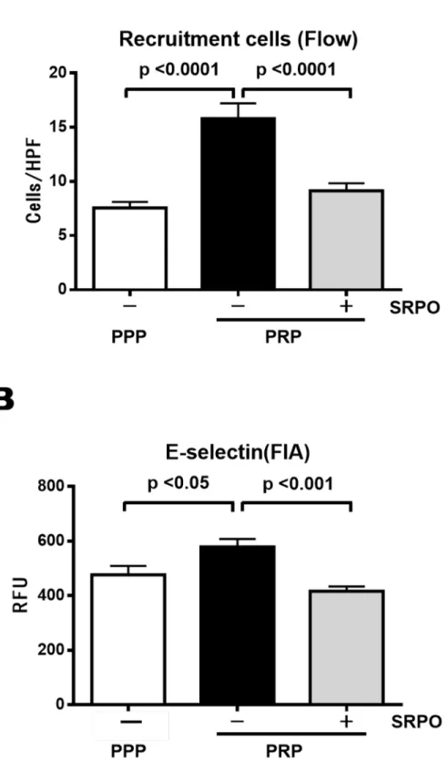
cm2). As shown inFig 3A, PRP increased THP-1 cell adhesion to activated HUVECs (PPP,
7.6 ± 0.6/HPF vs. PRP, 15.5 ± 1.4/HPF;P<0.001). Pre-incubation of PRPs with SRPO reduced
their adhesion (PRP + SRPO, 9.2 ± 0.7/HPF;P<0.001) (n = 20 in each group).
Cell surface E-selectin expression was upregulated significantly by PRP (PPP, 476.7 ± 32.6 RFU vs. PRP, 578.0 ± 29.6 RFU;P<0.05), but this upregulation was reduced by
pre-incuba-tion with SRPO (PRP + SRPO, 416.3 ± 17.4 RFU;P<0.001) (Fig 3B).
Effect of sarpogrelate hydrochloride on phorbol 12-myristate
13-acetate-induced leukocyte adhesion to activated human umbilical vein
endothelial cells
in vitro
To directly assess the molecular mechanisms of the anti-inflammatory effects of SRPO, we
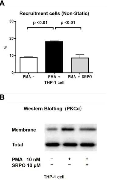
con-ducted a THP-1-HUVEC adhesion assayin vitrounder non-static rotational conditions. PKCα
Fig 1. Effects of SRPO on HFFD-induced obesity.(A) Body weight (n = 17, 28, 29). (B) Epididymal fat weight (n = 13, 18, 18). (C) Liver weight (n = 17, 23, 25). Values are the mean±SE in each group.
(Abbreviations: NC = normal chow; HFFD = high-fat high-fructose diet; VEH = vehicle; SRPO = Sarpogrelate hydrochloride.)
significantly increased their adhesion to HUVECs under non-static conditions (P<0.001). By
contrast, pre-incubation of THP-1 cells with SRPO (10μM, 1 h) reduced the PMA-induced
THP-1 cell adhesion to PMA-activated HUVECs (10 nM, 8 h;P<0.01) (Fig 4A).
In monocytes, PKCαis one of the key modulators of the inflammatory process, including
leukocyte-endothelial interactions [22]. To explore the molecular mechanisms of the anti-adhesive action of SRPO, we examined the effect of SRPO on the PKCαactivity levels in THP-1 cells. As shown inFig 4B, the results of the immunoblotting analyses indicated that PMA
increased the amount of PKC-αprotein in THP-1 cell membrane fractions [PMA (-),
9.1 ± 0.3% vs. PMA (+), 18.2 ± 0.4%;P<0.01], whereas the increase in PKC-αprotein was reduced by SRPO (PMA + SRPO, 8.7 ± 1.9%;P<0.01) (n = 3 in each group).
Discussion
The results of this study demonstrate that the administration of SRPO reduced leukocyte-endothelial cell interactions through its effects on platelets, monocytes, and adipose tissue both in vitroandin vivo. SRPO is a 5-HT2AR antagonist, which is used clinically as an antiplatelet
drug, and is not known to antagonize 5-HT2Breceptor and others [23]. 5-HT2AR is widely
expressed in a variety of cells, including platelets [24], monocytes [10,25], adipocytes [7,26], and smooth muscle cells [27]. 5-HT2R is a phospholipase C stimulator and a member of the GPCR family. Several studies have shown that the 5-HT2AR signaling pathway includes
diacyl-glycerol, PKC, MAPK, AP-1, and NF-κB [4–7], and thus GPCR activation can theoretically affect multiple signaling pathways [28,29]. However, inverse agonism is a recently discovered feature of GPCR systems [30,31]. Interestingly, 5-HT2AR has also been reported to exhibit
constitutive activity, and SRPO has been reported to exhibit a potent inverse agonist activity against 5-HT2AR [32]. Based on the GPCR cross-talk theory, an inverse agonist of one GPCR
may result in the inhibition of multiple signaling pathways [33]. Therefore, SRPO can act as an antiplatelet drug but also as an inverse agonist of 5-HT2AR in a variety of cells, which may
explain the pleiotropic atheroprotective effects of SRPO. In this study, we examined the pleio-tropic effects of SRPO on leukocyte-endothelial cell interactions bothin vivoandin vitro.
First, we investigated the effect of SRPO on HFFD-induced leukocyte-endothelial interac-tions in the femoral artery using an IVM technique, which we had developed to directly moni-tor leukocyte interactions with atheroprone arteriesin vivo[18]. Chronic administration of SRPO significantly reduced the body weight, visceral fat weight, serum MCP-1 levels, and leu-kocyte-endothelial cell interactions in the HFFD + VEH group.
It has been proposed that fat mass growth in obesity is a result of adipocyte hypertrophy and the recruitment of new adipocytes from pre-adipocytes [34]. 5-HT2AR is expressed in
adi-pocytes [7,26] and 5-HT has been shown to accelerate adipocyte differentiation, whereas 5-HT2AR antagonists reduce adipogenesis [26]. Uchida-Kitajima et al. reported that the
expres-sion of 5-HT2AR increased during adipocyte differentiation in hypertrophic adipocytes, and
the 5-HT2AR signaling pathway stimulated MAPK in adipocytes [7]. Other studies have shown
that 5-HT2AR inhibition induced reductions in body weight [1,2] and the visceral fat weight
[2] in high-fat diet-fed rats. Thus, SRPO may have inhibited adipocyte proliferation and differ-entiation via the 5HT2AR signaling pathway in the HFFD-fed obese mice, thereby resulting in
decreased body weight and visceral fat weight (Fig 1).
In obesity, the adipokines secreted by adipose tissue have proinflammatory and atherogenic effects [35]. In particular, MCP-1 is a potent chemokine that is upregulated in obesity and it
mean±SE (n = 13, 13, 11). Serum MCP-1 level of the HFFD + VEH group was higher than in the NC group, and SRPO prevented the increase in serum MCP-1 level on the HFFD + VEH group.
Fig 3. THP-1 cell adhesion to HUVECs (Flow chamber assay) and expression of E-selectin in HUVECs.
plays a key role in leukocyte-endothelial cell interactions [22], which comprise the crucial ini-tial step in the pathogenesis of atherosclerosis [36]. Adipose tissue is an important source of
MCP-1 [37], and the MCP-1 mRNA and protein levels have been reported to be upregulated
in the adipose tissue of high-fat diet-fed obese mice [38]. MCP-1 gene expression is regulated
in Materials and Methods. Data are the mean±SD of three independent experiments in each group. (B) SRPO significantly reduced the expression of PRP-induced E-selectin in HUVECs. E-selectin expression was determined by FIA as described in Materials and Methods. Data are the mean±SD of 6 independent experiments in each group.
doi:10.1371/journal.pone.0147929.g003
Fig 4. THP-1 cell adhesion to HUVECs (Non-static rotational assay) and expression of PKCαin THP-1
cells.(A) SRPO significantly reduced PMA-triggered THP-1 cell adhesion to the HUVECs. THP-1 cells were pretreated with SRPO (10μM) for 1 h and stimulated with PMA (10 nM) for 10 min before the assay. Preliminary experiments with trypan blue staining demonstrated that THP-1 cells were not damaged by SRPO (10μM, 1h) treatment (data not shown). Adhesion assays were performed as described in Materials and Methods. Data are the mean±SD of three independent experiments in each group. (B) SRPO attenuated PMA-induced PKCαactivation in THP-1 cells. THP-1 cells were incubated in the presence or absence of SRPO (10μM) for 1 h and stimulated with PMA (10 nM) for 10 min before the assay, and membrane proteins and total PKCαprotein were detected by immunoblotting. Data are representative of three independent experiments.
by the MAPK, AP-1, and NF-κB signaling pathways [39]. The MCP-1 secreted by adipocytes adheres to glycosaminoglycans on the surfaces of endothelial cells [40] and it attracts leuko-cytes to vascular endothelial cells by activating endothelial cells and monoleuko-cytes. MCP-1 acti-vates THP-1 cells by activating the PKCαsignaling pathway, which results in the upregulation ofα4 andβ2 integrins [22].
The results of the present study indicate that increases in the serum MCP-1 levels in HFFD-fed obese mice were accompanied by increases in the visceral fat volume and in leukocyte-endothelial cell interactions, whereas all of these increases were prevented by SRPO. 5-HT2AR
is also expressed in adipocytes [7,26] and monocytes [10,25]. SRPO may indirectly inhibit MCP-1 production by reducing the adipose tissue volume and directly inhibit MCP-1
produc-tion via its inverse agonist acproduc-tion on the 5HT2AR, thereby reducing MAPK, AP-1, and NF-κB
activation. Given the inverse effects of SRPO on GPCR signaling cross-talk, SRPO may also reduce the effects of MCP-1 via the 5-HT2AR/PKC signaling pathway. Overall, the inverse
effects of SRPO on the MAPK/NF-κB or PKC signaling pathways may inhibit both MCP-1
production and the effects of MCP-1, thereby reducing leukocyte-endothelial cell interactions in HFFD-fed obese mice (Fig 2).
SRPO was originally used as an antiplatelet drug, so the influence of antiplatelet effects of SRPO must also be considered. Obesity is associated with increases in platelet activation [41] and the parameters that reflect platelet activation, such as the mean platelet volume, soluble P-selectin, and soluble CD40 ligand (sCD40L), are also increased in the blood of obese patients [42]. Platelet activation is induced by interactions between several agonists and GPCRs that recognize thrombin, adenosine diphosphate, thromboxane A2, platelet activating factor,
epi-nephrine, 5-HT, and chemokines, including MCP-1 [43]. The platelets activated by the GPCR/
PKC signaling pathway amplify inflammatory processes through pleiotropic interactions with vascular cells, blood cells, adipocytes, and adipokines. For example, activated platelets induce MCP-1 secretion [44] and the expression of E-selectin on endothelial cells via an NF-κ B-dependent signaling pathway [45]; and sCD40L is released by activated platelets to initiate vari-ous inflammatory responses [13], including E-selectin expression [46] and the release of
che-mokines including MCP-1 [46]. Incubation of human adipocytes with recombinant CD40L
induces the expression of MCP-1 and adipogenesis via the MAPK/NF-κB signaling pathway
[47]. Incubation of monocytes with CD40L activates PKC or the MAPK/NF-κB signaling
path-way [48,49]. 5-HT2AR is expressed in platelets [24], monocytes [10,25], and adipocytes [7, 26]; thus, SRPO may reduce leukocyte-endothelial cell interactions in HFFD-fed obese mice by inhibiting the effects of activated platelets.
We also investigated the detailed molecular mechanisms using anin vitroleukocyte adhe-sion assay system with PPP or PRP. We identified a potential role for endothelial E-selectin in
PRP-mediated THP-1 adhesion to HUVECs (Fig 3). E-selectin is a member of the selectin
5-HT2AR/G protein/PKC pathway in monocytes [10]. 5-HT2AR is expressed in monocytes [10,
25], so our data suggest that SRPO at least partly reduces PMA-induced monocyte adhesion to
activated HUVECs by modulating PKCαactivation in THP-1 cells. The potential mechanisms
underlying this process appear to involve the inverse agonist activity of SRPO against the
5-HT2AR/PKC signaling pathway.
Conclusion
In summary, SRPO inhibited leukocyte-endothelial interactions enhanced by platelets in vitro. Potential mechanism seems to involve PKCa activation in leukocytes. We demonstrated that SRPO reduced leukocyte adhesion to femoral artery in HFFD-treated mice in vivo. SRPO also reduced visceral fat weight and serum MCP-1 level in these mice. These data indicate novel anti-inflammatory role of SRPO in metabolic syndrome.
Supporting Information
S1 Video. NC (Leukocyte-endothelial Interactions: Intravital microscopy).
(MP4)
S2 Video. HFFD + VEH (Leukocyte-endothelial Interactions: Intravital microscopy).
(MP4)
S3 Video. HFFD + SRPO (Leukocyte-endothelial Interactions: Intravital microscopy).
(MP4)
Author Contributions
Conceived and designed the experiments: HK MO. Performed the experiments: HK YA MD. Analyzed the data: HK YA MD. Contributed reagents/materials/analysis tools: HK YA MD MO. Wrote the paper: HK MO MY. Advised the experiments: KN.
References
1. Shen KP, Lin HL, Hsieh SL, Kwan AL, Chen IJ, Wu BN. Eugenosedin-A prevents hyperglycaemia, hyperlipidaemia and lipid peroxidation in C57BL/6J mice fed a high-fat diet. J Pharm Pharmacol 2009; 61:517–525. doi:10.1211/jpp/61.04.0015PMID:19298700.
2. Takishita E, Takahashi A, Harada N, Yamato M, Yoshizumi M, Nakaya Y. Effect of sarpogrelate hydro-chloride, a 5-HT2 blocker, on insulin resistance in Otsuka Long-Evans Tokushima fatty rats (OLETF rats), a type 2 diabetic rat model. J Cardiovasc Pharmacol 2004; 43:266–270. PMID:14716215.
3. Xu YJ, Zhang M, Ji L, Elimban V, Chen L, Dhalla NS. Suppression of high lipid diet induced by athero-sclerosis sarpogrelate. J Cell Mol Med 2012; 16:2394–2400. doi:10.1111/j.1582-4934.2012.01554.x PMID:22348587.
4. Machida T, Ohta M, Onoguchi A, Iizuka K, Sakai M, Minami M, et al. 5-Hydroxytryptamine induces cyclooxygenase-2 in rat vascular smooth muscle cells: Mechanisms involving Src, PKC and MAPK activation [corrected]. Eur J Pharmacol 2011; 656:19–26. PMID:21262218.
5. Ito T, Ikeda U, Shimpo M, Yamamoto K, Shimada K. Serotonin increases interleukin-6 synthesis in human vascular smooth muscle cells. Circulation 2000; 102:2522–2527. PMID:11076827.
6. Grewal JS, Mukhin YV, Garnovskaya MN, Raymond JR, Greene EL. Serotonin 5-HT2A receptor induces TGF-beta1 expression in mesangial cells via ERK: proliferative and fibrotic signals. Am J Phy-siol 1999; 276:F922–930. PMID:10362781.
7. Uchida-Kitajima S, Yamauchi T, Takashina Y, Okada-Iwabu M, Iwabu M, Ueki K, et al. 5-Hydroxytrypta-mine 2A receptor signaling cascade modulates adiponectin and plasminogen activator inhibitor 1 expression in adipose tissue. FEBS Lett 2008; 582:3037–3044. doi:10.1016/j.febslet.2008.07.044 PMID:18675814.
9. Inoue M, Okazaki T, Kitazono T, Mizushima M, Omata M, Ozaki S. Regulation of antigen-specific CTL and Th1 cell activation through 5-Hydroxytryptamine 2A receptor. Int Immunopharmacol 2011; 11:67– 73. PMID:20971187.
10. Suguro T, Watanabe T, Kanome T, Kodate S, Hirano T, Miyazaki A, et al. Serotonin acts as an up-regu-lator of acyl-coenzyme A:cholesterol acyltransferase-1 in human monocyte-macrophages. Atheroscle-rosis 2006; 186:275–281. doi:10.1016/j.atherosclerosis.2005.08.007PMID:16157345.
11. Lin OA, Karim ZA, Vemana HP, Espinosa EV, Khasawneh FT. The antidepressant 5-HT2A receptor antagonists pizotifen and cyproheptadine inhibit serotonin-enhanced platelet function. PLoS One 2014; 9:e87026. PMID:24466319; PubMed Central PMCID: PMCPmc3900701. doi:10.1371/journal.pone. 0087026
12. Kuckleburg CJ, Yates CM, Kalia N, Zhao Y, Nash GB, Watson SP, et al. Endothelial cell-borne platelet bridges selectively recruit monocytes in human and mouse models of vascular inflammation. Cardio-vasc Res 2011; 91:134–141. doi:10.1093/cvr/cvr040PMID:21285294.
13. Poggi M, Jager J, Paulmyer-Lacroix O, Peiretti F, Gremeaux T, Verdier M, et al. The inflammatory receptor CD40 is expressed on human adipocytes: contribution to crosstalk between lymphocytes and adipocytes. Diabetologia 2009; 52:1152–1163. doi:10.1007/s00125-009-1267-1PMID:19183933.
14. Kajiwara I, Soejima H, Miyamoto S, Ogawa H. Effects of additional treatment of sarpogrelate to aspirin therapy on platelet aggregation and plasma plasminogen activator inhibitor activity in patients with sta-ble effort angina. Thromb Res 2011; 128:547–551. doi:10.1016/j.thromres.2011.06.001PMID: 21722942.
15. Ogawa S, Mori T, Nako K, Ishizuka T, Ito S. Reduced albuminuria with sarpogrelate is accompanied by a decrease in monocyte chemoattractant protein-1 levels in type 2 diabetes. Clin J Am Soc Nephrol 2008; 3:362–368. doi:10.2215/cjn.03450807PMID:18235151; PubMed Central PMCID: PMCPmc2390947.
16. Nomura S, Shouzu A, Omoto S, Nishikawa M, Iwasaka T. 5-HT2A receptor antagonist increases circu-lating adiponectin in patients with type 2 diabetes. Blood Coagul Fibrinolysis 2005; 16:423–428. PMID: 16093733.
17. Nonogaki K, Nozue K, Oka Y. Increased hypothalamic 5-HT2A receptor gene expression and effects of pharmacologic 5-HT2A receptor inactivation in obese Ay mice. Biochem Biophys Res Commun 2006; 351:1078–1082. doi:10.1016/j.bbrc.2006.10.173PMID:17097612.
18. Osaka M, Hagita S, Haraguchi M, Kajimura M, Suematsu M, Yoshida M. Real-time imaging of mechani-cally injured femoral artery in mice reveals a biphasic pattern of leukocyte accumulation. Am J Physiol Heart Circ Physiol 2007; 292:H1876–H1882. doi:10.1152/ajpheart.00708.2006PMID:17172278.
19. Yoshida M, Sawada T, Ishii H, Gerszten RE, Rosenzweig A, Gimbrone MA Jr, et al. Hmg-CoA reduc-tase inhibitor modulates monocyte-endothelial cell interaction under physiological flow conditions in vitro: involvement of Rho GTPase-dependent mechanism. Arterioscler Thromb Vasc Biol 2001; 21:1165–1171. PMID:11451746.
20. Nagel T, Resnick N, Atkinson WJ, Dewey CF Jr, Gimbrone MA Jr. Shear stress selectively upregulates intercellular adhesion molecule-1 expression in cultured human vascular endothelial cells. J Clin Invest 1994; 94:885–891. doi:10.1172/jci117410PMID:7518844; PubMed Central PMCID:
PMCPmc296171.
21. Yoshida M, Westlin WF, Wang N, Ingber DE, Rosenzweig A, Resnick N, et al. Leukocyte adhesion to vascular endothelium induces E-selectin linkage to the actin cytoskeleton. J Cell Biol 1996; 133:445– 455. PMID:8609175; PubMed Central PMCID: PMCPmc2120789.
22. Kojima C, Kawakami A, Takei T, Nitta K, Yoshida M. Angiotensin-converting enzyme inhibitor attenu-ates monocyte adhesion to vascular endothelium through modulation of intracellular zinc. J Pharmacol Exp Ther 2007; 323:855–860. doi:10.1124/jpet.107.127944PMID:17878405.
23. Muntasir HA, Hossain M, Bhuiyan MA, Komiyama T, Nakamura T, Ozaki M, et al. Identification of a key amino acid of the human 5-HT(2B) serotonin receptor important for sarpogrelate binding. J Pharmacol Sci 2007; 104:274–277. PMID:17609583.
24. Uchitomi Y, Kugaya A, Akechi T, Nakano T, Inagaki M, Matsuoka Y, et al. Lack of association between suicidal ideation and enhanced platelet 5-HT2A receptor-mediated calcium mobilization in cancer patients with depression. Biol Psychiatry 2002; 52:1159–1165. PMID:12488061.
25. Durk T, Panther E, Muller T, Sorichter S, Ferrari D, Pizzirani C, et al. 5-Hydroxytryptamine modulates cytokine and chemokine production in LPS-primed human monocytes via stimulation of different 5-HTR subtypes. Int Immunol 2005; 17:599–606. doi:10.1093/intimm/dxh242PMID:15802305.
27. Nishihira K, Yamashita A, Tanaka N, Moriguchi-Goto S, Imamura T, Ishida T, et al. Serotonin induces vasoconstriction of smooth muscle cell-rich neointima through 5-hydroxytryptamine2A receptor in rab-bit femoral arteries. J Thromb Haemost 2008; 6:1207–1214. doi:10.1111/j.1538-7836.2008.02996.x PMID:18435827.
28. Werry TD, Wilkinson GF, Willars GB. Mechanisms of cross-talk between G-protein-coupled receptors resulting in enhanced release of intracellular Ca2+. Biochem J 2003; 374:281–296.
29. Selbie LA, Hill SJ. G protein-coupled-receptor cross-talk: the fine-tuning of multiple receptor-signalling pathways. Trends Pharmacol Sci 1998; 19:87–93. PMID:9584624.
30. Latek D, Modzelewska A, Trzaskowski B, Palczewski K, Filipek S. G protein-coupled receptors—recent advances. Acta Biochim Pol 2012; 59:515–529. PMID:23251911.
31. Bond RA, Ijzerman AP. Recent developments in constitutive receptor activity and inverse agonism, and their potential for GPCR drug discovery. Trends Pharmacol Sci 2006; 27:92–96. doi:10.1016/j.tips. 2005.12.007PMID:16406086.
32. Muntasir HA, Bhuiyan MA, Ishiguro M, Ozaki M, Nagatomo T. Inverse agonist activity of sarpogrelate, a selective 5-HT2A-receptor antagonist, at the constitutively active human 5-HT2A receptor. J Pharmacol Sci 2006; 102:189–195. PMID:17031071.
33. Bouaboula M, Perrachon S, Milligan L, Canat X, Rinaldi-Carmona M, Portier M, et al. A selective inverse agonist for central cannabinoid receptor inhibits mitogen-activated protein kinase activation stimulated by insulin or insulin-like growth factor 1. Evidence for a new model of receptor/ligand interac-tions. J Biol Chem 1997; 272:22330–22339. PMID:9268384.
34. Giri S, Rattan R, Haq E, Khan M, Yasmin R, Won JS, et al. AICAR inhibits adipocyte differentiation in 3T3L1 and restores metabolic alterations in diet-induced obesity mice model. Nutr Metab (Lond) 2006; 3:31. PMID:16901342; PubMed Central PMCID: PMCPmc1564022.
35. Zhang H, Cui J, Zhang C. Emerging role of adipokines as mediators in atherosclerosis. World J Cardiol 2010; 2:370–376. doi:10.4330/wjc.v2.i11.370PMID:21179304; PubMed Central PMCID:
PMCPmc3006473.
36. Gosling J, Slaymaker S, Gu L, Tseng S, Zlot CH, Young SG, et al. MCP-1 deficiency reduces suscepti-bility to atherosclerosis in mice that overexpress human apolipoprotein B. J Clin Invest 1999; 103:773– 778. doi:10.1172/jci5624PMID:10079097; PubMed Central PMCID: PMCPmc408147.
37. Sartipy P, Loskutoff DJ. Monocyte chemoattractant protein 1 in obesity and insulin resistance. Proc Natl Acad Sci U S A 2003; 100:7265–7270. doi:10.1073/pnas.1133870100PMID:12756299; PubMed Central PMCID: PMCPmc165864.
38. Yu R, Kim CS, Kwon BS, Kawada T. Mesenteric adipose tissue-derived monocyte chemoattractant pro-tein-1 plays a crucial role in adipose tissue macrophage migration and activation in obese mice. Obesity (Silver Spring) 2006; 14:1353–1362. doi:10.1038/oby.2006.153PMID:16988077.
39. Roebuck KA, Carpenter LR, Lakshminarayanan V, Page SM, Moy JN, Thomas LL. Stimulus-specific regulation of chemokine expression involves differential activation of the redox-responsive transcription factors AP-1 and NF-kappaB. J Leukoc Biol 1999; 65:291–298. PMID:10080530.
40. Proudfoot AE, Handel TM, Johnson Z, Lau EK, LiWang P, Clark-Lewis I, et al. Glycosaminoglycan bind-ing and oligomerization are essential for the in vivo activity of certain chemokines. Proc Natl Acad Sci U S A 2003; 100:1885–1890. doi:10.1073/pnas.0334864100PMID:12571364; PubMed Central PMCID: PMCPmc149928.
41. Santilli F, Vazzana N, Liani R, Guagnano MT, Davi G. Platelet activation in obesity and metabolic syn-drome. Obes Rev 2012; 13:27–42. doi:10.1111/j.1467-789X.2011.00930.xPMID:21917110.
42. Angelico F, Alessandri C, Ferro D, Pignatelli P, Del Ben M, Fiorello S, et al. Enhanced soluble CD40L in patients with the metabolic syndrome: Relationship with in vivo thrombin generation. Diabetologia 2006; 49:1169–1174. doi:10.1007/s00125-006-0222-7PMID:16570157.
43. Offermanns S. Activation of platelet function through G protein-coupled receptors. Circ Res 2006; 99:1293–1304. doi:10.1161/01.res.0000251742.71301.16PMID:17158345.
44. Gawaz M, Neumann FJ, Dickfeld T, Koch W, Laugwitz KL, Adelsberger H, et al. Activated platelets induce monocyte chemotactic protein-1 secretion and surface expression of intercellular adhesion mol-ecule-1 on endothelial cells. Circulation 1998; 98:1164–1171. PMID:9743506.
45. Proenca-Ferreira R, Brugnerotto AF, Garrido VT, Dominical VM, Vital DM, Ribeiro Mde F, et al. Endo-thelial activation by platelets from sickle cell anemia patients. PLoS One 2014; 9:e89012. doi:10.1371/ journal.pone.0089012PMID:24551209; PubMed Central PMCID: PMCPmc3923877.
47. Missiou A, Wolf D, Platzer I, Ernst S, Walter C, Rudolf P, et al. CD40L induces inflammation and adipo-genesis in adipose cells—a potential link between metabolic and cardiovascular disease. Thromb Hae-most 2010; 103:788–796. doi:10.1160/th09-07-0463PMID:20174757.
48. Yan JC, Wu ZG, Kong XT, Zong RQ, Zhang LZ. Effect of CD40-CD40 ligand interaction on diacylgly-cerol-protein kinase C signal transduction pathway and intracellular calcium in cultured human mono-cytes. Acta Pharmacol Sin 2003; 24:687–691. PMID:12852836.
49. Inoue Y, Otsuka T, Niiro H, Nagano S, Arinobu Y, Ogami E, et al. Novel regulatory mechanisms of CD40-induced prostanoid synthesis by IL-4 and IL-10 in human monocytes. J Immunol 2004; 172:2147–2154. PMID:14764680.
50. Ferri C, Desideri G, Valenti M, Bellini C, Pasin M, Santucci A, et al. Early upregulation of endothelial adhesion molecules in obese hypertensive men. Hypertension 1999; 34:568–573. PMID:10523328.
51. Considine RV, Nyce MR, Allen LE, Morales LM, Triester S, Serrano J, et al. Protein kinase C is increased in the liver of humans and rats with non-insulin-dependent diabetes mellitus: an alteration not due to hyperglycemia. J Clin Invest 1995; 95:2938–2944. doi:10.1172/jci118001PMID:7769136; PubMed Central PMCID: PMCPmc295982.