Zélia Licínia Ferreira Gouveia
Dissertation presented to obtain the Ph.D degree in Biochemistry
Instituto de Tecnologia Química e Biológica António Xavier | Universidade Nova de Lisboa
Targeting Heme with Single
Domain Antibodies
Zélia Licínia Ferreira Gouveia
Dissertation presented to obtain the Ph.D degree in Biochemistry
Instituto de Tecnologia Química e Biológica António Xavier | Universidade Nova de LisboaOeiras, Junho, 2015
Targeting Heme with Single
Domain Antibodies
Zélia Licínia Ferreira Gouveia
Supervisor:
Dr. Miguel P.Soares
Instituto Gulbenkian da Ciência
Oeiras, Junho, 2015
Targeting Heme with Single
Domain Antibodies
Co-Supervisor:
Dr. Smilja Todorovic
Instituto de Tecnologia Química e Biológica
I
Firstly and primarily I would like to express my most sincere gratitude to my Ph.D supervisor Dr. Miguel P. Soares for accepting me as his Ph.D student and to trust me this Ph.D project. I want to thank him for his help, advices and for non-stop teaching and extremely helpful and constructive scientific discussions. Today, I am the scientist that you help to build, thank you very much! I extremely grateful to Dr. Miguel P. Soares for his understanding and support, in a particular period of my life in which I had to take several weeks of my work time to be and support my family as we were having a extremely difficult time in our life. For all of this and more, thank you very much!
I am grateful to Dr. Smilja Todorovic, for giving me the opportunity to join in her laboratory and for accepting me as Ph.D student, for her advices, support and precious and fundamental help in Resonance Raman spectroscopy.
I also want to thank to all the present and past members of Inflammation groups for their help, support, discussion and the most diverse moments spent together. A special thanks to Sofia, for her advices, support and many other things. To Viktória Jeney, for all the help and teach in the begining of this project and for the beautiful words and advices. Thanks the wonderful person, Ramus, for showing me that the comprehension and the calm can be your best friends in the moments that everything seems to be under tumult. Thanks to Raffi, for all your help. Sílvia, thank you very much for been always so friendly. To Rita Carlos, Susana Ramos and Sebastian Weiss thanks for the comments and the correction of the introduction part of the thesis.
II
To my good friend Ana Rita Mateus, my thanks for the support and for always been presence in the good or less good moments. Ana Regalado, my sincere and most adorable friend, thank you very much for being always present and willing to listen and share ideas and advices.
I am also grateful to the people involved in the many collaborations that support this work (chapter 2), including
Dr. João Gonçalves from Faculdade de Farmácia e Instituto de Medicina Molecular, for receiving me in his research group for almost two years and to supervise my work in the development of single domain antibodies against heme using phage display technology. Thanks also to all his group members, for their help, support and time.
Dr. Frederico Aires-da-Silva, from Technophage for providing me many useful suggestions about Phage display technology and many other things. Also, for all the scientific discussions and for being always available to take time to teach me and sharing his scientific knowledge.
Dr. Claúdio Gomes from Instituto tecnologia Química e Biológica, for his kindness, availability and all his scientific help to performed spectroscopic techniques
Dr. Olga Iranzo from Instituto tecnologia Química e Biológica, for her precious help in the biotinylation of Heme.
Dr. Iqbal Hamza from the University of Maryland, for his kindness, availability and discussion on this work. Also, for receiving me his laboratory and providing me all the material and support required to perform HPR assay. To all his group members and in special Tamika Samuel and Carine White for their kindness, availability and scientific or non scientific help, as well as Jason Sinclair and Xiaojing Yuan for their kindness and availability.
And to all the other collaborators, my thanks.
III
I want to thank to Instituto Gulbenkian da Ciência and all IGC community, for providing an amazing scientific environment.
All this work was only possible because I have amazing people supporting me at all the moments, my loved companion and my family and of course my good friends.
I want to thank my beloved family and specially my mother, Felismina Pereira Ferreira and my father, José da Assunção Gouveia, for so many things that there is no space in this thesis to mention them. My most and important thank you. Ao meu querido pai, José da Assunção Gouveia, o meu Muito Obrigada para todo o sempre! Se hoje defendo esta tese é sem dúvida graça a ti e à mãe!
To my companion Pierre Crozet, first I would like to thank you for being such an amazing friend and for all the love demonstrated and shared. Thank you for all the support, help and comments/discussions in non and scientific fields. Thanks for helping me in my thesis, correcting/commenting it. I am also grateful to his amazing and adorable family to receive me like family. Merci à tous, et surtout à Joёlle Crozet-Vaugelade, Jean Jeacques Crozet et Elisabeth Crozet.
I would like to acknowledge the Fundação para a Ciência e Tecnologia (Apoio financeiro da FCT e do FSE no âmbito do Quadro Comunitário de apoio, BD: SFRH/BD/44828/2008), Instituto Gulbenkia da Ciência and Inflammation Group of Dr. Miguel P. Soares, who have finantially supported my Ph.D Work.
I would like to apologize in advance to the many people that I might not have mention in this section but for sure help me along these years to develop this work. For all of them, my sincere thanks.
I would like to finalize with these two popular proverbs “Não importa o tamanho da
IV
the clouds pass, the sun shines) and by dedicating this thesis to my family, but specially to my beloved father José da Assunção Gouveia, who will always be with us and to my beloved mother Felismina Pereira Ferreira, without them this thesis could not be possible.
My eternal thanks!
V
[wav(x), wav(y); Eg]; Waving
5c; Five coordinate
6c; Six coordinate
aa; amino acids
Ab(s); Antibodie (s)
ABCB10 or ABC-me; ATP-binding cassette
sub-family B member 10
ABCB6; ATP-binding cassette sub-family B
member 6
ABCG2; ATP-binding cassette transporter
G2
ACN; Acetonitrile
ACTH; Adrenocorticotropic hormone
Ala, A; Alanine
ALA;δ-aminolevulinic acid
ALAS;δ -aminolevulinate synthase
ALAS-E;δ -aminolevulinate synthase
erythroid-specific (=ALAS2)
ALAS-N; Delta-aminolevulinate synthase
nonspecific (=ALAS 1)
ANOVA; Analysis of variance
ApoE; Apolipoprotein E
Arg, R; Arginine
Asp, N; Asparagine
ATP; Adenoside triphosphate
ATPase; Adenylpyrophosphatase
ATR-FTIR; Attenuated total reflection Fourier
transform infrared
BACH1; Basic leucine zipper transcription
factor 1
Bach2; Basic leucine zipper transcription
factor 2
BCP; Breast cancer resistance protein
Biotin; N-[-5- (Hydrazinocarboxy)
pentyl]-D-biotinamide
Blimp-1; B lymphocyte-induced maturation
protein-1
BR; Bilirubin
BV; Biliverdin
BVR; Biliverdin reductase
BW; Body weight
CCD; Charge coupled device
ccm; System I cytochrome c maturation
CcmE; Cytochrome c maturation E
CD; Circular dichroism
CD14; Cluster of differentiation 14
CD163; Cluster of differentiation 163
cDNA; Complementary DNA
CDR; Complementarity-determining regions
cGMP; Cyclic guanosine monophosphate
CH; Heavy chain constant domain
ChEBI; Chemical entities of biological
interest
Cl; Chloride
CL; Liht chain constant domain
CLP; Cecal ligation and puncture
CN; Cysteine‑ asparagine
CO; Carbon monoxide
CO2; Carbon dioxide
CoPP; Cobalt protoporphyrin IX
COPRO III; Coproporphyrinogen III
VI CORMs; Monoxide releasing molecules
CPOX; Coproporphyrinogen oxidase
CS; Cysteine-serine
Ctrl; Control sdAb
CY; Cyanine dyes
Cys, C; Cysteine
Cytc; Cytochrome c
DAPI; 4',6-diamidino-2-phenylindole
DETAPAC; Diethylenetriaminepentaacetic
acid
DeutP; Iron deuteroporphyrin IX
DHB; 2,5-dihydroxybenzoic acid
DMEM; Dulbecco's modified eagle medium
DMF; Dimethylformamide
DMSO; Dimethylsulfoxide
DNA; Deoxyribonucleic acid
dom, A2u; Doming
E.coli;Escherichia cole
EDTA; Ethylenediaminetetraacetic acid
EEA; Early endosomes
ELISA; Enzyme-linked immunosorbent
assay
ER; Endoplasmatic reticulum
ERK-2; Extracellular signal-regulated
kinases 2
Fab(s); Antigen binding fragment(s)
FABP; Fatty acid binding protein
FBS; Fetal bovine serum
Fc; Constant fragment region
FcgRs; Fc receptors
FcRn; Neonatal Fc receptor
Fe; Iron
Fe2 (SO4)3; Iron (III) sulfate
Fe2+; Ferrous ion
Fe3+; Ferric ion
FECH; Ferrochelatase
FePP; Hemin
FePPCH3; Iron protoporphyrin IX dimethyl
ester chloride
FF; Fast flow
FITC; Fluorescein isothiocyanate
FLVCR2; Feline leukemia virus subgroup C
receptor 2
FR; Framework region
Ft; Ferritin
FtH; Heavy/heart chain
FtL; Light/liver chain h
Fv; Variable fragment domain
Gly, G; Glycine
GaPP; Gallium protoporphyrin IX
GATA1; GATA binding protein 1 (globin
transcription factor 1)
GSH; Reduced glutathione
GSSG; Oxidized glutathione
GST; Glutathione S-transferase B
H2O2; Hydrogen peroxide
H2SO4; Sulfuric acid
HA; Human influenza hemagglutinin
HABA; 4'-hydroxyazobenzene-2-carboxylic
acid
HATU; 2-
(1H-7-azabenzotriazol-1-yl)-
1,1,3,3-tetramethyluroniumhexafluorophosphate
methanaminium
Hb; Hemoglobin
HBC; Heme buffering capacity
HbC; Hemoglobin C
VII HBM; Heme-binding motifs
HBP23; Heme-binding protein 23 kDa
HbS; Hemoglobin S
HCl; Hydrochloric acid
HCP-1; Heme carrier protein
HD; Heme depleted serum medium
HDL; High density lipoprotein
HEK293; Human embryonic kidney 293
Heme; Protoheme IX
Hep2; Human epithelial type 2
HEPES;
4-(2-hydroxyethyl)-1-piperazineethanesulfonic acid
HIF-1; Hypoxia inducible factor-1
His, H; Histidine,
HMB; Hydroxymethylbilane
HMBS; Hydroxylmethylbilane synthase
HO-1; Heme oxygenase 1
HOCl; Hypochlorous acid
Hp; Haptoglobin
HPLC; High performance liquid
chromatography
HPX; Hemopexin
HRG-1; Heme-responsive gene-1
HRM; Heme regulatory motifs
HRP; Horseradish peroxidase
HSA; Human serum Albumin
HSAH; Human serum albumin bound to
heme
HSP90; Heat-shock protein 90
ICAM; Intercellular adhesion molecule
IFNβ; Interferon beta
Ig; Immunoglobulin
IL; Interleukine
Ile, I; Isoleucine
iNOS; Inducible nitric oxide synthase
IPTG; Isopropyl β-ᴅ-1-thiogalactopyranoside
IκB; Inhibitor of κB
JNK; c-Jun N-terminal Kinase
K+; Potassium ion
KC; keratinocyte derived chemokine
KD; Dissociation constant
LB; Luria Broth
LDL; Low density lipoprotein
Leu, L; Leucine
LIPR; Leucine-isoleucine-proline-arginine
LRP; Low-density lipoprotein
receptor-related protein/CD91
LS; Low spin
LTB4; Leukotriene B4
Lys, K; Lysine
m/z; Mass-to-charge ratio
mAb; Monoclonal Ab
MALDI; Matrix-assisted laser
desorption/ionization
MAPKs; Mitogen-activated protein kinases
Mb; Myoglobin
MEL; Murine erythroleukemia
Met, M; Methionine
MetHb; Methemoglobin
MFRN1; Mitoferritin 1
MgCl2; Magnesium chloride
Mn2+; Manganese ion
Mø(s); Macrophage(s)
MPO; Myeloperoxidase
mRNA; Messenger ribonucleic acid
MS; Mass spectrometry
VIII MyD88; Myeloid differentiation primary
response gene 88
NAC; N-acetylcysteine
NaCl; Sodium chloride
NADPH; Nicotinamide adenine dinucleotide
phosphate
NaHCO3; Sodium hydrogen carbonate
NaOH; Sodium hydroxide
NAR; (IgNAR) variable domain
NETs; Neutrophil extracellular traps
NF-κB; Nuclear factor kappa-light-chain-enhancer of activated B cells
NLRP3; Leucine rich repeat containing
family pyrin domain containing 3
inflammasome
NO; Nitric oxide
NOD; Nucleotide-binding oligomerization
domain receptor
NOX; NADPH Oxidase
NPAS2; Neuronal PAS domain-containing
protein 2
NRF2; Nuclear factor (erythroid-derived
2)-like 2
O2; Dioxygen
O2-; Superoxide ion
ON; Overnight
p22HBP; Heme-binding protein 22
PBG; Porphobilinogen
PBS; Phosphate buffered saline
PCD; Programmed cell death
PCR; Polymerase in chain reaction
PEG; Polyethylene Glycol
PG; Prostaglandins
PGBS; Porphobilinogen synthase
Phe, F; Phenylalanine
PHZ; Phenylhydrazine
PI3K; Phosphoinositide 3-kinase
PKB or Akt; Protein kinase B
PKC; Protein kinase C
pNPP; para-Nitrophenylphosphate
PP IX; Metal free protoporphyrin IX
PP; protoporphyrin
PPG IX; Protoporphyrinogen IX
PPIX; Protoporphyrin IX
PPOX; Protoporphyrin oxidase
Pro, P; Proline
pro,A1u; Propellering
PRR; Pattern recognition receptor
Prx I; Peroxiredoxin
RBC; Red blood cells
RIP; Receptor-interacting protein
RIPK1; Receptor-interacting serine/threonine
kinase 1
ROS; Reactive oxygen species
RR; Resonance Raman
RT; Room temperature
RU; Resonance units
ruf, B1u; Ruffling
Ser, S; Serine
S/N; Sinal to noise ratio
SA; Succinylacetone
sad, B2u; Saddling
SCD; Sickle cell disease
scFv; Single chain Fv
SD; Standard deviation
sdAb(s); Single domain Ab(s)
SDS-PAGE; Sodium dodecyl
IX SEM; Standard error of the mean
Ser; S; Serine
sGC; Guanylyl cyclase
SIRS; Systemic inflammatory response
syndrome
SnPP; Tin protoporphyrin IX
SOD; Superoxide dismutase
SOUL; Heme-binding protein 2
SPR; Surface plasmon resonance
SyK; Spleen tyrosine kinase
TBS; Tris-buffered saline
TGF-β; Transforming growth factor β
Thr, T; Threonine
TLR; Toll like receptor
TMB; 3,3’,5,’-Tetramethylbenzidine
TNF; Tumor necrosis factor
TOFMS; Time-of-flight mass spectrometry
Trp, W; Tryptophan
TRIF; TIR (Toll–IL-1
receptor)-domain-containing adapter-inducing interferon
Tyr, Y; Tyrosine
t-α1m; Truncated form of α -microglobulin
URO III; Uroporphyrinogen III
UROD; Uroporphyrinogen III decarboxylase
UROS; Uroporphyrinogen III co-synthase or
isomerase
Val, V; Valine
VH; Heavy chain variable domain
VHH; heavy-chain-only antibody (Ab) V(H)
VL; Light chain variable domain
V-NAR; Variable NAR domain from shark
VWF; Willebrand factor
WBP; Weibel-Palade bodies
XIAP; X-chromosome-linked inhibitor of
apoptosis
ZnPP; Zinc protoporphyrin IX
α1m; α -microglobin
X
This thesis “Targeting heme using single domain antibodies” aimed to develop methodologies that would overcome the current limitations in the detection and
quantification of “free heme”, i.e. redox active heme that is loosely bound to proteins or to other biological molecules. Here, we described the generation of single domain antibodies specific against heme using phage display technology and characterized them by different techniques such as affinity determination, antigen binding specificity, structural analysis (spectroscopy techniques), in vitro and ex-vivo actions. These sdAbs were further used to develop methodologies for the
quantitative and qualitative analysis of “free heme” and its cellular detection as well as to modulate heme reactivity.
This research work was performed mainly in Instituto Gulbenkian de Ciência under the scientific supervision of Dr. Miguel P. Soares. While, the development of the sdAbs was performed in collaboration and under the supervision of Dr. João Gonçalves in Faculdade de Farmácia da Universidade de Lisboa and Dr. Frederico Aires-da-Silva in Technophage from Instituto de Medicina Molecular da Universidade de Lisboa. Spectroscopy work was performed in the Instituto tecnologia Química e Biológica. These include Resonance Raman spectroscopy under the scientific supervision of the co-supervisor Dr. Smilja Todorovic and CD and FTIR spectroscopy in collaboration and under the scientific supervision of Dr. Claúdio Soares. The sdAb affinity measurements were also performed in Instituto tecnologia Química e Biológica with the supervision of Dr. Frederico Aires-da-Silva. The flow cytometry and immunofluorescence assay was performed by Ana Rita Carlos, IGC and ascorbate oxidation designed Roland Stocker, University of New South Wales, Australia.
The thesis is organized in three chapters, preceded by a Portuguese abstract translated in English. In the chapter 1, general introductory review on the subject is provided. In the chapter 2 the general and most significant results obtained during the research period are shown and discussed. In the chapter 3 a general discussion
XI
integrating all work and future perspectives are presented. In the appendix is presented and discussed supplementary information about a preliminary work to generate constructs, namely dimers of sdAbs and sdAbs containing an additional region of heme recognition (CDR), based on the sdAbs developed in chapter 2, namely sdAbs 1A6 and 2H7.
During my PhD project was able to participate in the elaboration of a review
manuscript entitled “Heme Cytotoxicity And The Pathogenesis of Immune Mediated Inflammatory Diseases.” (DOI:10.3389/fphar.2012.00077) from Rasmus Larsen,
XII
Hemo, i.e. ferro (Fe) contido na porfirina IX, funciona como grupo prostético numa variedade de hemoproteins que exercem funções biológicas vitais e portanto, essenciais para sustentar a vida. Hemo é uma molecula extremamente reactiva, podendo participar em reacções redox e presumivelmente por esse motivo deve ser sequestrado no interior de um bolso hémico nas hemoproteínas, para controlo da sua reactividade. Contudo, sob condições de stress biológico, as hemoproteínas podem libertar o seu grupo prostético formando “hemo livre” que liga-se fracamente às proteínas, ou outras moléculas adquirindo presumivelmente actividade redox descontrolada. Para além disso, um crescente número de evidências suporta a
noção de que o “hemo livre” pode comportarse como um agente vasoativo, pro -inflamatório e citotóxico quando libertado de um subconjunto de hemoproteínas como por exemplo, a extracelular hemoglobina gerada durante condições hemolíticas. Assim sendo, eventualmente o “hemo livre” contribui para a patogénese de doenças inflamatórias imuno-mediadas associadas com hemólise. As actuais limitações das abordagens metodológicas que possibilitem a análise quantitativa e qualitativa do “hemo livre” impedem a determinação inequívoca dos seus efeitos patológicos, tal como o seu eventual tratamento terapêutico. Para superar estas limitações, usamos a technologia de exibição de fagos para gerar um painel de anticorpos de domínio único (‘single domain antibodies’, sdAbs), nomeadamente cadeia variável leve (VL), que reconhecem específicamente “hemo
livre” após libertação das respectivas hemoproteínas.
O sdAb 2H10 foi selecionado, uma vez que é o mais específico para o “hemo livre” e a sua ligação ao hemo foi caracterizada usando as várias técnicas espectroscopicas (absorção no UV-visível, Raman ressonante, Dicroísmo circular). Estas técnicas demonstraram a presença de um hemo férrico seis coordenado de baixo ‘spin’, com uma cisteína como quinto ligando e, possívelmente uma lisina ou tirosina como o sexto ligado. Para além disso, a estructura secondária do sdAb,
composta principalmente por folhas β antiparalelas, sofre alterações
XIII
conformacionais para acomodar o hemo. Foi também demonstrado que estes sdAbs podem ser usados para detectar “hemo livre” em células humanas atravês de técnicas de imunofluorescência ou citometria de fluxo, tal como no plasma utilizando um ensaio ‘enzyme linked immunoabsorbant assay’ (ELISA), desenvolvido a partir dos sdAbs.
Para verificar a existência do “hemo livre” no plasma, utilizámos um modelo experimental previamente estabelecido usando a fenilhidrazina (PHZ) para induzir hemólise aguda em murganhos. Neste ensaio o “hemo livre” não é detectado no plasma, mesmo quando os níveis de hemo total e de absorção celular do hemo aumentam, enquanto que a capacidade do plasma para ligar o hemo diminui transitoriamente. Eventualmente, a elevada capacidade de tampão do plasma é assegurada por proteínas, ou outras moléculas biológicas, proporcionando um mecanismo preciso e regulador para a manutenção da homeostase sistémica do hemo. Demonstrámos ainda que a ligação de sdAb 2H10 ao hemo reduz a sua actividade redox, ainda mais eficientemente do que o seu eliminador plasmático, albumina, uma acção que possivelmente possibilitará a inhibição dos efeitos patológicos do hemo.
Em resumo, nós demonstramos que estes sdAbs podem ser usados para ultrapassar as actuais limitações relativas à análise quantitativa e qualitativa do
“heme livre” em condições hemolíticas, ou noutras condições biológicas associadas presumivelmente com a libertação do hemo das hemoproteinas. Propõe-se a utilização dos sdAbs para fins terapêuticos, nomeadamente do sdAb
XIV
Heme, i.e. iron (Fe) protoporphyrin IX, functions as a prosthetic group in a variety of hemoproteins that participate in vital biologic functions essential to sustain life. Heme is a highly reactive molecule, participating in redox reactions, and presumably for this reason it must be sequestered within the heme pockets of hemoproteins, controlling its reactivity. However, under biological stress conditions, hemoproteins can release their prosthetic groups, generating “free heme”, which binds loosely to proteins or to other molecules and presumably acquires unfettered redox activity. Moreover, a growing body of evidence supports the notion that “free
heme” can act in a vasoactive, pro-inflammatory and cytotoxic manner when released from a subset of these hemoproteins, such as extracellular hemoglobin, generated during hemolytic conditions. Presumably, “free heme” contributes to the pathogenesis of immune mediated inflammatory diseases associated with hemolysis. Current limitations in methodological approaches allowing a qualitative and quantitative analyzes of “free heme”, hinder the capability of determining unequivocally its pathologic effects as well as its eventual therapeutic targeting. To overcome this, we use phage display technology to generate a panel of single domain antibodies (sdAbs), namely variable light (VL), recognizing specifically “free
heme”. We selected the sdAb 2H10, as being the most specific for “free heme”, characterized heme binding to this sdAb using spectroscopic techniques (UV-Visible absorption, Resonance Raman and Circular Dichroism). This revealed the presence of a 6 coordinate low spin ferric heme probably, with a cysteine as the fifth ligand and possibly a lysine or tyrosine as the sixth axial ligand. Furthermore, the secondary structure of the sdAb, composed predominantly by anti-parallel β -sheets, undergoes conformational changes to accommodate heme. We also demonstrate that these sdAb can be used to detect “free heme” in human cells using immunofluorescent techniques or flow cytometry as well as in plasma using sdAb based sandwich enzyme linked immunoabsorbant assay (ELISA).
XV
To verify the existence of “free heme” in plasma, we used a well-established experimental model of phenylhydrazine induced acute hemolysis in mice. This assay revealed that no “free heme” is detected in plasma even though total heme levels and cellular heme uptake increased while the heme binding capacity of plasma decreased transiently. Presumably, the high buffer capacity of plasma provided by proteins or other biological molecules provides a tight regulatory mechanism for the maintenance of systemic heme homeostasis. We also demonstrated that the binding of sdAb 2H10 inhibits the heme redox activity, presumably contributing for the prevention of the deleterious effect of heme.
Acknowledgements I
Abbreviations V
Preface X
Sumário XII
Abstract XV
Chapter 1:
General Introduction 1
1. The Biology of Heme 2
1.1. Discovery of Heme Molecule 2
1.2. Structure and Biochemistry of Heme 3
1.2.1. Heme Interaction with Proteins 5
1.2.1.1 Heme Pocket 6
1.2.1.2 Heme Binding Motifs 10
1.2.1.3 Non-planar Heme Structure 11
2. Heme Biosynthesis 12
3. Heme Transporters 15
3.1. Heme Importers 15
3.2. Heme Exporters 18
4. Heme Binding Proteins/Chaperones 21
4.1. Intracellular Heme Binding Proteins 23
4.2. Extracellular Heme Binding Proteins 25
5. Heme Catabolism 30
5.1. Enzymatic Mechanism of Heme Catabolism – Heme oxygenase 31 5.2. Non-Enzymatic Mechanism of heme Catabolism 33
5.2.1 Hydrogen Peroxide 33
5.2.2 Hydrochlorous 33
6. Heme as a Pathophysiological Molecule 34
6.1. Pro-Oxidant Activity of Heme 35
6.2. Pro-Inflammatory Properties of Heme 38
7. Heme in the Pathogenesis of Immune Mediated Inflammatory
Diseases 42
7.1. Heme and Malaria 43
7.2. Heme and Sepsis 44
7.3. Heme and Sickle Cell Disease 46
8. Targeting Heme in the Treatment of Immune Mediated Inflammatory
Diseases 48
8.1. Pharmacologic Administration of Gasotransmitters 49
8.2. Induction of HO-1 51
8.3. Hemoglobin and Heme Scavengers 53
8.4. Heme Transporters 54
8.5. Therapeutic Application of Antibodies, Abs 54
8.5.1. Abs Structure and Function 56
8.5.1.1. Ab Fragments 58
8.5.1.2. Single Domain Abs 60
8.5.2. Ab Production using Phage Display 61 8.5.2.1. Selection and Screening of Abs using Phage Display
Technology 63
8.5.2.2. Libraries used in Phage display Technology 65
9. Aims of the Thesis 67
10. References 68
Chapter 2:
Targeting Heme with Single Domain Antibodies 87
1. Abstract 91
2. Introduction 92
3. Results 94
3.2. Heme sdAb specificity 96 3.3. Structural characterization of sdAbs/heme complexes 100
3.4. Detection of cellular heme 104
3.5. Quantitative analysis of circulating heme after acute hemolysis in
vivo 107
3.6. Modulation of heme biologic activity by sdAb 109
4. Discussion 112
5. Experimental Procedures 117
6. Acknowledgements 130
7. References 131
8. Supplementary figure 134
Chapter 3:
General Discussion 137
References 161
Appendix:Supplementary Information
Engineering of sdAbs: Dimerization of sdAbs against heme and
generation of sdAbs containing CDR3 167
1. Abstract 170
2. Introduction 170
3. Results 172
3.1. Generation of different constructs of based on previously described
sdAbs 172
3.2. Constructs expression in different bacteria strains and their
purification 173
3.3. Heme Binding 175
3.4. Specificity of the sdAbs constructs 177
4. Discussion 180
6. Acknowledgements 187
Chapter 1:
General Introduction
Figure 1. Hemoproteins containing the most common heme types 4 Figure 2. Structure of the different heme groups existent in hemoproteins
from a variety of species 5
Figure 3. Hemoproteins with their respective heme pockets 7 Table 1. Selected representatives of mammalian hemoproteins and
heme transient binding proteins. 8
Figure 4. Non-planar distortions in the porphyrin macrocycle in different
hemoproteins 11
Figure 5. Heme biosynthesis 13
Figure 6. Extracellular and intracellular heme transport 16 Table 2: Putative heme and/or porphyrin transporters 17
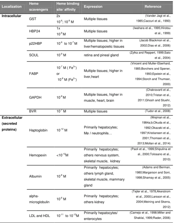
Table 3. Described heme scavengers 22
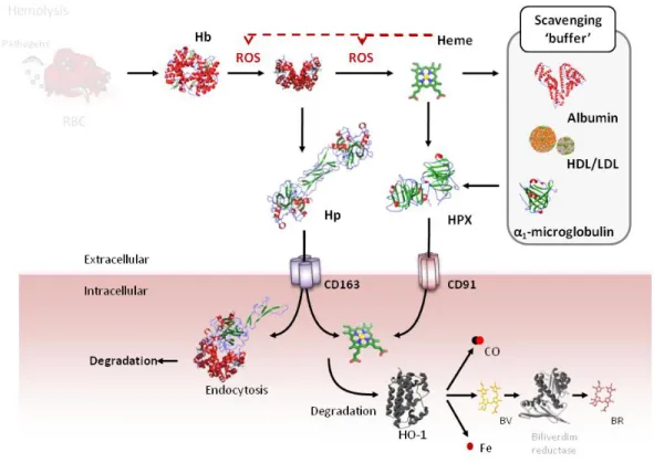
Figure 7. Heme scavengers 28
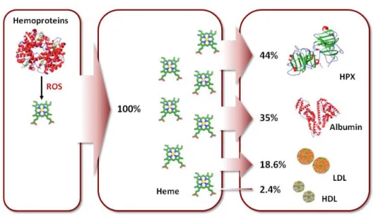
Figure 8. Predicted heme distribution in plasma 29
Figure 9. Heme catabolism. 30
Figure 10. Pathophysiologic effect of heme. 34 Figure 11. Pro-oxidant effect of heme. 37 Figure 12. Pro-inflammatory effect of heme. 41
Figure 13. Targeting “free heme” 49
Figure 14. Hybridoma and Phage display technology for the
development of Abs. 55
Figure 15. Schematic representation of the five different classes of Abs
Figure 18. Schematic representation of a filamentous phage expressing sdAb on their surface fused with pIII protein and the structure and
aminoacid sequence of sdAbs. 62
Figure 19. Schematic representation of a phage display cycle, also
named phage display panning, applied in the selection of Abs. 64
Chapter 2:
Targeting Heme with Single Domain Antibodies
Figure 1. Selection of heme binding sdAbs 95 Figure 2. SdAbs heme binding specificity 97 Figure 3. SdAbs heme binding specificity to other heme analogs and
hemoproteins 99
Table 1. Summary of the sdAbs binding specificity against different
tetrapyrroles 100
Figure 4. Characterization of sdAb binding to heme 103 Figure 5. Detection of intracellular heme by sdAbs 105 Figure 6. Detection of heme in different cellular organelles 106 Figure 7. Heme accumulation in plasma during acute hemolysis in mice 109 Figure 8. Targeting heme biologic activity with sdAbs 111 Suppl. Figure 1. MALDI-TOF/TOF zoom analysis of biotinylated heme 134 Suppl. Figure 2. Sensogram of heme binding to sdAb 2H10 135 Suppl. Figure 3. Characterization of heme binding to sdAb 1A6 and 2H7
by UV-Visible spectrophotometry 135
Suppl. Figure 4. Sandwich ELISA for heme detection 136
Chapter 3:
General Discussion
Figure 1.3D model structure of 2H10 sdAbs 150 Figure 2. Model of heme distribution upon hemolysis according to the
Appendix:Supplementary Information
Engineering of sdAbs: Dimerization of sdAbs against heme and generation of sdAbs containing CDR3
Figure 1. Generation of sdAbs constructs 172 Figure 2. Protein expression in different bacteria strains and their
purification 174
Figure 3. Binding to heme 176
Figure 4. Structure of the different heme analogs 178 Figure 5. CDR1CDR3 2H7-2H7 heme binding specificity to heme, its
Acknowledgements I
Abbreviations V
Preface X
Sumário XII
Abstract XV
Chapter 1:
General Introduction 1
1. The Biology of Heme 2
1.1. Discovery of Heme Molecule 2
1.2. Structure and Biochemistry of Heme 3
1.2.1. Heme Interaction with Proteins 5
1.2.1.1 Heme Pocket 6
1.2.1.2 Heme Binding Motifs 10
1.2.1.3 Non-planar Heme Structure 11
2. Heme Biosynthesis 12
3. Heme Transporters 15
3.1. Heme Importers 15
3.2. Heme Exporters 18
4. Heme Binding Proteins/Chaperones 21
4.1. Intracellular Heme Binding Proteins 23
4.2. Extracellular Heme Binding Proteins 25
5. Heme Catabolism 30
5.1. Enzymatic Mechanism of Heme Catabolism – Heme oxygenase 31 5.2. Non-Enzymatic Mechanism of heme Catabolism 33
5.2.1 Hydrogen Peroxide 33
5.2.2 Hydrochlorous 33
6. Heme as a Pathophysiological Molecule 34
6.1. Pro-Oxidant Activity of Heme 35
6.2. Pro-Inflammatory Properties of Heme 38
7. Heme in the Pathogenesis of Immune Mediated Inflammatory
Diseases 42
7.1. Heme and Malaria 43
7.2. Heme and Sepsis 44
7.3. Heme and Sickle Cell Disease 46
8. Targeting Heme in the Treatment of Immune Mediated Inflammatory
Diseases 48
8.1. Pharmacologic Administration of Gasotransmitters 49
8.2. Induction of HO-1 51
8.3. Hemoglobin and Heme Scavengers 53
8.4. Heme Transporters 54
8.5. Therapeutic Application of Antibodies, Abs 54
8.5.1. Abs Structure and Function 56
8.5.1.1. Ab Fragments 58
8.5.1.2. Single Domain Abs 60
8.5.2. Ab Production using Phage Display 61 8.5.2.1. Selection and Screening of Abs using Phage Display
Technology 63
8.5.2.2. Libraries used in Phage display Technology 65
9. Aims of the Thesis 67
10. References 68
Chapter 2:
Targeting Heme with Single Domain Antibodies 87
1. Abstract 91
2. Introduction 92
3. Results 94
3.2. Heme sdAb specificity 96 3.3. Structural characterization of sdAbs/heme complexes 100
3.4. Detection of cellular heme 104
3.5. Quantitative analysis of circulating heme after acute hemolysis in
vivo 107
3.6. Modulation of heme biologic activity by sdAb 109
4. Discussion 112
5. Experimental Procedures 117
6. Acknowledgements 130
7. References 131
8. Supplementary figure 134
Chapter 3:
General Discussion 137
References 161
Appendix:Supplementary Information
Engineering of sdAbs: Dimerization of sdAbs against heme and
generation of sdAbs containing CDR3 167
1. Abstract 170
2. Introduction 170
3. Results 172
3.1. Generation of different constructs of based on previously described
sdAbs 172
3.2. Constructs expression in different bacteria strains and their
purification 173
3.3. Heme Binding 175
3.4. Specificity of the sdAbs constructs 177
4. Discussion 180
6. Acknowledgements 187
Chapter 1:
General Introduction
Figure 1. Hemoproteins containing the most common heme types 4 Figure 2. Structure of the different heme groups existent in hemoproteins
from a variety of species 5
Figure 3. Hemoproteins with their respective heme pockets 7 Table 1. Selected representatives of mammalian hemoproteins and
heme transient binding proteins. 8
Figure 4. Non-planar distortions in the porphyrin macrocycle in different
hemoproteins 11
Figure 5. Heme biosynthesis 13
Figure 6. Extracellular and intracellular heme transport 16 Table 2: Putative heme and/or porphyrin transporters 17
Table 3. Described heme scavengers 22
Figure 7. Heme scavengers 28
Figure 8. Predicted heme distribution in plasma 29
Figure 9. Heme catabolism. 30
Figure 10. Pathophysiologic effect of heme. 34 Figure 11. Pro-oxidant effect of heme. 37 Figure 12. Pro-inflammatory effect of heme. 41
Figure 13. Targeting “free heme” 49
Figure 14. Hybridoma and Phage display technology for the
development of Abs. 55
Figure 15. Schematic representation of the five different classes of Abs
Figure 18. Schematic representation of a filamentous phage expressing sdAb on their surface fused with pIII protein and the structure and
aminoacid sequence of sdAbs. 62
Figure 19. Schematic representation of a phage display cycle, also
named phage display panning, applied in the selection of Abs. 64
Chapter 2:
Targeting Heme with Single Domain Antibodies
Figure 1. Selection of heme binding sdAbs 95 Figure 2. SdAbs heme binding specificity 97 Figure 3. SdAbs heme binding specificity to other heme analogs and
hemoproteins 99
Table 1. Summary of the sdAbs binding specificity against different
tetrapyrroles 100
Figure 4. Characterization of sdAb binding to heme 103 Figure 5. Detection of intracellular heme by sdAbs 105 Figure 6. Detection of heme in different cellular organelles 106 Figure 7. Heme accumulation in plasma during acute hemolysis in mice 109 Figure 8. Targeting heme biologic activity with sdAbs 111 Suppl. Figure 1. MALDI-TOF/TOF zoom analysis of biotinylated heme 134 Suppl. Figure 2. Sensogram of heme binding to sdAb 2H10 135 Suppl. Figure 3. Characterization of heme binding to sdAb 1A6 and 2H7
by UV-Visible spectrophotometry 135
Suppl. Figure 4. Sandwich ELISA for heme detection 136
Chapter 3:
General Discussion
Figure 1.3D model structure of 2H10 sdAbs 150 Figure 2. Model of heme distribution upon hemolysis according to the
Appendix:Supplementary Information
Engineering of sdAbs: Dimerization of sdAbs against heme and generation of sdAbs containing CDR3
Figure 1. Generation of sdAbs constructs 172 Figure 2. Protein expression in different bacteria strains and their
purification 174
Figure 3. Binding to heme 176
Figure 4. Structure of the different heme analogs 178 Figure 5. CDR1CDR3 2H7-2H7 heme binding specificity to heme, its
1
C
HAPTER1
2
1 T
HEB
IOLOGY OFH
EMEHeme is an iron (Fe)-containing protoporphyrin IX molecule involved in vital biological processes in numerous organisms, including mammals. It is incorporated as prosthetic group of hemoproteins that have crucial roles in biological processes, such as gas storage and transport, enzymatic catalysis and regulation of gene expression, among others (Chapman et al., 1997;Poulos, 2007b).
The distribution of heme varies among different organisms, tissues and cells. In humans around 80% of heme remains in red blood cells (RBC) while 15% is found in the liver and the remaining 5% in other tissues (Zhang, 2011). Nevertheless, all mammalian cells require basal levels of heme used for the synthesis and correct function of crucial hemoproteins.
1.1 Discovery of The Heme Molecule
The circulatory system sustains blood transit through the organism and as such, is critical to maintain homeostasis. For instance, it transports nutrients (such as amino acids; aa and electrolytes), oxygen (O2), carbon dioxide (CO2), hormones, proteins as well as RBC and immune cells. RBCs are the most common cell type in blood, accounting for around 35-40% of the total blood volume (5x10⁶ RBC/µm3 of blood). The remaining volume corresponds to plasma, composed by water containing around 70 g/L proteins (e.g. albumin, globulins) and electrolytes (e.g. sodium, calcium, magnesium), glucose, hormones and other compounds as well as other cell types (Edsall, 1972b).
3
globin, the two assembling to form hemoglobin (Edsall, 1972a). It was only in 1929 however, that the chemical structure of heme was revealed by Fischer, who synthesized hemin, i.e. chloride (Cl) Fe-protoporphyrin (PP) IX molecule, and confirmed its structure, as previously proposed by Piloty and Küster (Fischer and Zeile, 1929). This discovery was awarded with the Nobel Prize in Chemistry in 1930 in recognition of “researches into the constitution of haemin and chlorophyll and especially for his synthesis of haemin” (Fischer and Zeile, 1929;Garcia-Sanchez et al., 2013).
1.2 Structure and Biochemistry of Heme
Heme is a hydrophobic metallo-compound composed by four methane-bridged pyrroles (tetrapyrrole ring) bound to a central Fe atom through their nitrogen atoms (Fig. 1) (Kumar and Bandyopadhyay, 2005;Tsiftsoglou et al., 2006;Poulos, 2007b). The Fe atom can acquire different oxidative states, most commonly ferrous (Fe2+) with a neutral chemical charge or ferric (Fe3+), positively charged and optimized for anion binding (Kumar and Bandyopadhyay, 2005). Although ferrous and ferric are the most common, other oxidation heme states from Fe2+ to Fe5+ can occur, as observed in cytochrome c oxidase or peroxidases (Ogura et al., 1996). Additionally, the planar conformation of heme can suffer small distortions within proteins, which are very common and functionally relevant (Shelnutt et al., 1998).
4
release from hemoproteins (Allen et al., 2003). Other heme variants can be found in a variety of organisms such as bacteria, namely heme d (Murshudov et al., 1996), heme d1 (Sjogren and Hajdu, 2001), heme o (Abramson et al., 2000), heme P460 (Igarashi et al., 1997) and siroheme (Oliveira et al., 2008) (Fig. 2).
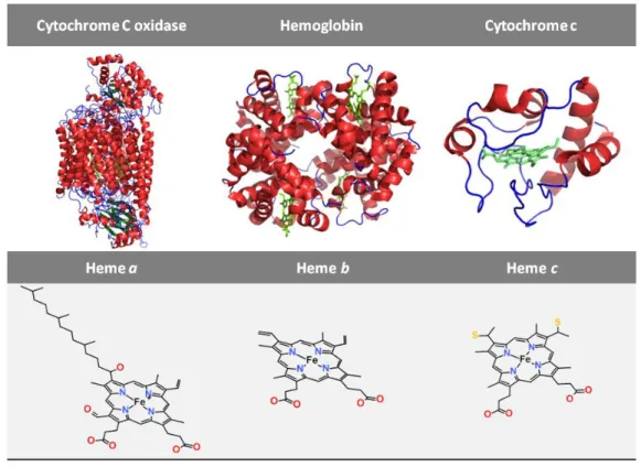
Figure 1: Hemoproteins containing the most common heme types. Crystalographic structure
of bovine heart cytochrome C oxidase (PDB ID: 1OCC), human hemoglobin (PDB ID: 2W6V) and
horse heart cytochrome c (PDB ID: 1HRC) (upper) and their prosthetic group, heme type a
(ChEBI: 83281), heme b group (ChEBI: 55376) and heme c group (ChEBI: 60562) (lower),
5 1.2.1 Heme Interaction with Proteins
Heme is essential for the proper folding and function of hemoproteins (Chapman et al., 1997;Poulos, 2007b) that perform a wide range of biological functions, including transport (e.g hemoglobin) and storage (e.g. myoglobin) of diatomic gaseous molecules such as oxygen (O2), nitric oxide (NO) and CO (Evans and Brayer,
Figure 2: Structure of the different heme groups existent in hemoproteins from a variety of
species. Heme types: a (ChEBI: 83281), b (ChEBI: 55376), c (ChEBI: 60562), d (ChEBI: 62802),
d1(ChEBI: 61147), P460(ChEBI: modified 60562), o (ChEBI: 36301), siroheme (ChEBI: 28599) and
myeloperoxidase (also known as heme m, ChEBI: 55376) are found in hemoproteins, such as
cytochrome c oxidase, hemoglobin, cytochrome c, catalase hydroperoxidase II, cytochrome cd1
nitrite reductase, hydroxylamine oxidoreductase, ubiquinol oxidase, sulfite reductase and
myeloperoxidase, respectively. These structures were manipulated according to their ChEBI
(Chemical Entities of Biological Interest; http://www.ebi.ac.uk/chebi/), using the software Acceryls
Draw. Table shows the relative abundance of the heme type amongst all hemoproteins described in
6
1990;Aono, 2008;Kakar et al., 2010b). Some hemoproteins are endowed with enzymatic activity (e.g. peroxidases, catalase and synthases) (Reid et al., 1981;Crane et al., 1997;Zederbauer et al., 2007b;Capitanio et al., 2011) and are involved in key metabolic processes (e.g. cytochromes P-450, peroxidases) (Bushnell et al., 1990;Guengerich, 2001). These include the respiratory electron transport chain in the mitochondria (e.g. cytochromes c, cytochrome c oxidase) (Gray and Winkler, 1996;Capitanio et al., 2011), gene transcription (e.g. heme binding to the transcription regulatory protein basic leucine zipper transcription factor 1, BACH1) (Youn et al., 2006;Hira et al., 2007), micro-RNA processing (e.g RNA-binding protein DiGeorge critical region-8, DGCR8) (Weitz et al., 2014), regulation of circadian rhythms (e.g. Neuronal PAS domain-containing protein 2, NPAS2)(Kaasik and Lee, 2004) and signal transduction pathways (e.g.BACH1) (Zenke-Kawasaki et al., 2007) or ion-channel functions (e.g. sodium channel) (Horrigan et al., 2005;Wang et al., 2009).
1.2.1.1 Heme Pocket
The “heme pockets” of hemoproteins (Fig. 1 and 3; Table 1) are characterized by a high abundance of aromatic amino acids (aa), i.e. as phenylalanine (Phe, F), tyrosine (Tyr, Y) or tryptophan (Trp, W) and by few non-charged aa (Li et al., 2011), conferring the hydrophobicity required to stabilize heme and its binding to protein matrix. Furthermore, hemoproteins contain frequently five highly conserved aa in their heme pockets, including histidine (His, H), methionine (Met, M), cysteine (Cys, C), Tyr and lysine (Lys, K) that can act as axial Fe-heme ligands (Li et al., 2011). Beside these, other aa can bind to Fe-heme, including proline (Pro, P) through its N-terminus amino group (Lanzilotta et al., 2000;Clark et al., 2004) or asparagine
7
also establish interactions with the heme porphyrin, whereas arginine (Arg, R) and other positively charged aa can interact with the negatively charged heme propionate groups (Fig. 3) (Schneider et al., 2007).
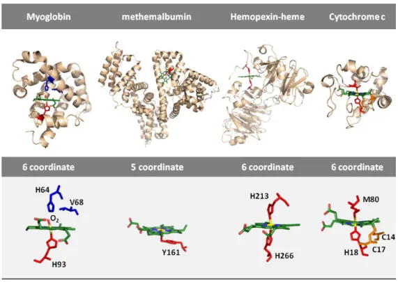
Figure 3: Hemoproteins with their respective heme pockets. Structure of human sperm whale
oxymyoglobin (PDB ID: 1MBN), methemalbumin (PDB ID: 1N5U), rabbit heme scavenger
hemopexin bound to heme (PDB ID: 1QJS) and horse heart cytochrome c (PDB ID: 1HRC) (upper)
with their heme group highlighted. Detail view of heme group of the corresponding hemoproteins
(lower). The heme-Fe in oxymyoglobin is a six coordinate (6c) low spin (LS) with a histidine (H93)
and a molecule of oxygen (O2) axial ligands. Upon oxygen dissociation, myoglobin acquires a five
coordinate (5c) high spin (HS) heme state. Heme in albumin is a 5cHS with a tyrosine (Y161) amino
acid bound to Fe. In hemopexin, heme has two histidine (H213 and H266) coordinated to its Fe
atom and therefore a 6cLS state. Unlike in heme b type hemoproteins, heme in cytochrome c is
covalently attached to the protein through two cysteines (C14 and C17), as thus defined as heme c;
its Fe is coordinated with a proximal Histidine (H18) and a distal methionine (M80), forming a 6c LS
8
Table 1:Selected representatives of mammalian hemoproteins and heme transient binding
proteins. Proteins were classified according to their function and the number and type of heme
were indicated as well as the heme axial ligands.
Function Representative Proteins
Heme
Number/ Type
Axial
ligand(s) Reference
Electron
transport
Cytochrome c 1/ c-type His/Met (Mirkin et al., 2008)
Cytochrome b5 1/ b-type His/ His (Wirtz et al., 2000)
Cytochrome c oxidase (complex IV)
2/ a-type His His/His
(Tsukihara et al., 1995)
Cytochrome c reductase/
cytochrome bc1
2/ b-type 1/ c-type
His/His His/His
His/Met
(Xia et al., 1997;Zhang et al., 1998)
Gas binders
and carriers
Hemoglobin 4 / b-type His (Park et al., 2006)
Myoglobin 1 / b-type His (Evans and Brayer,
1988;Vojtechovsky et al., 1999) Catalytic
activity - enzyme
Catalase I 1/ b-type Tyr (Putnam et al., 2000)
Cytochrome P450 (2D6) 1/ b-type Cys (Rowland et al., 2006)
Indoleamine
2,3-dioxygenase
1/ b-type His/ligand (Sugimoto et al., 2006)
NO synthase 2/ b-type Cys (Crane et al., 1997) Cystathione β-synthase 1/ b-type Cys/His (Meier et al., 2001) Tryptophan 2,3
dioxygenase
4/ b-type His (Zhang et al., 2007)
Heme degradation
Heme Oxygenase 1/ b-type His/ H2O (Lad et al., 2003)
Heme binding and transport
Hemopexin 1/ b-type His/His (Paoli et al., 1999)
Albumin 1/ b-type Tyr (Wardell et al., 2002) α1-microglobulin 1/b-type His/Cys (Allhorn et al., 2003;Larsson
et al., 2004;Meining and Skerra, 2012)
Heme-sensing
/heme
regulation
NPAS2 2/b-type His/Cys or
His/His
His/?
(Dioum et al., 2002a;Uchida et al., 2005)
Bach 1 5/b-type 4x Cys
1xCys/His
(Hira et al., 2007)
Iron regulatory protein 2 2/b-type Cys/? His/His
(Jeong et al., 2004a;Jeong et al., 2004b;Ishikawa et al., 2011) Soluble guanylyl cyclase 1/ b-type His (Pellicena et al., 2004;Martin et al., 2005;Ma et al., 2007)
9
The axial coordination of Fe in heme is directly correlated with protein function. Fe establishes primarily four bonds with the pyrrole nitrogens of protoporphyrin IX and one or two additional bounds with aa of the heme pocket (i.e. the axial ligands) constituting the five (5c) or six coordinate (6c) heme, respectively (Fig. 3). Usually, in gas transporter proteins such as myoglobin and hemoglobin, heme is in a 5c state with His as the fifth axial ligand while the remaining binding site is vacant for the binding of small molecules such as oxygen (Vojtechovsky et al., 1999;Park et al., 2006). In other proteins, namely enzymes with a 5c, one of the axial ligands can be either His or Tyr while the sixth axial is ligand-free and available for substrate binding (Reid et al., 1981;Rowland et al., 2006). Cytochromes involved in electron transfer reactions typically display six coordinated heme, in which both axial ligands are usually occupied by a bi-His or by His-Met (Mirkin et al., 2008).
10
also bind proteins to regulate their biologic function, as it is the case for heme driven inhibition of the transcriptional repressor BACH1 (Hira et al., 2007).
1.2.1.2 Heme Binding Motifs
Hemoproteins possess characteristic sequences of aa, contained inside heme pockets, referred as heme-binding motifs (HBM) or heme regulatory motifs (HRM). There are two HBM that bind heme heme c covalently, namely CXXCH and CXXCK in which ”X” refers to any aa (Hartshorne et al., 2007;Fufezan et al., 2008;Smith et al., 2010;Li et al., 2011). Other HBM, such as CXXXCH and CXXXXCH, present in multi-hemic cytochromes also bind heme c (Aragao et al., 2003). The AXXCH and FXXCH HBM bind heme c covalently through a single thioether bond (Ginger et al., 2012). In myeloperoxidases, heme c is bound covalently by two thioether bonds as well as by a unique methionyl-sulfonium bond involving a Met (Kooter et al., 1999). In cytochrome P460, heme c is also bound covalently through a CXXCH motif by an additional ligation to other aa, namely a Lys in cytochrome P460 (Pearson et al., 2007) or a His in hydroxylamine oxidoreductase (Igarashi et al., 1997).
Heme a, b, d (Murshudov et al., 1996), d1 (Sjogren and Hajdu, 2001), o (Abramson et al., 2000) and siroheme (Oliveira et al., 2008) (Fig. 2) create non-covalent hydrophobic and electrostatic interactions with hemoproteins. For instance, heme b is mainly associated to a HBM with the aa GX[H/R]XC[P/L/ A/V]G, i.e. Glycine (Gly, G); X; His or Arg; X; Cys; Pro or Leu or Alanine (Ala, A) or Val and
Gly, where X can be any aa (Li et al., 2011). Another related FXXGXXCXG motif, i.e Phe; X; X; Gly; X; X; Cys; X and Gly, was described in the mammalian
11 1.2.1.3 Non-planar Heme Structure
Heme is classically represented in its planar conformation, which is the most favorable thermodynamically (Fig. 1 and 2). However, in the heme pocket of proteins, heme is exposed to several forces exerted by the surrounding aa, which induce distortions in the porphyrin structure (Shelnutt et al., 1998). This has been observed in several proteins with similar biological functions and in different organisms (Fig. 4), suggesting that alterations to the nonplanar structures of heme
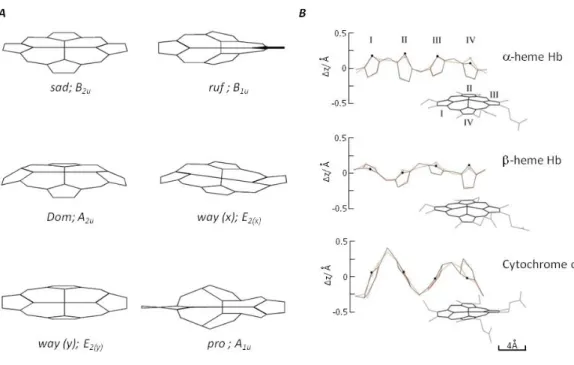
Figure 4: Non-planar distortions in the porphyrin macrocycle in different hemoproteins.
Although heme is usually represented as a planar structure; it acquires different non-planar
distortions in proteins. A. There are six lowest frequency out-of-plane normal coordinate
deformations of the porphyrin macrocycle used to decomposed the non planar distortions of heme
in proteins. These are referred to as saddling (sad, B2u), ruffling (ruf, B1u), doming (dom, A2u),
waving [wav(x), wav(y); Eg], and propellering (pro,A1u). B. Non-planar distortion of the heme
12
modulate hemoproteins function (Shelnutt et al., 1998). To understand this observation, Shelnutt and co-workers developed a computational procedure named “normal-coordinate structural decomposition method”, that allows determining and comparing non-planar distortions of heme, as assessed in crystal structures of hemoproteins. These include six lowest frequency out-of-plane normal coordinate deformations, namely: saddling (sad, B2u), ruffling (ruf, B1u), doming (dom, A2u), waving [wav(x), wav(y); Eg], and propellering (pro,A1u) (Fig. 4) (Jentzen et al., 1998;Shelnutt et al., 1998). The biggest heme distortions were found in type-c cytochromes and peroxidases that present mainly saddled distortions, while in myoglobin (type-b), heme was predominantly found in a domed conformation (Jentzen et al., 1998;Shelnutt et al., 1998).
2 H
EMEB
IOSYNTHESIS13
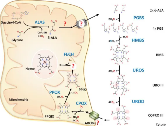
Figure 5: Heme biosynthesis. The first step of heme biosynthesis occurs in the mitochondrial matrix with the condensation of glycine and succinyl-CoA by the mitochondrial δ-aminolevulinate synthase (ALAS) to generate δ-aminolevulinic acid (ALA). ALA is then translocated to the cytosol by unknown mechanism where the next four steps of heme biosynthesis occur. In the cytosol, two
molecules of ALA react with pyridoxal phosphate to form a porphobilinogen (PBG) through the
catalyst porphobilinogen synthase (PGBS). Four molecules of PBG are condensed by
hydroxylmethylbilane synthase (HMBS), generating a molecule of hydroxymethylbilane (HMB) that
is converted into uroporphyrinogen III (URO III) by uroporphyrinogen III co-synthase or isomerase
(UROS). The last step in the cytosol is the conversion of decarboxylation of URO III into
coproporphyrinogen III (COPRO III) by uroporphyrinogen III decarboxylase (UROD). COPRO III is
then translocated into the mitochondrial intermembranal space by a proposed transporter, ABCB6
and then by oxidative decarboxylation it is converted into protoporphyrinogen IX (PPG IX) by the
coproporphyrinogen oxidase (CPOX). This is followed by PPG IX oxidation by protoporphyrin
oxidase (PPOX) into protoporphyrin IX (PPIX) that is translocated to the mitochondrial matrix, for
ferrous iron cation (Fe2+) incorporation by the enzyme ferrochelatase (FECH) into the macrocycle of
14
Both ALAS proteins harbor two putative HBM in the mitochondrial targeting peptide at the N-termini, and a third HBM near the N-terminus of the mature protein (Dailey et al., 2005). Heme binding to the HBM of the immature protein presumably blocks mitochondrial import and consequently heme synthesis, thus avoiding the accumulation of intracellular heme that would be otherwise deleterious (Munakata et al., 2004). This level of regulation occurs only on the ALAS1 and not on the ALAS2 isoform (Munakata et al., 2004). ALAS1 gene expression is down-regulated by heme itself, while ALAS2 expression is controlled at the transcriptional level by different erythroid specific factors, such as GATA binding protein 1 (globin transcription factor 1; GATA1) (Surinya et al., 1997).
15
consists at the incorporation of a ferrous Fe cation (Fe2+) into the macrocycle of PPIX to form protoheme IX (heme), by the enzyme ferrochelatase (FECH) (E.C. 4.99.1.1) in the mitochondrial matrix (Wu et al., 2001). The de novo synthesized heme is finally transported to the cytosol where it can either be incorporated into apoproteins to form hemoproteins or be directly involved in other biological functions (e.g. transcription, translation, cellular differentiation).
3 H
EMET
RANSPORTERSHeme transport is tightly regulated so that heme is delivered to different compartments where it is used in diverse biological processes. The hydrophobic nature of heme determines its insolubility in aqueous solutions, making it unlikely that heme could be transported as a free molecule and suggesting the existence of heme binding partners that assist its transport from one cell compartment to another. Additionally, its hydrophobic nature should allow heme diffusion through cellular membranes, although this was shown to occur rather slowly, partially due to the hydrophilicity conferred by the propionate groups of heme (Light and Olson, 1990). This dual nature of heme and its limitations to diffuse freely through membranes imposes the need for heme transporters, and several have been described (Fig. 6; Table 2). Considering that “free heme” is cytotoxic (Gozzelino et al., 2010;Larsen et al., 2010;Gozzelino and Soares, 2011) “carriers” and/or chaperone proteins transport heme to specific apoproteins or to be degraded in different cell compartments and/or tissues.
3.1 Heme Importers
16
The mammalian HRG-1 ortholog is highly expressed in the brain, heart, kidney and muscle and to a lesser extent in liver, lung, placenta and intestine (Rajagopal et al.,
Figure 6: Extracellular and intracellular heme transport. As “free heme” is highly cytotoxic,
intracellular heme is maintained bound to a heme carrier/chaperone, providing a support for heme
transfer between the different compartments and transporters. Intracellular heme transport is
mediated by at least three importers, i.e. Haem carrier protein 1 (HCP-1), feline leukemia virus subgroup C cellular receptor 2 (FLVCR2) and Heme-responsive gene 1 protein (HRG-1) and three
cellular membranar exporters, i.e. ATP-binding cassette, sub-family B 6 (ABCB6), feline leukemia virus subgroup C cellular receptor 1a (FLVCR1a) and ATP-binding cassette sub-family G member 2
(ABCG2). HCP-1 has been described to be responsible for heme uptake in the intestine and
FLVCR2 from a wide range of mammalian cells. HRG-1 was initially described to export heme from
the lysosome/endosome compartment; however, HRG-1 can travel to the plasma membrane and
import heme (Rajagopal et al., 2008). ABCB6 is localized in the plasma membrane as well as in the
lysosome/endosome compartment (Kiss et al., 2012). FLVCR1b is responsible for heme export from
the mitochondria into the cytosol. ABCB10 forms a complex with MFRN1 and FECH, probably
17
2008). HRG-1 is localized in the membranes of endosomes and lysosomes and mediates heme transport from these acidic compartments into the cytoplasm (Fig. 6). More recently, HRG-1 was identified in the plasma membranes and lysosomes of non-polarized human epithelial type 2 (Hep2) cells as well as coupled to the vacuolar ATPase (adenylpyrophosphatase; EC 3.6.1.3) proton pump, which induces endosomal acidification, increasing HRG-1 affinity towards heme.
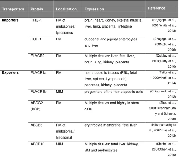
Table 2: Putative heme and/or porphyrin transporters. The transporters were classified
according to their functions and then localization and expression.
Transporters Protein Localization Expression Reference
Importers HRG-1 PM of
endosomes/
lysosomes
brain, heart, kidney, skeletal muscle,
liver, lung, placenta, intestine
(Rajagopal et al., 2008;White et al., 2013)
HCP-1 PM duodenal and jejunal enterocytes
and liver
(Shayeghi et al., 2005;Qiu et al., 2006) FLVCR2 PM Multiple tissues: liver, fetal liver,
brain, lung, kidney ,placenta
(Quigley et al., 2004;Duffy et al., 2010) Exporters FLVCR1a PM hematopoietic tissues (PBL, fetal
liver, spleen, Lymph node),
pancreas, kidney, placenta
(Tailor et al., 1999;Vinchi et al., 2014)
FLVCR1b MIM progenitors of the hematopoetic cells (Chiabrando et al., 2012) ABCG2
(BCP)
PM Multiple tissues and highly in stem
cells
(Zhou et al., 2001;Krishnamurth
y and Schuetz, 2005) ABCB6 PM of
endosomal/ lysosomal
erythrocyte membrane, fetal liver (Krishnamurthy et al., 2007;Kiss et al., 2012)
ABCB10 MIM Multiple tissues: fetal liver, kidney, BM and erythrocytes
18
HCP-1 is a low affinity and non-exclusive heme cellular importer (e.g. it can also translocate folate), expressed mainly in the liver and in duodenum enterocytes and, to a lesser extent in jejunum enterocytes (Fig. 6). HCP-1 is responsible for heme absorption from food (Shayeghi et al., 2005;Qiu et al., 2006;Le Blanc et al., 2012).
FLVCR2 is a heme cellular importer expressed in several mammalian non-hematopoietic tissues including brain, placenta, lung, liver, and kidney as well as in hematopoietic tissues, including fetal liver, spleen, lymph nodes, thymus, leukocytes, and bone marrow (Duffy et al., 2010)(Fig. the FLVCR2 6). Mutations in gene were linked to Fowler syndrome, a vascular disorder of the brain, but the association of its cellular function as a heme cellular importer with the disease still remains to be elucidated (Duffy et al., 2010).
3.2 Heme Exporters
After the last step of de novo heme synthesis, newly generated heme must be exported from the mitochondria so that it can be incorporated into apoproteins, generating a variety of hemoproteins. Two heme mitochondrial exporters have been described, namely, the Feline Leukemia Virus Subgroup C Receptor 1 (FLVCR1) (Quigley et al., 2004) and the ATP-binding cassette transporter G2 (ABCG2)(Krishnamurthy and Schuetz, 2005).
19
via a direct interaction between the transporter and the scavenger (Yang et al.,
2010b). The two FLVCR1 isoforms seem to have different functions. FLVCR1b is essential for erythroid differentiation, while FLVCR1a is required for intracellular heme homeostasis among different cells types, with an important impact on heme synthesis and consequently, on hemoprotein function (Chiabrando et al., 2012).
The FLVCR1 transporter is required for erythroid maturation (Quigley et al., 2004), a notion supported by the fact that FLVCR1-null mice lack mature erythrocytes, as they develop severe macrocytic anemia and proerythroblast apoptosis (Quigley et al., 2004). This is not observed in humans carrying point mutations in this transporter, which presumably lead to alterations in its transmembrane domains (Yanatori et al., 2012), but do not lead to overt defect in erythropoiesis (Yanatori et al., 2012). This questions whether FLVCR1 is essential for heme transport and erythropoiesis in humans.
ABCG2 is another heme cellular exporter also known as breast cancer resistance protein (BCP), which was discovered based on its role in conferring drug resistance of tumor cells (Doyle et al., 1998;Miyake et al., 1999). ABCG2 is not specific for heme, as it can bind other protoporphyrins (PP) (e.g. Zinc PP, Cobalt PP, empty PP), preventing their accumulation within cells and contributing to PP homeostasis (Krishnamurthy et al., 2004;Desuzinges-Mandon et al., 2010). Even though ABCG2 binds to a wide range of PP, its highest binding affinity is for heme (Desuzinges-Mandon et al., 2010). Similarly to FLVCR1, it delivers heme to heme-binding proteins such as albumin (Desuzinges-Mandon et al., 2010). ABCG2 is expressed in a wide variety of cells, including hematopoetic stem cells, being particularly expressed at high levels in the early stages of hematopoiesis (Zhou et al., 2001), contrarily to FLVCR1 that is mainly expressed during erythropoiesis (Quigley et al., 2004).