Published: March 31, 2011
pubs.acs.org/biochemistry
Low-Spin Heme
b
3
in the Catalytic Center of Nitric Oxide Reductase
from
Pseudomonas nautica
Cristina G. Tim
oteo,
†Alice S. Pereira,*
,†Carlos E. Martins,
†Sunil G. Naik,
‡Am
erico G. Duarte,
†Jos
e J. G. Moura,
†Pedro Tavares,
†Boi Hanh Huynh,
‡and Isabel Moura*
,††
Requimte, Centro de Química Fina e Biotecnologia, Departamento de Química, Faculdade de Ci^encias e Tecnologia,
Universidade Nova de Lisboa, Quinta da Torre, 2829-516 Monte de Caparica, Portugal
‡
Department of Physics, Emory University, Atlanta, Georgia 30322, United States
b
S Supporting InformationD
enitrifying bacteria can be found in a wide variety ofenvironments ranging from streams, soils, to human in-testines.Ps. nautica617 is a halofile bacterium found in marine sediments contaminated with effluents from oil refineries.1,2It is able to reduce nitrate to dinitrogen by utilizing the catalytic functions of four specific enzymes:3The Mo-pterin containing
nitrate reductase, the cytochrome cd1 nitrite reductase, the
cytochrome bc complex nitric oxide reductase, and the
Cu-containing nitrous oxide reductase. The nitrate reductase, cyto-chromecd1, and nitrous oxide reductase fromPs. nauticahave been subjected to extensive biochemical and structural studies,47 while detailed characterization of nitric oxide reductase (NOR) fromPs. nauticahas not been reported.
There are three classes of NOR identified in bacteria, all
membrane bound. Thefirst class, called cNOR, uses a soluble
cytochromecor a cupredoxin as its electron donor, whereas the two other classes, qNOR and qCuNOR, are quinol oxidases. The cNOR’s are composed of two subunits: a small subunit (NORC,
∼17 kDa) that contains a c-type heme and a large subunit
(NORB,∼56 kDa) that contains twob-type hemes and a
non-heme iron. Enzymes belonging to this class have been isolated from four denitrifying bacteria: Ps. stutzeri,8,9Paracoccus (Pa.)
Received: October 4, 2010 Revised: March 31, 2011
ABSTRACT:Respiratory nitric oxide reductase (NOR) was purified from membrane extract ofPseudomonas(Ps.)nautica cells to homogeneity as judged by polyacrylamide gel electro-phoresis. The purified protein is a heterodimer with subunits of molecular masses of 54 and 18 kDa. The gene encoding both subunits was cloned and sequenced. The amino acid sequence shows strong homology with enzymes of the cNOR class. Iron/
heme determinations show that one hemecis present in the
small subunit (NORC) and that approximately two hemeband
one non-heme iron are associated with the large subunit
(NORB), in agreement with the available data for enzymes of the cNOR class. M€ossbauer characterization of the as-purified, ascorbate-reduced, and dithionite-reduced enzyme confirms the presence of three heme groups (the catalytic hemeb3and the electron transfer hemeband hemec) and one redox-active non-heme Fe (FeB). Consistent with results obtained for other cNORs, hemecand hemebinPs. nauticacNOR were found to be low-spin while FeBwas found to be high-spin. Unexpectedly, as opposed to the presumed high-spin state for hemeb3, the M€ossbauer data demonstrate unambiguously that hemeb3is, in fact, low-spin in both
ferric and ferrous states, suggesting that heme b3 is six-coordinated regardless of its oxidation state. EPR spectroscopic
measurements of the as-purified enzyme show resonances at theg∼6 and g∼23 regions very similar to those reported
previously for other cNORs. The signals atg= 3.60, 2.99, 2.26, and 1.43 are attributed to the two charge-transfer low-spin ferric hemecand hemeb. Previously, resonances at theg∼6 region were assigned to a small quantity of uncoupled high-spin FeIIIheme
b3. This assignment is now questionable because hemeb3is low-spin. On the basis of our spectroscopic data, we argue that the g= 6.34 signal is likely arising from a spinspin coupled binuclear center comprising the low-spin FeIIIhemeb3and the high-spin
FeBIII. Activity assays performed under various reducing conditions indicate that hemeb3has to be reduced for the enzyme to be active. But, from an energetic point of view, the formation of a ferrous heme-NO as an initial reaction intermediate for NO reduction is disfavored because heme [FeNO]7is a stable product. We suspect that the presence of a sixth ligand in the FeII-hemeb3may weaken its affinity for NO and thus promotes, in thefirst catalytic step, binding of NO at the FeBIIsite. The function of hemeb3 would then be to orient the FeB-bound NO molecules for the formation of the NN bond and to provide reducing equivalents for
denitrificans,10,11Halomonas (H.) halodenitrificans,12,13and more recentlyPs. aeruginosa.14The enzymes of the qNOR class consist of only one subunit of 84.5 kDa. Similar to the NORB subunit of
cNOR, each molecule of qNOR contains twob-type hemes and
one non-heme iron. With the exception of the qNOR isolated from the denitrifying bacteria Ralstonia eutropha,15 all other purified qNOR’s reported so far were identified in pathogenic microorganisms lacking the other denitrifying enzymes.16,17The qCuNOR has been isolated from the Gram-positive denitrifying Bacillus azotoformans.18It is a heterodimer, composed of a small subunit of 16 kDa containing a CuAcenter and a large subunit of 57 kDa containing twob-type hemes and a non-heme iron center, similar to those in qNOR and in NORB of cNOR.
The NORs of all three classes catalyze the two-electron reduction of nitric oxide to nitrous oxide, a reaction that involves
the formation of a NN bond:
2NOþ2eþ2Hþ fN2OþH2O
Biochemical and spectroscopic investigations of cNORs, to-gether with site-directed mutagenesis studies, have provided
evidence for a binuclear catalytic site, comprising a heme b
(hemeb3) and a non-heme iron center (FeB).1921In the resting state of the enzyme (oxidized form), both Fe sites are suggested to be high-spin ferric and antiferromagnetically coupled, most probably, through a μ-oxo bridge,22 resulting in a diamagnetic ground state and consequently EPR silent. The mechanism of NO reduction remains a subject of intense interest and has been
reviewed recently.2326 Currently two alternative mechanisms
are being considered, namely, thecisandtransmechanism. In the cismechanism, one of the two iron sites of the binuclear center binds both NO molecules, and the other Fe site plays the role of electron transfer and/or assist in abstracting the O atom from
one of the NO molecules. In the trans mechanism, one NO
molecule is bound to each one of the iron sites of the binuclear center. Ultrarapid freeze quench EPR studies under single turn-over conditions showed simultaneous appearance of EPR signals
that are characteristics of ferrous hemeNO and non-heme
ferrousNO complexes at 0.5 ms after introduction of NO,14
providing evidence for the proposedtransmechanism. A recent spectroscopic study of a functional model complex also shows a reaction intermediate that exhibits both aS= 3/2 [FeBNO]7and
aS= 1/2 heme Fe(II)NO EPR signal, supporting thetrans
mechanism.27On the other hand, ferrous hemeNO complexes
are highly stable, making it improbable for ferrous hemeNO to
be a reaction intermediate. Also, a redox titration study of thePa.
denitrificanscNOR shows that the midpoint redox potential of
hemeb3is more than 200 mV lower than that of FeB,28indicating the presence of a thermodynamic barrier to prevent the reduc-tion of hemeb3and thus avoid the formation of a highly stable,
probably inhibitory, ferrous heme b3NO complex. The
pre-sence of such a thermodynamic barrier has led to the proposal that the three-electron-reduced cNOR with an oxidized hemeb3 is the active form of the enzyme.28In support of this proposal, studies of cyanide binding to various redox states of the enzyme indicated that affinity for CN
for the three-electron-reduced state is 1000-fold higher than that for the fully reduced or oxidized state.29But, interestingly, more recent studies withH. halodenitrificanscNOR12showed that NO can bind to both heme b3and FeBin their oxidized as well as reduced states and that the key determining factor in preventing NO binding to the binuclear center is the presence of theμ-oxo bridge. Either a
weakening or a rupturing of the oxo-bridge would permit the binding of NO irrespective of the redox state of the hemeb3FeB
center. Here we report a detailed biochemical and spectroscopic characterization of cNOR isolated from the denitrifying bacter-iumPs. nautica. In contrast to previously assumed high-spin state for hemeb3in cNORs isolated from other organisms, our results indicate that, forPs. nauticacNOR, hemeb3is low-spin in both oxidized and reduced states and that hemeb3has to be reduced for the enzyme to be active.
’EXPERIMENTAL PROCEDURES
Cell Growth and NOR Purification.Pseudomonas nautica617 cells were grown as described in Prud^encio et al.6 For 57Fe growth, FeSO4was replaced by the57Fe isotope salt. Cells were harvested by centrifugation at the end of the stationary phase. The yield in wet weight was 1 g of cells per liter of culture medium. Cells were ressuspended in 100 mM Tris-HCl pH = 7 buffer and disrupted on a French Press. The crude extract was centrifuged at 8000gfor 20 min to separate the intact cells and only then ultracentrifuged twice at 125000gfor 90 min to recover
the membrane fraction, which was frozen at80°C. Membranes
were washed as described in Girsh et al.,19 except for the
sonication step that was done in 500 mL portions, in four cycles of 10 min in ice. All ultracentrifugation steps were performed at 180000gfor 90 min. Purification of nitric oxide reductase was carried out at 4°C, following a modified procedure from the one described in Girsh et al.19Proteolysis inhibitors (1 mM PMSF and 10 mM benzamidine) were added to the supernatant of the extraction with 0.6% of DDM. The percentage of detergent used was judged to be effective as no significant NOR activity remained in the membrane fraction. This supernatant was applied to a
DEAE-Biogel A column (Bio-Rad, 5.525.5 cm, 140 mL of resin per
gram of protein) equilibrated with 50 mM Tris-HCl pH = 8, 0.02% DDM, 0.01% (v/v) 2-phenylethanol. The column was washed with 460 mL of the same buffer. A linear gradient between 0 and 500 mM NaCl was used, with a total volume of 2 L. The presence of NOR was assessed by activity assays and UV/vis spectroscopy. The fractions with specific activity higher than 40%, eluted between 250 and 400 mM NaCl, were pooled. After a concentration step in a Diaflo apparatus (Amicon) with a YM30 membrane, the buffer was exchanged to 10 mM potassium phosphate buffer (KPB) pH 7, 400 mM NaCl, and the fraction was frozen in 340 mg aliquots of protein. Each aliquot was then further purified in a Macro-prep ceramic hydroxyapatite CHT-I 20μm column (Bio-Rad, 2.627 cm) coupled to aAKTA basic::
HPLC system (Amersham Biosciences). A linear gradient was
applied using NaCl (0.40 M) and potassium phosphate buffer
(KPB, 0.0101.50 M), pH 7, with a flow of 2 mL/min for 3 h. A
Tricine Sodium Dodecyl Sulfate Electrophoresis. SDS-PAGE was performed using a method developed by Shagger and von Jagow,30which uses tricine instead of glicine buffer and has the advantage of conveying the same degree of separation as in the Laemmli method with lower acrylamide concentrations. To avoid smearing of the NOR bands and aggregation, samples were incubated at 40°C for 30 min before loading.
Spectroscopic Methods.UV/vis spectra were recorded in a Shimadzu UV-265 split-beam spectrophotometer connected to an IBM PC compatible computer. Low-temperature EPR mea-surements were performed in an X-band Bruker EMX spectro-meter equipped with an Oxford Instruments liquid helium flow
cryostat. Weak-field M€ossbauer spectra were recorded on a
spectrometer equipped with a Janis 8DT variable-temperature cryostat. The M€ossbauer spectrometer operates at a constant acceleration mode in transmission geometry. The zero velocity refers to the centroid of a room temperature spectrum of a metallic iron foil. Analysis of the M€ossbauer spectra was per-formed with the program WMOSS (SEE Co.).
Enzymatic Assay.NO reductase activity was measured with an ISO-NO Mark II electrode with a 2 mm sensor in an appro-priate reaction vessel (World Precision Instruments). NO aqu-eous solution, 100μM at 20°C,19was prepared from a 5% NO/ 95% He mixture (Air liquide). The NO/He mixture was previously washed by bubbling in a 5 M KOH solution and through nonbuffered water, at pH 3, to prevent NO2
formation. The reaction vessel, with the electrode fitted on top, was degassed prior to addition of solution. Degassed solution of 20 mM KPB, pH 6, 0.02% (w/v) DDM containing the electron donor was introduced. NO aqueous solution was then added, and the assay was initiated by enzyme addition (70 nM final protein concentration). ISO-NO Mark II electrode was coupled to a Quad-16/EFA-400, and data acquisition/treatment was performed by Data-Trax Data Acquisition software (World Precision Instruments). Different cytochromes fromPs. nautica, purified as described by Saraiva et al.,31,32were used as electron donors, each at a concentration of 3μM in the assay solution.
Protein and Metals Determination.Protein determination was performed using the BCA method (Sigma) or by the Lowry
method33 using bovine heart cytochrome cas standard. Total
iron content was determined by inductive coupled plasma
emis-sion analysis. Heme b and c content was determined by the
pyridine hemocromogen method. The results were analyzed
according to Berry and Trumpower.34Non-heme iron content
was determined by the TPTZ method.35Amino acid analysis was performed as described by Moore et al.36after a 24 or 48 h
hydro-lysis with 6 N HCl at 110 °C with a Pharmacia Alpha Plus
Aminoacid analyzer.
NOR Gene Coding Sequence Determination.Ps. nautica 617 cells were grown for 16 h, at 30°C, 220 rpm, in the absence of nitrate. Genomic DNA was isolated using a standard CTAB
extraction protocol.37 To obtain the sequence of the
NOR-encoding gene, a first PCR was performed, using degenerate primers, designed by comparison of known NORB protein sequences. PCR was performed in a thermal cycler (Biometra). A product of∼980 bp was obtained, purified by gel extraction
using the QIAquick Gel Extraction Kit (QIAGEN) and ligated into the pGEM-TEasy commercial vector (Promega) according to the manufacturer’s instructions. The ligation product was used to transformEpicurian coliXL1-Blue competent cells (Stratagene). Positive clones (white colonies) were grown overnight in Luria
Broth medium with 100 μg/mL ampicilin at 37 °C. Plasmid
DNA was isolated and sequenced in a Amersham Pharmacia Biosciences ALF Express II automated sequencer. The remaining sequence of NORB subunit and the sequence of the NORC subunit were obtained applying the Universal Genome Walker Kit (Clontech) according to the manufacturer’s instructions. All primers were synthesized by Sigma-Genosys.
Redoxt Titration. Redox titrations were carried out in an anaerobic chamber. The redox potential was measured directly in the cell using a combined silver/silver chloride and platinum electrode (Crison) calibrated with quinhydrone at controlled pH. Several different dye mediators at 0.10μM (anthraquinone-2-sulfonate, 2-hydroxy-1,4-naphthoquinone, phenazine, duro-quinone, 50-hydroxy-1,4-naphthoquinone, 1,4-naphthoquinone,
phenazine metasulfate, 1,2-naphthoquinone, tetramethyl-p-phenyl-enediamine, 1,2-naphthoquinone-4-sulfonic acid, potassium fer-ricyanide, phenazine ethasulfate, and 2,5-dimethylbenzoquinone) were added to the protein sample, selected in order to mini-mize their interference at wavelengths of interest. The reaction mixture was buffered with the mix described above. Reduction was accomplished by adding aliquots of concentrated solutions (1525 mM) of sodium ascorbate or dithionite. Visible spectra
(395900 nm) were monitored with a J&M Tidas diode array
spectrophotometer.
’RESULTS
General Biochemical and Physical Properties.The DNA
sequence of the gene that codifies Ps. nautica cNOR was
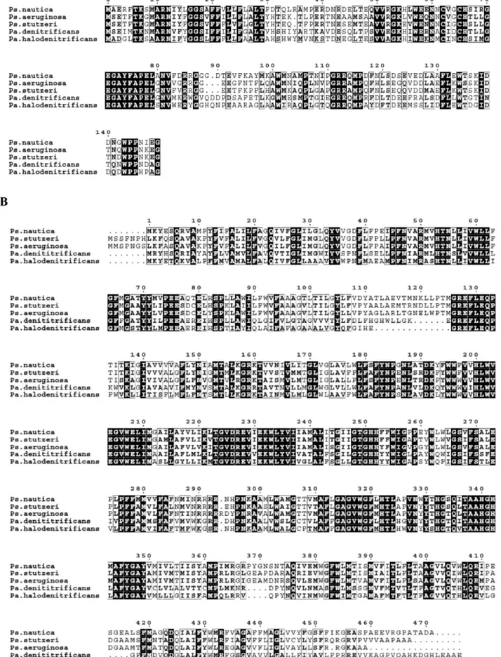
determined (see Figure S1, Supporting Information). Figure 1
compares thePs. nauticacNOR primary sequence, deduced from
the gene sequence, with those of other bacterial cNORs. The histidine residues that have been identified as ligands of the heme
iron and non-heme iron atoms (H53 and H342 for heme b;
H340 for heme b3; H200, 251, and 252 for FeB) and the
catalytically essential glutamate residues (E128, E131, E204,
E208, and E273)3841 are all conserved inPs. nauticacNOR.
In addition, two other histidine (H322 and H332) and five other glutamate residues (E75, E76, E80, E228, and E231) are also conserved. From the deduced amino acid composition and Fe
cofactor content (one hemecin NORC; two hemebmoieties
and a non-heme Fe in NORB), molecular masses of 17 590 and 54 380 Da were calculated respectively forPs. nauticaNORC and
NORB subunits. On SDS-PAGE gel, purifiedPs. nauticacNOR
shows two bands with apparent molecular masses of 17 kDa (NORC subunit) and 36 kDa (NORB subunit). The apparent molecular mass of NORC subunit determined from SDS-PAGE is similar to that calculated from its gene-coding primary sequence. The apparent molecular mass of NORB determined from SDS-PAGE is substantially different from that deduced from the amino acid composition, however. This discrepancy had been reported for other cNORs and was attributed to the
hydrophobic character of NORB.9,13,19 Heme staining
metho-dology revealed that only the low molecular weight band (i.e., NORC subunit) had a covalently ligated heme. A total iron
content of 3.5(0.5 Fe/cNOR molecule was obtained by plasma
emission analysis. Calcium content was determined to be 0.7(
0.2 Ca/cNOR. No other transition metal was detected. Values of
1.0(0.1 Fe and 1.7(0.2 Fe per NORB subunit were obtained
for non-heme iron and hemeb, respectively, while a value of
1.0(0.1 Fe/NORC subunit was obtained for hemec, consistent
UV/vis Absorption Spectra. The as-purified Ps. nautica cNOR, at pH 7, displays a UV/vis spectrum (Figure 2A, blue line) typical of heme-containing proteins and very similar to those of other purified cNORs. The spectrum shows a protein peak at 280 nm, a Soret band at 411 nm, and a broad band at around 550 nm with two small absorption peaks at 530 and 560 nm in theβandRregions, respectively. The appearance of
the small absorption peaks indicates the presence of a small
amount of reduced heme, in accord with the M€ossbauer data
presented below. The absorbance ratio of the Soret band at 411 nm to the protein peak at 280 nm was determined to be 1.4. Protein determinations by both Lowry and BCA methods revealed interference from detergent present in the buffer. Thus, protein quantifications derived from amino acid composition were used to estimate an extinction coefficient of 295 mM1cm1 at 411 nm for the oxidized cNOR. This value was then used to estimate protein concentration in all assays. It is important to
note that while cNORs isolated from Pa. denitrificans andH.
halodenitrificansexhibit an absorption band around 595 nm,19,42 such a band is absent from thePs. nauticacNOR. On the basis of a resonance Raman investigation, which suggests the presence of a μ-oxo group bridging hemeb3and FeBin the oxidized cNOR,22 the 595 nm band has been attributed to a ligand-to-metal charge transfer band associated with a high-spin ferric hemeb3without the proximal His ligand.43The absence of a 595 nm band in the as-purifiedPs. nauticacNOR therefore suggests a different spin
state and/or a different coordination environment for hemeb3 (see below). A low-intensity broad band at around 662 nm is observed but shows no redox dependence, indicating minor presence of inactive protein.
Reducing the enzyme with ascorbate red-shifts the maximum of the Soret band to 418 nm and sharpens the optical bands in the
R/βabsorption region with maxima at 524 and 552 nm and a
shoulder at 558 nm (Figure 2A, red line), consistent with reduction of low-spin heme groups. The absorption band at 552 nm has been assigned to hemecand the shoulder at 558 nm to hemeb.11,42,44Thus, the optical data indicate both hemeband
hemecare reduced by ascorbate. Upon reduction with sodium
dithionite, the Soret maximum is further shifted to 422 nm and theRandβbands become more intense, indicating reduction of
more heme groups (Figure 2A, green line). Figure 2B displays a difference spectrum (black line) between that of the dithionite-and ascorbate-reduced enzyme. On the basis of the following M€ossbauer data, which indicate that only hemeb3is oxidized in the ascorbate-reduced enzyme and that it can be reduced by
dithionite, this difference spectrum represents absorption
changes arising from the reduction of hemeb3 and indicates
clearly that the reduced hemeb3is low-spin. Reoxidation of the ascorbate- or dithionite-reduced enzyme produces a spectrum identical to that of the as-isolated enzyme, indicating reversible oxidationreduction processes. In the presence of PMS,
ascor-bate can reduce the enzyme to a redox state equivalent to that of the dithionite-reduced cNOR as judged by optical spectroscopy (Figure S2).
Optical Oxidation/Reduction Titrations.The UV/vis
spec-trum of the as-purified Ps. nautica cNOR showed no pH
dependence in the range of pH 59. To determine the redox
properties of the metallocofactors, redox potentiometric titra-tions monitored by optical absorptitra-tions were performed at two pH values 6 and 7. The resulting spectra as a function of the redox potential at pH 7 (Figure 3A) were globally fitted (SPECFIT, Spectrum Software Associates) and spectral components decon-voluted. Good fits of the data were obtained by using two independent Nernst equations of different midpoint redox potentials that differ by∼200 mV. At pH 7, the midpoint redox
potentials were determined to be EI = þ215 ( 10 mV and
EII=38(10 mV, and at pH 6,EI =þ232(10 mV and
EII=16(10 mV. To illustrate the goodness of our fits, in
Figure 3B, we plotted the absorbance at 551 nm as a function of redox potential and compared the experimental data (circles) with the theoretical values (solid line) calculated by using the parameters obtained from the fits. On the basis of the M€ossbauer and EPR data presented below,EIis assigned to both hemecand hemebandEIIis assigned to hemeb3. As no absorption features are attributed to FeB, the redox potential of FeBis not determined from this measurement. Optical redox titration has also been performed onPa. denitrificanscNOR.28Although the midpoint potentials determined for the heme groups inPa. denitrificans enzyme are about 100 mV higher than those reported here,
the midpoint redox potentials for hemec(310 mV) and hemeb
(345 mV) in thePa. denitrificansenzyme are also found to be
comparable and are more than 200 mV above that of hemeb3
(60 mV). In other words, the large redox potential difference
between the reactive hemeb3and the electron-transfer heme
groups, a functionally important factor, is conserved among cNOR’s.
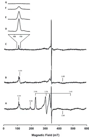
EPR Results. The as-purified Ps. nauticacNOR exhibits an EPR spectrum (Figure 4A) that is similar to those reported for Figure 2. (A) UV/vis spectra ofPs. nauticacNOR in 100 mM pH 7
KPB, 0.02% dodecyl maltoside, 0.01% PE (as-isolated, blue line; ascorbate-reduced, red line; dithionite-reduced, green line). (B) Difference
other cNOR’s.19,21,42Two sets of low-spin ferric heme signals are clearly visible. The prominent set of signals, withgvalues of 2.99, 2.26, and 1.43, have been attributed to the histidinehistidine
ligated hemebof the NORB subunit.21The highly anisotropic set of signals, from which only the gmax value of 3.60 can be
observed, have been attributed to the heme c of the NORC
subunit.45Spin quantification of these two low-spin ferric heme
signals were done using the Aasa and Vanngard method46
modified according to De Vries and Albracht,47 together with spectral simulation. Approximately 0.9 spin/molecule and
0.7 spin/molecule were determined for the hemeband heme
c signals, respectively. (In our spin quantification estimations, gmid= 1.43 andgmin= 1.00 were assumed for hemec.) In addition, a broad signal atg= 1.60, two derivative type signals atg= 2.01 and 2.05, and two overlapping signals at aroundg= 6.30 region are detected. The signals atg= 2.05 and 1.60, although not yet
assigned, are present in other purified enzymes such as Pa.
denitrificansandPs. aeruginosaenzymes.14,26The current assump-tion is that in the oxidized enzyme the catalytic center is
composed of a high-spin ferric heme b3 antiferromagnetically
coupled to a high-spin ferric FeB, resulting in a diamagneticS= 0 ground state that yields no EPR resonances. The signals at the g= 6.30 region have been assigned to a small quantity of decoupled
ferric high-spin hemeb3and the signal atg= 2.01 to FeBor an organic radical.19,42The M€ossbauer data, presented below, reveal that in the as-purifiedPs. nauticaNOR hemeb3is in fact low-spin. Consequently, a re-evaluation of the above assignment for signals at theg= 6.3 region is required because a decoupled ferric low-spin hemeb3would not produce signals at theg= 6 region.
Reduction ofPs. nauticacNOR with sodium ascorbate (1 mM)
leads to the disappearance of the ferric low-spin signals (g= 3.60, 2.99, 2.26, and 1.45), while the signals aroundg= 6.30, 2.05, 2.01, and 1.60 are retained (Figure 4B). The disappearance of the ferric low-spin signals is consistent with the optical data (presented
above) and the M€ossbauer results (presented below), which
show that hemecand hemebcan be readily reduced by ascorbate to diamagnetic low-spin FeIIstates.
Further reduction with sodium dithionite eliminates the derivative-type EPR signal at theg= 6 region, but the sharp absorption-type signal at g = 6.34 is still clearly detectable (Figure 4C). Temperature dependence of this signal is charac-teristic of an electronic ground state system (Figure 4DG). The
M€ossbauer data (presented below) show that in the ascorbate-and dithionite-reduced enzyme significant portions of FeB and Figure 3. (A) UV/vis spectra ofPs. nauticacNOR as a function of redox
potential at pH 7. (B) Absorbance at 551 nm as a function of redox potential at pH 7 (100 mM pH 7 KPB, 0.02% dodecyl maltoside, 0.01% PE). The solid line is the result calculated by using the parameters obtained from globallyfitting the spectra shown in (A).
Figure 4. EPR spectra at 9.653 GHz of the as-isolated (A), ascorbate-reduced (B), and dithionite-ascorbate-reduced (C)Ps. nauticacNOR (265μM in 100 mM KPB, pH 7, 0.02% DDM, 0.01% PE). Experimental conditions of spectra AC: temperature = 12 K, microwave power = 0.2 mW,
modulation frequency = 100 kHz, modulation amplitude = 0.5 mT, receiver gain = 1105, conversion time = 163.84 ms, and time constant
= 81.92 ms and of spectra D and E: microwave power = 6.3 mW, modulation frequency = 100 kHz, modulation amplitude = 0.4 mT, receiver gain = 2105, conversion time = 81.92 ms, and time constant =
hemeb3remain in their oxidized states. It is therefore possible that the g= 6.34 signal could be associated with the oxidized hemeb3-FeBcenter.
Mossbauer Characterization.€ M€ossbauer spectroscopy was used to further characterize the electronic states of the Fe centers and to provide direct and quantitative measurements on the compositions of the oxidation states of the Fe centers in various
redox forms of the enzyme. Figure 5 shows the M€ossbauer
spectra of the as-purified (A), ascorbate-reduced (B), and dithio-nite-reduced (C) samples recorded at 180 K in a 50 mT magnetic field applied parallel to the γ-radiation. At this temperature, the electronic relaxation of Fe centers in proteins are generally fast in comparison with the57Fe nuclear precession, causing cancellation of the internal magnetic field at the 57Fe nucleus and resulting in quadrupole doublets for the Fe centers. Thus, these spectra are composed of only quadrupole doublets arising from different Fe species, facilitating the analysis of the spectra. On the other hand, there are eight possible redox states for the four Fe sites in cNOR. Consequently, analysis of these spectra remains challenging, and a global analysis including all three spectra is applied.
First, the absorption peak at∼2.5 mm/s (indicated by a dotted
line) can be assigned to the high-energy line of the quadrupole doublet arising from the high-spin ferrous FeB. Since this peak is well resolved from the rest of the absorptions, it allows for a reliable quantification on the amount of reduced Fe
Bin the sample. It is therefore interesting to note that all three forms of the enzyme contain detectable amounts of reduced FeB, includ-ing the as-purified sample. Second, the most prominent feature observed in the spectrum of the as-purified sample (Figure 5A) is a broad quadrupole doublet (line positions indicated by two
vertical lines) with apparent parameters (ΔEQ∼2 mm/s and
δ∼0.2 mm/s) that are indicative ofS= 1/2 low-spin ferric heme
species. A rough estimate of its absorption area indicates that this doublet accounts for∼70% of the total Fe in the sample. Since
hemeband hemeccan contribute up to a maximum of only 50%, the remaining contribution has to arise from hemeb3. In other words,the oxidized heme b3in Ps. nautica cNOR is a low-spin ferric
heme, in contrast to the high-spin ferric states reported for heme
b3 of cNOR’s from other organisms.42,43 Upon addition of
sodium ascorbate, the intensity of this low-spin ferricheme
doublet decreases to about 25% of the total Fe absorption, and a new doublet (indicated by two vertical dashed lines), accounting
for∼50% of the Fe absorption, is detected (Figure 5B). The
apparent parameters for this doublet (ΔEQ ∼1.1 mm/s and
δ∼0.4 mm/s) are characteristics for low-spin ferrous hemes. Taking into consideration the midpoint redox potentials deter-mined for the three heme groups (see above), only the electron-transfer hemecand hemebare reducible by ascorbate. Thus, this new doublet is assigned to the reduced hemeband hemec. With this assignment, it becomes obvious that in the spectrum of the as-purified enzyme (Figure 5A) there are absorptions arising
from these reduced hemes. In other words, hemeband hemec
are partially reduced in the as-purified enzyme, in agreement with the optical data described above. Observation of partial reduction of hemeband hemecin the as-purified enzyme is consistent with
the high midpoint redox potential (215230 mV) determined
for these heme groups. Addition of sodium dithionite further decreases the intensity of the low-spin ferric heme doublet and increases the intensity of the low-spin ferrous heme doublet (Figure 5C), indicating that the active site hemeb3is partially
reduced by dithionite and that the reduced heme b3 is also
low-spin.
With the above understanding, it is possible to analyze these three spectra simultaneously using one set of parameters. To reduce the number of variable parameters, the following rational assumptions were made: (1) the total absorption intensity (oxidized plus reduced) for each one of the four Fe sites is 1/4 of the total Fe absorption, (2) the line widths of a quadrupole doublet arising from a heme group are the same, and (3) for each sample, the [oxidized]/[reduced] ratio of hemeband hemecis the same. (This assumption is based on the observed similar
midpoint redox potentials for hemeband hemec; see above.)
During the analysis, we also realized that the electronic relaxation rate of ferric FeBis not much faster than the57Fe precession rate, resulting in a broad and featureless spectrum. A broad quadru-pole doublet was therefore used to approximate the spectrum of ferric FeB. The results of the least-squaresfit are presented in Figure 5 and Table 1. The parameters obtained are consistent with high-spin states for the oxidized and reduced FeB, as expected for most non-heme Fe cofactors in proteins, and low-spin states for all three heme groups regardless of their redox states, suggesting 6-coordinate environments for both ferric and Figure 5. M€ossbauer spectra of the as-purified (A), ascorbate-reduced
(B), and dithionite-reduced (C)Ps. nauticacNOR (805μM in 100 mM KPB, pH 7, 0.02% DDM, 0.01% PE). The spectra (vertical bars) were collected at 180 K in a weak magneticfield of 50 mT. The color lines
displayed above the data are the deconvoluded spectra for the for Fe cofactors in the enzyme: FeBIII, orange; FeBII, green; FeIII-hemeb3, red;
FeII-hemeb
3, blue; FeIII-hemeb, magenta; FeII-hemeb, cyan; FeIII-heme
c, purple; FeII-hemec, blue-green. The back solid lines overlaid with the
experimental spectra are composite spectra simulated with the para-meters and percent absorptions listed in Table 1. The dotted vertical line indicates the position of the high-energy line of the FeBIIdoublet. The
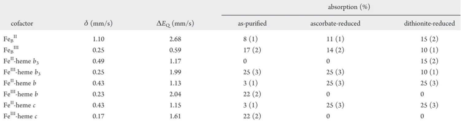
ferrous hemes. The M€ossbauer percent absorptions determined for the quadrupole doublets (Table 1) provide a quantitative assessment of the redox status of all four Fe cofactors in the three different oxidation states of the enzyme: hemeband hemecare partially reduced (3/25 = 12%) in the as-purified enzymes and
can be completely reduced by ascorbate. Heme b3 is fully
oxidized in the as-purified enzyme and cannot be reduced by
ascorbate alone. This observation is consistent with the 200 mV redox potential difference detected between hemeb3and the two electron transfer hemeband hemec. In the as-purified enzyme, FeB is significantly reduced (∼30% reduction), and yet in the dithionite-reduced enzyme, it is only partially reduced. The level of reduction (60%), interestingly, is identical to that of hemeb3, suggesting the presence of interactions between FeBand hemeb3 which could complicate their redox behaviors. The M€ossbauer
results, taken together with results obtained from enzymatic activity studies (presented below), provide information for determining the“active”redox-state of the enzyme, information that is essential for understanding enzyme mechanism.
Enzymatic Activities.As a reference, specific activity of the as-purified enzyme (Table 2) was first determined by using the established assay developed by Girsh et al.,19in which the assay
solution contains 10 mM sodium ascorbate, 100μM PMS, and
20μM horse heart cytochromecin 20 mM KPB at pH 6. The
specific activity values obtained are comparable to reported values forPa. denitrificansandPs. aeruginosaenzymes at this pH.28,51
Examinations on the effects of various electron donors
re-vealed that horse heart cytochrome c had no effect on the
enzymatic activity and was ineffective as an electron donor. Presence of ascorbate and PMS in the assay solution was sufficient to produce full activity of the enzyme. Interestingly, Ps. nauticacytochromec552was found to be an effective electron donor and could produce fully active enzymes (Table 2). In an effort to obtain further evidence to supportc552as the physio-logical electron donor forPs. nauticacNOR, a set of enzymatic assays was performed with six different low-spin cytochromes obtained from Ps. nautica (cytochromes c551, c552, c459, c553, c553(548) and membrane bound cytochrome c4).31,32,48 In this series of experiments, due to limits in the amounts of some of the purified cytochromes, the experiments were carried out with 3 μM of cytochromes. Only the periplasmatic cythocromec552was found to show a significant value of specific activity. All other cytochromes were ineffective in providing electrons toPs. nautica cNOR (last six rows, Table 2).
It is interesting to note that the midpoint redox potential ofPs. nautica c552is aboutþ250 mV at pH 7.6,32which is more than
200 mV higher than the reduction potential for hemeb3. From a purely thermodynamical consideration, this suggests that c552 may not be a good electron donor for cNOR. But, as presented above, we have shown thatc552is the only small cytochrome that is capable to catalyze the reduction of NO. In fact, the same concern can also be raised for hemecand hemebbecause their midpoint redox potentials are also much higher than that of heme b3. Taking these observations into consideration, we suggest that the initial reductive activation of hemeb3 and the transfer of electrons from cytochromec552, hemeb, and hemecto the diiron
Table 1. M€ossbauer Parameters and Percent Absorptions of Fe Cofactors in the As-Purified, Ascorbate-Reduced, and Dithionite-Reduced cNOR fromPs. nauticaa
absorption (%)
cofactor δ(mm/s) ΔEQ(mm/s) as-purified ascorbate-reduced dithionite-reduced
FeBII 1.10 2.68 8 (1) 11 (1) 15 (2)
FeBIII 0.25 0.59 17 (2) 14 (2) 10 (1)
FeII-hemeb
3 0.49 1.17 0 0 15 (2)
FeIII-hemeb
3 0.25 1.99 25 (3) 25 (3) 10 (1)
FeII-hemeb 0.43 1.13 3 (1) 25 (3) 25 (3)
FeIII-hemeb 0.23 2.04 22 (2) 0 0
FeII-hemec 0.43 1.15 3 (1) 25 (3) 25 (3)
FeIII-hemec 0.17 1.61 22 (2) 0 0
a
The values in parentheses give the uncertainties in the last significant digits estimated for the percent absorptions. Because of the complexity of the
spectra, the M€ossbauer parameters (δandΔEQ) werefixed during the least-squaresfittings. The line widths of the quadrupole doublets used in ourfits
are 0.28 mm/s for FeII-hemeband FeII-hemec, 0.4 mm/s (left line) and 0.3 mm/s (right line) for FeIII-hemeb, 0.3 mm/s (left) and 0.5 mm/s (right) for FeIII-hemec, 0.28 mm/s (left) and 0.35 mm/s (right) for FeII-hemeb3, 0.36 mm/s (left) and 0.43 mm/s (right) for FeIII-hemeb3, 0.35 mm/s (left) and
0.3 mm/s (right) for FeBII.
Table 2. Specific Activity ofPs. nauticacNOR as a Function of Electron Donor and Assay Conditionsa
electron donor
specific activity (μmol min1mg1)
none
ascorbateþPMSþoxidized horse heart cyt.c 3.8
ascorbateþPMS 4.3
PMSþreduced horse heart cyt.c 0.7
ascorbate 0.7
PMS <0.1
reduced horse heart cyt.c <0.1 reducedPs. nauticacyt.c552 3.5 reducedPs. nauticacyt.c552 0.6b reducedPs. nauticacyt.c549 <0.1b reducedPs. nauticacyt.c551 <0.1b reducedPs. nauticacyt.c553 <0.1b reducedPs. nauticacyt.c553(548) <0.1b reducedPs. nauticacyt.c4 <0.1b aErrors between duplicates are approximately(20%. Concentrations
used in the assay solutions are [cNOR] = 70 nM, [ascorbate] = 10 mM, [PMS] = 100μM, and [cyt.] = 20μM.b[cyt.] = 3μM in the assay
active site for NO reduction are separate events that could have distinct electron transfer pathways.
’DISCUSSION
Spin State of Hemeb3.We have purifiedPs. nauticacNOR to
homogeneity and characterized the purified enzyme in detail by using biochemical and spectroscopic methods. The purified enzyme exhibits an optical spectrum with an absorbance ratio ofA411/A280= 1.4, one of the highest purity preparations ever reported for cNOR. Heme and Fe content determinations show
a cofactor stoichiometry of 1:2:1 for hemec:hemeb:non-heme
iron per molecule of cNOR, indicating full occupancy of metal
centers in the purified enzyme. Regarding metal content, Ps.
nauticacNOR was found to contain Ca. While the value obtained is substoichiometric, the presence of calcium was also observed in the X-ray crystallographic structure of thePs. aeruginosaenzyme,51 indicating that Ca presence might be a common feature between anaerobic and microaerobic respiratory enzymes. As shown in Figure 1, arginine residues 50 and 433 (Ps. nauticanumbering) are conserved in the fivePseudomonasandParacoccussequences. These residues are also present in Ca binding incbb3cytochrome oxidase fromPs. stutzeri.59
Consistent with results reported for cNORs isolated from other organisms, the spectroscopic data indicate that the spin states of the electron transfer hemecand hemebare low-spin, and for the non-heme iron FeB, it is high-spin. Contrary to
the conventionally assumed high-spin state for heme b3, the
M€ossbauer data revealed that the spin state of the catalytic heme b3is in fact low-spin. The UV/vis difference spectrum between the spectra of ascorbate-reduced and dithionite-reduced enzyme (Figure 2B), which displays the difference arising from changes in the oxidation state of hemeb3, supports the low-spin designation and indicates further that at room temperature the reduced heme b3remains low-spin. In support of ourfinding, a recent X-ray crystallographic structure of thePs. aeruginosaenzyme, published online by Hino and co-workers51while this manuscript was in the
reviewing process, shows that heme b3 is indeed
hexacoordi-nated. It is axialy coordinated to a His residue (H347,Ps. stutzeri numbering) and aμ-oxo (or a possibleμ-hydroxo) bridge to FeB. On the basis of temperature-dependent MCD and low-tempera-ture EPR data, Cheesman et al.21concluded that the spin state of hemeb3in oxidizedPs. stutzericNOR underwent a spin transi-tion from high spin at room temperature to low spin at 4.2 K. Our spectroscopic analysis clearly shows that hemeb3is low-spin in the ferric state between 4.2 and 180 K and in the ferrous active form from 4.2 K up to room temperature.
Reconsideration of the g = 6.34 Assignment. In the oxidized enzyme, the ferric hemeb3is bridging to the ferric FeBvia an oxo group.22,51Prior to this investigation, hemeb3 was assumed to be high-spin ferric42,45antiferromagnetically
coupled to the ferric high-spin (S = 5/2) FeB, forming a
diamagnetic (S= 0) ground state. Thus, no EPR signal was
expected to arise from the oxidized catalytic diiron center. The
signals at the g = 6 region were then assigned to a small
quantity of uncoupled heme b3. Also, previously, a g = 4.3
signal was detected and had been assigned to a minor quantity
of the uncoupled ferric FeB. For the as-purified Ps. nautica
cNOR, however, thisg= 4.3 signal is almost nonobservable
(Figure 4A). We therefore proposed that the previously
detected g = 4.3 signal may not arise from an uncoupled
FeBbut, rather, represent minor adventitiously bound ferric
impurity, which is not present in our as-purifiedPs. nautica cNOR.
Here, we present spectroscopic data establishing unambigu-ously that hemeb3is low-spin at low temperature (4.2180 K).
Spinspin coupling of a low-spin ferric (S= 1/2) hemeb3with a
high-spin ferric (S= 5/2) FeBwould yield an integer-spin ground state withS= 2 orS= 3 that may not be EPR silent.49On the basis of the following observations, we propose that theg= 6.34 signal may arise from the oxidized binuclear hemeb3-FeBsite. (1) The absorption-type signal detected atg= 6.34 for cNORs is distinct from the derivative-typeg= 6 signal expected for a high-spin ferric heme (Figure 4DG). (2) In the as-purified enzyme, there are four Fe cofactors that could be detected by EPR. The
electron-transfer ferric low-spin hemec and hemebare
com-pletely accounted for by the two sets of low-spin ferric EPR signals. Thus, theg= 6.34 signal has to be arising from either hemeb3or FeBor both. Because an isolated low-spin ferric heme
b3 cannot yield signals at the g = 6 region and because no
additional signals are detected that may be assigned to an isolated low-spin ferric hemeb3or to an isolated ferric high-spin FeB, a probable explanation would be that hemeb3and FeBare spin coupled to form an integer-spin system that yields theg= 6.34 signal. And, (3) the g = 6.34 signal is detected in all three oxidation states of the enzyme (as purified, ascorbate-reduced, and ditionite-reduced). According to the M€ossbauer data (Table 1), the only common oxidation state for all the Fe cofactors that is present in all three oxidation states of the enzyme and may be
detectable by EPR is the FeIIIheme b
3FeBIII site. Taken together, these observations argue favorably for the g = 6.34 signal to arise from a spin-coupled oxidized hemeb3FeBcenter. A definitive assignment for theg= 6.34 signal and a complete
understanding of the nature of the proposed heme b3FeB
spinspin coupling, however, will require further detailed
spec-troscopic investigations, such as parallel mode EPR studies coupled with low-temperature-high-field M€ossbauer measure-ments. Finally, it is important to point out that theg= 6.34 signal
have been observed for all as-purified cNORs that have been
characterized by EPR. Consequently, if the g= 6.34 signal is indeed arising from a spin-coupled binuclear center comprising a low-spin ferric hemeb3and a high-spin ferric FeB, then hemeb3is most likely low-spin in all cNORs in the experimental conditions studied.
Catalytic Site Coordination. In general, high-spin heme complexes are five-coordinated, while low-spin heme
com-plexes are six-coordinated, except for the low-spin [FeNO]7
heme complexes, which can be either five- or six-coordinated.
The fact that hemeb3 is low-spin in both its oxidized and
reduced states implies that it is six-coordinated, rather than five-coordinated as previously assumed. On the basis of amino acid sequence analysis and spectroscopic data comparison, we
believe that H340 (Ps. nauticanumbering) and aμ-oxo bridge
to FeB(for ferric hemeb3states) or a hydroxide anion (in the fully reduced enzyme) are responsible for the observed low-spin state of hemeb3. The presence of a sixth ligand for heme
b3 is consistent with fast kinetic measurements for CO
binding,60where it was found that the initial binding of CO
to FeIIheme b3 is ∼3 orders of magnitude slower than
photolysis-induced CO recombination, presumably reflecting the need to displace a blocking axial ligand. By comparison
with the known ligands of FeB in Ps. aeruginosa NOR, we
Redox State of the Active Enzyme.An important informa-tion that is absolutely required for understanding enzyme mechanism is the redox state of the active enzyme. For cNOR, both the three-electron reduced state (hemeb, hemec, and FeB are reduced while hemeb3is oxidized)28and the fully reduced state14,19,50have been proposed to be the active redox state. By exploiting the large midpoint redox potential differences between
heme b3 and the electron transfer heme c and heme b, we
attempted to address this important question by preparing a stable three-electron-reduced enzyme for activity assay. We reduced the as-purified enzyme by using a mild reductant, ascor-bate, and characterized the oxidation states of the cofactors by using M€ossbauer spectroscopy. The results show that hemeband hemecare completely reduced while hemeb3remains oxidized (Table 1). Approximately half of FeBis reduced. Activity assay indicates that the ascorbate-reduced cNOR is inactive (Table 2). The PMS and ascorbate (that have been used by several authors to reduce the enzyme in the activity assay) has been proved by UV/vis spectroscopy to yield a redox state similar to the one achieved by dithionite reduction (Figure S2). We therefore conclude that hemeb3has to be reduced for the enzyme to be active and that the fully reduced state is likely the active state of the enzyme.
Mechanistic Consideration.Currently, there are two
pro-posed mechanisms for NO reduction by cNOR: the cis- and
trans-mechanism (see Introduction). A major distinction be-tween these two mechanisms is at the initial step of coordinating two NO molecules at the binuclear center. In thecis-mechanism, both NO molecules are coordinated either to the reduced FeBor to hemeb3forming a FeIIdinitrosyl complex.28 In the trans-mechanism, one NO molecule is bound to each reduced Fe site
of the binuclear center forming a [FeNO]72 complex.14 The
cis-mechanism with two NO molecules bound to a reduced hemeb3has been questioned based on the fact that these types of complexes are too stable for turnover.
To prevent the binding of NO to a FeIIhemeb3forming a
stable [FeNO]7heme complex in the initial NO binding step, it
has been proposed that the three-electron reduced state with an
oxidized heme b3 is the active state of the enzyme.28 Our
conclusions that the fully reduced state is the active form of
the enzyme and that heme b3 is six-coordinated suggest an
alternative possibility that favors NO binding to FeBin the initial catalytic step: The presence of a sixth ligand weakens the affinity
of NO binding to heme b3 and promotes the formation of a
FeIIdinitrosyl complex at FeB. In such a scenario, the initial
function of heme b3 could be (1) to assist in adjusting the
orientation of the FeB-bound NO molecules and subsequent
formation of the NN bond and (2) to provide the essential
electron for the reduction of NO. Without further kinetic data, we cannot exclude that at a later mechanistic step hemeb3will somehow bind to NO (or to a reaction intermediate). Recently,
Lachmann and co-workers52 demonstrated that CO binds to
reduced hemeb3(probably displacing the sixth ligand initially
present) and that flash-induced dissociation causes heme b3
oxidation and catalysis. It is also known that for the CO-bound enzyme NO can still bind to the FeBsite.14Both observations support the possibility of an initial binding of NO to FeB. It is
important to comment that we believe that thecis-mechanism
(with ligation of NO to FeB) is energetically favorable. The NO dissociation rate constants for reducedb-type ferrous porphyrin complexes are in the order of 103105s1, demonstrating the
stability of this type of nitrosyl complex.5356To our knowledge
very few hemic proteins form unstable/transient nitrosyl com-plexes: NO synthase, cytochromecd1-type nitrite reductase, and cytochrome P450 all exhibit fast NO dissociation rate constants (2100 s1). One should mention that even with NO synthase,
an enzyme that catalyzes the formation of NO, the release of NO is only productive from a FeIIIhemeNO catalytic
intermedi-ate and very slow from the reduced FeIIhemeNO form.57In
the case of cytochromecd1 nitrite reductase from denitrifying bacteria the fast NO dissociation from the ferrous heme form is thought to be associated with the involvement of hemed1. Fungal NOR, a member of the cytochrome P450 family, contains a heme
bwith axial cysteinate coordination. The NO substrate binds
to the ferric state of heme b, leading to the formation of a
6-coordinated ferrichemenitrosyl first intermediate. The
FeIIINO complex is unstable, and the dissociation of NO is
due to the presence of the cysteinyl ligand and to the ferric state of the heme, such as in NO synthase.
It is worth noting thatPs. nauticaNOR is also able to reduce molecular oxygen.58 Although it is not established how wide-spread such bifunctionality is among cNOR, the fact that it was not reported for all characterized enzymes might be an indication that an alternative mechanism cannot be excluded.
’ASSOCIATED CONTENT
b
S Supporting Information. Figure S1 showing the DNA and deduced amino acid sequence ofPs. nauticaNOR and Figure S2 comparing the UV/vis spectrum of dithionite-reduced cNOR with that of the enzyme reduced by ascorbate plus PMS. This material is available free of charge via the Internet at http:// pubs.acs.org.’AUTHOR INFORMATION
Corresponding Author
*E-mail: [email protected] (A.S.P.), [email protected] (I.M.). Phone: 351-212948345. Fax: 351-212948550.
Funding Sources
This work was supported in part by funds from PDTC/QUI/ 64638/2006 grant (I.M.), SFRH/BPD/14863/2003 (C.G.T.), SFRH/BD/17840/2004 (C.E.M.), SFRH/BD/39009/2007 (A.G.D.), and National Institutes of Health grant GM 47295 (B.H.H.).
’ACKNOWLEDGMENT
We thank Marcia Guilherme for her help in the purification of
Ps. nauticaNOR.
’ABBREVIATIONS
NOR, nitric oxide reductase; EPR, electron paramagnetic
reso-nance; PMSF, phenylmethylsulfonylfluoride; DDM, sodium
n-dodecylR-D-maltoside; DEAE, diethylaminoethyl cellulose;
’REFERENCES
(1) Bonin, P., Bertrand, J. C., Giordano, G., and Gilewicz, M. (1987) Specific sodium dependence of a nitrate reductase in a marine bacterium.
FEMS Microbiol. Lett. 48, 5–9.
(2) Bonin, P., Gilewicz, M., Denis, M., and Bertrand, J. C. (1989)Salt requirements in the denitrifying bacteriumPseudomonas nautica617. Res. Microbiol. 140, 159–169.
(3) Tavares, P., Pereira, A. S., Moura, J. J., and Moura, I. (2006) Metalloenzymes of the denitrification pathway.J. Inorg. Biochem. 100
2087–2100.
(4) Brown, K., Djinovic-Carugo, K., Haltia, T., Cabrito, I., Saraste, M., Moura, J. J., Moura, I., Tegoni, M., and Cambillau, C. (2000) Revisiting the catalytic CuZ cluster of nitrous oxide (N2O) reductase.
Evidence of a bridging inorganic sulfur.J. Biol. Chem. 275, 41133–41136. (5) Lopes, H., Besson, S., Moura, I., and Moura, J. J. (2001)Kinetics of inter- and intramolecular electron transfer ofPseudomonas nautica cytochrome cd1 nitrite reductase: regulation of the NO-bound end
product.J. Biol. Inorg. Chem. 6, 55–62.
(6) Brown, K., Tegoni, M., Prudencio, M., Pereira, A. S., Besson, S., Moura, J. J., Moura, I., and Cambillau, C. (2000)A novel type of catalytic copper cluster in nitrous oxide reductase.Nat. Struct. Biol. 7, 191–195. (7) Correia, C., Besson, S., Brondino, C. D., Gonzalez, P. J., Fauque, G., Lampreia, J., Moura, I., and Moura, J. J. (2008)Biochemical and spectroscopic characterization of the membrane-bound nitrate reduc-tase fromMarinobacter hydrocarbonoclasticus617.J. Biol. Inorg. Chem. 13, 1321–1333.
(8) Heiss, B., Frunzke, K., and Zumft, W. G. (1989)Formation of the N-N bond from nitric oxide by a membrane-bound cytochrome bc complex of nitrate-respiring (denitrifying)Pseudomonas stutzeri.J. Bacteriol. 171, 3288–3297.
(9) Kastrau, D. H., Heiss, B., Kroneck, P. M., and Zumft, W. G. (1994)Nitric oxide reductase fromPseudomonas stutzeri, a novel cyto-chromebccomplex. Phospholipid requirement, electron paramagnetic resonance and redox properties.Eur. J. Biochem. 222, 293–303.
(10) Hoglen, J., and Hollocher, T. C. (1989)Purification and some
characteristics of nitric oxide reductase-containing vesicles from Para-coccus denitrificans.J. Biol. Chem. 264, 7556–7563.
(11) Carr, G. J., and Ferguson, S. J. (1990)The nitric oxide reductase ofParacoccus denitrificans.Biochem. J. 269, 423–429.
(12) Sakurai, T., Nakashima, S., Kataoka, K., Seo, D., and Sakurai, N. (2005)Diverse NO reduction byHalomonas halodenitrificansnitric oxide reductase.Biochem. Biophys. Res. Commun. 333, 483–487.
(13) Sakurai, T., Sakurai, N., Matsumoto, H., Hirota, S., and Yamauchi, O. (1998)Roles of four iron centers inParacoccus halodenitrificansnitric oxide reductase.Biochem. Biophys. Res. Commun. 251, 248–251.
(14) Kumita, H., Matsuura, K., Hino, T., Takahashi, S., Hori, H., Fukumori, Y., Morishima, I., and Shiro, Y. (2004)NO reduction by nitric-oxide reductase from denitrifying bacteriumPseudomonas aerugi-nosa: characterization of reaction intermediates that appear in the single turnover cycle.J. Biol. Chem. 279, 55247–55254.
(15) Cramm, R., Pohlmann, A., and Friedrich, B. (1999)Purification
and characterization of the single-component nitric oxide reductase fromRalstonia eutrophaH16.FEBS Lett. 460, 6–10.
(16) Hendriks, J., Oubrie, A., Castresana, J., Urbani, A., Gemeinhardt, S., and Saraste, M. (2000)Nitric oxide reductases in bacteria.Biochim. Biophys. Acta 1459, 266–273.
(17) Householder, T. C., Fozo, E. M., Cardinale, J. A., and Clark, V. L. (2000)Gonococcalnitric oxide reductase is encoded by a single gene,norB, which is required for anaerobic growth and is induced by nitric oxide.Infect. Immun.(68), 5241–5246.
(18) Suharti, Heering, H. A., and de Vries, S. (2004)NO reductase fromBacillus azotoformansis a bifunctional enzyme accepting electrons from menaquinol and a specific endogenous membrane-bound
cyto-chromec551.Biochemistry 43, 13487–13495.
(19) Girsch, P., and de Vries, S. (1997)Purification and initial kinetic
and spectroscopic characterization of NO reductase fromParacoccus denitrificans.Biochim. Biophys. Acta 1318, 202–216.
(20) Hendriks, J., Warne, A., Gohlke, U., Haltia, T., Ludovici, C., Lubben, M., and Saraste, M. (1998)The active site of the bacterial nitric oxide reductase is a dinuclear iron center.Biochemistry 37, 13102–13109. (21) Cheesman, M. R., Zumft, W. G., and Thomson, A. J. (1998)The MCD and EPR of the heme centers of nitric oxide reductase from Pseudomonas stutzeri: Evidence that the enzyme is structurally related to the heme-copper oxidases.Biochemistry 37, 3994–4000.
(22) Moenne-Loccoz, P., Richter, O. M. H., Huang, H. W., Wasser, I. M., Ghiladi, R. A., Karlin, K. D., and de Vries, S. (2000)Nitric oxide reductase fromParacoccus denitrificanscontains an oxo-bridged heme/ non-heme diiron center.J. Am. Chem. Soc. 122(38), 9344–9345.
(23) Moenne-Loccoz, P. (2007)Spectroscopic characterization of heme iron-nitrosyl species and their role in NO reductase mechanisms in diiron proteins.Nat. Prod. Rep. 24(3), 610–620.
(24) Zumft, W. G. (2005)Nitric oxide reductases of prokaryotes with emphasis on the respiratory, heme-copper oxidase type.J. Inorg. Biochem. 99, 194–215.
(25) Watmough, N. J., Field, S. J., Hughes, R. J., and Richardson, D. J. (2009)The bacterial respiratory nitric oxide reductase. Biochem. Soc. Trans. 37, 392–399.
(26) Field, S. J., Thorndycroft, F. H., Matorin, A. D., Richardson, D. J., and Watmough, N. J. (2008)The respiratory nitric oxide reductase (NorBC) fromParacoccus denitrificans.Methods Enzymol. 437, 79–101. (27) Collman, J. P., Dey, A., Yang, Y., Decreau, R. A., Ohta, T., and Solomon, E. I. (2008)Intermediates Involved in the Two Electron Reduc-tion of NO to N(2)O by a FuncReduc-tional Synthetic Model of Heme Containing Bacterial NO Reductase.J. Am. Chem. Soc. 130, 16498–16499. (28) Gronberg, K. L., Roldan, M. D., Prior, L., Butland, G., Cheesman, M. R., Richardson, D. J., Spiro, S., Thomson, A. J., and Watmough, N. J. (1999)A low-redox potential heme in the dinuclear center of bacterial nitric oxide reductase: implications for the evolution of energy-conserving heme-copper oxidases.Biochemistry 38, 13780–13786.
(29) Gronberg, K. L., Watmough, N. J., Thomson, A. J., Richardson, D. J., and Field, S. J. (2004)Redox-dependent open and closed forms of the active site of the bacterial respiratory nitric-oxide reductase revealed by cyanide binding studies.J. Biol. Chem. 279, 17120–17125.
(30) Schagger, H., and von Jagow, G. (1987)Tricine-sodium dodecyl sulfate-polyacrylamide gel electrophoresis for the separation of proteins in the range from 1 to 100 kDa.Anal. Biochem. 166, 368–379.
(31) Saraiva, L. M., Besson, S., Moura, I., and Fauque, G. (1995) Purification and preliminary characterization of three c-type
cyto-chromes fromPseudomonas nauticastrain 617. Biochem. Biophys. Res.
Commun. 212, 1088–1097.
(32) Saraiva, L. M., Fauque, G., Besson, S., and Moura, I. (1994) Physico-chemical and spectroscopic properties of the monohemic cytochrome C552 from Pseudomonas nautica 617. Eur. J. Biochem.
224, 1011–1017.
(33) Lowry, O. H., Rosebrough, N. J., Farr, A. L., and Randall, R. J. (1951)Protein Measurement with the Folin Phenol Reagent.J. Biol. Chem. 193, 265–275.
(34) Berry, E. A., and Trumpower, B. L. (1987)Simultaneous determination of hemesa,b, andcfrom pyridine hemochrome spectra. Anal. Biochem. 161, 1–15.
(35) Fischer, D. S., and Price, D. C. (1964)A simple serum iron method using the new sensitive chromogen tripyridyl-S-triazine.Clin. Chem. 10, 21–31.
(36) Moore, S., and Stein, W. H. (1963)Chromatographic determi-nation of amino acids by the use of automatic recording equipment. Methods Enzymol. 6, 819–831.
(37) Ausubel, F. M., Brent, R., Kingston, R. E., Seidman, J. G., Smith, J. A., and Struhl, K., (Eds.) (1989)Current Protocols in Molecular Biology, Greene Pub. Associates and Wiley-Interscience, New York.
(38) Butland, G., Spiro, S., Watmough, N. J., and Richardson, D. J. (2001)Two conserved glutamates in the bacterial nitric oxide reductase are essential for activity but not assembly of the enzyme.J. Bacteriol. 183, 189–199.
entry point in the bacterial respiratory nitric-oxide reductase. J. Biol. Chem. 283, 3839–3845.
(40) Thorndycroft, F. H., Butland, G., Richardson, D. J., and Watmough, N. J. (2007)A new assay for nitric oxide reductase reveals two conserved glutamate residues form the entrance to a proton-conducting channel in the bacterial enzyme.Biochem. J. 401, 111–119.
(41) Flock, U., Lachmann, P., Reimann, J., Watmough, N. J., and
::
Adelroth, P. (2009)Exploring the terminal region of the proton pathway in the bacterial nitric oxide reductiase.J. Inorg. Biochem. 103, 845–850. (42) Sakurai, N., and Sakurai, T. (1997)Isolation and characteriza-tion of nitric oxide reductase fromParacoccus halodenitrificans. Biochem-istry 36, 13809–13815.
(43) Field, S. J., Prior, L., Roldan, M. D., Cheesman, M. R., Thom-son, A. J., Spiro, S., Butt, J. N., Watmought, N. J., and RichardThom-son, D. J. (2002)Spectral properties of bacterial nitric-oxide reductase - Resolu-tion of pH-dependent forms of the active site hemeb(3).J. Biol. Chem. 277(23), 20146–20150.
(44) Fujiwara, T., and Fukumori, Y. (1996)Cytochrome cb-type nitric oxide reductase with cytochromecoxidase activity fromParacoccus denitrificansATCC 35512.J. Bacteriol. 178, 1866–1871.
(45) Oubrie, A., Gemeinhardt, S., Field, S., Marritt, S., Thomson, A. J., Saraste, M., and Richardson, D. J. (2002)Properties of a soluble domain of subunitCof a bacterial nitric oxide reductase.Biochemistry 41, 10858–10865.
(46) Aasa, R., and Vanngard, T. (1975)EPR signal intensity and powder shapes: a reexamination.J. Magn. Reson. 19, 308–315.
(47) De Vries, S., and Albracht, S. P. J. (1979)Intensity of highly anisotropic low-spin heme EPR signals.Biochim. Biophys. Acta 546, 334–340. (48) Saraiva, L. M., Besson, S., Fauque, G., and Moura, I. (1994) Characterization of the dihemic cytochrome c549 from the marine
denitrifying bacteriumPseudomonas nautica617.Biochem. Biophys. Res.
Commun. 199, 1289–1296.
(49) Hendrich, M. P., and Debrunner, P. G. (1989)Integer-spin electron paramagnetic resonance of iron proteins.Biophys. J. 56, 489–506. (50) Moenne-Loccoz, P., and de Vries, S. (1998)Structural char-acterization of the catalytic high-spin hemebof nitric oxide reductase: A resonance Raman study.J. Am. Chem. Soc. 120, 5147–5152.
(51) Hino, T., Matsumoto, Y., Nagano, S., Sugimoto, H., Fukumori, Y., Murata, T., Iwata, S., and Shiro, Y. (2010)Structural basis of biological N2O generation by bacterial nitric oxide reductase.Science 330, 1666–1670.
(52) Lachmann, P., Huang, Y., Reimann, J., Flock, U., and Adelroth, P. (2010)Substrate Control of Internal Electron Transfer in Bacterial Nitric-oxide Reductase.J. Biol. Chem. 285, 25531–25537.
(53) Rinaldo, S., Arcovito, A., Giardina, G., Castiglione, N., Brunori, M., and Cruruzzolla, F. (2008)New insights into the activity ofPseudomonas aeruginosa cd1nitrite reductase.Biochem. Soc. Trans. 36, 1155–1159.
(54) Ford, P. C., and Lorkovic, I. M. (2002)Mechanistic aspects of the reaction of nitric oxide with transition-metal complexes.Chem. Rev. 102, 993–1017.
(55) Cooper, C. E. (1999)Nitric oxide and iron proteins.Biochim. Biophys. Acta 1411, 290–309.
(56) Goodrich, L. E., Paulat, F., Praneeth, V. K. K., and Lehnert, N. (2010)Electronic structure of heme-nytrosyls and its significance for
nitric oxide reactivity, sensing, transport, and toxicity in biological systems.Inorg. Chem. 49, 6293–6316 and references therein.
(57) Wang, Z.-Q., Wei, C.-C., and Stuehr, D. J. (2010)How does a valine residue that modulates heme-NO binding kinetics in inducible NO synthase regulate enzyme catalysis?J. Inorg. Biochem. 104, 349–356. (58) Cordas, C. M., Pereira, A. S., Martins, C. E., Timoteo, C. G., Moura, I., Moura, J. J. G., and Tavares, P. (2006)Nitric oxide reductase: Direct electrochemistry and electrocatalytic activity.ChemBioChem 7, 1878–1881. (59) Buschmann, S., Warkentin, E., Xie, H., Langer, J. D., Ermler, U., and Michel, H. (2010)The Structure of cbb(3) Cytochrome Oxidase Provides Insights into Proton Pumping.Science 329, 327–330.