Action of phosphorylated
mannanoligosaccharides
on immune and hematological
responses and fecal consistency of dogs experimentally infected with
enteropathogenic
Escherichia coli
strains
E.M.M.F. Gouveia
1, I.S. Silva
1, G. Nakazato
2, V.J.V. Onselem
3, R.A.C. Corrêa
3,
F.R. Araujo
4, M.R. Chang
11Programa de Pós-Graduação em Saúde e Desenvolvimento para a Região Centro-Oeste, Universidade
Federal de Mato Grosso do Sul, Campo Grande, MS, Brazil.
2Departamento de Microbiologia, Universidade Estadual de Londrina, Londrina, PR, Brazil. 3Departamento de Cirurgia, Universidade Federal de Mato Grosso do Sul, Campo Grande, MS, Brazil.
4Embrapa Gado de Corte, Campo Grande, MS, Brazil.
Submitted: July 30, 2011; Approved: September 10, 2012.
Abstract
The therapeutic action of phosphorylated mannanoligosaccharides (MOS) was investigated regard-ing its prebiotic activity on enteropathogenicEscherichia coli(EPEC). Diarrhea was induced in dogs
by experimental infection with EPEC strains. Then MOS was supplied once a day, in water for 20 days. Immunological (IgA and IgG), hematological (lymphocytes, neutrophils and monocytes) and bacteriological variables (PCR detection of theeaegene of EPEC recovered from stool culture),
as well as occurrence of diarrhea were evaluated. All strains caused diarrhea at 24, 48 and 72 h after infection. PCR results indicated thatE. coliisolated from stool culture of all infected animals had the eae gene. There was no significant difference among groups as to number of blood cells in the
hemogram and IgA and IgG production. MOS was effective in recovering of EPEC-infected dogs since prebiotic-treated animals recovered more rapidly from infection than untreated ones (p < 0.05). This is an important finding since diarrhea causes intense dehydration and nutrient loss. The use of prebiotics for humans and other animals with diarrhea can be an alternative for the treatment and pro-phylaxis of EPEC infections.
Key words:enteropathogenicE. coli,in vivoexperiment, dogs, phosphorylated 31 mannanoligo-saccharides, PCR.
Introduction
Phosphorylated mannanoliglaueosaccharides (MOS) act beneficially since they modulate the immune response of animals by presenting the pathogen to Peyer’s patches. Pathogens and toxins associated with MOS form a great blend that is easily identified by the immune system. Studies have reported higher production of serum immuno-globulin A (IgA) in dogs, in addition to higher neutrophil activity (Laue and Tucker, 2006). Bio-Mos®(AllTech) is a phosphorylated mannanoligosaccharide extracted from the cell walls of the yeastSacharomyces cerevisae. This
com-plex is rich in mannose and occupies the pathogen binding
site, preventing bacteria from binding to mannose receptors in the intestinal epithelium and colonizing the host cells (Collet, 2000).
Escherichia coliis a facultative anaerobic bacterium
that inhabits the intestine of humans and animals (Drasar and Hill, 1974). The following diarrheagenicE. coli
vari-ants are currently known to have acquired virulence:Shiga toxin-producing E. coli (STEC), enterotoxigenic E. coli
(ETEC), enteropathogenicE. coli(EPEC), enteroinvasive E. coli (EIEC), enterohemorrhagic E. coli (EHEC) and
enteroaggregative E. coli (EAEC) (Nataro and Kaper,
1998).
Send correspondence to E.M.M.F. Gouveia. Programa de Pós-Graduação em Saúde e Desenvolvimento para a Região Centro-Oeste, Universidade Fed-eral de Mato Grosso do Sul, Avenida das Bandeiras 1296, 79080-001, Campo Grande, MS, Brazil. E-mail: [email protected].
The lesions produced by EPEC infections in the intes-tinal epithelium are named attaching and effacing (A/E) le-sions. This phenomenon is characterized by intimate bacterial adherence to the intestinal epithelium. The pres-ence of A/E lesions is associated with a disarranging in the enzymatic system of digestive absorption, leading to malabsorption (Nataro and Kaper, 1998) and accumulation of actin and other cytoskeletal proteins, which results in the formation of pedestal-like structures (Cray and Moon, 1995). In the II International Symposium on EPEC, in 1995, researchers classified EPEC samples into two cate-gories (Kaper, 1996): typical EPECs, which cause A/E le-sions and have theeaegene and the EAF plasmid but do not
have thestxgene; and atypical EPECs, which cause A/E
le-sions and have theeaegene but do not have thestxgene and
the EAF plasmid (Nataro and Kaper, 1998). Thus, studies on virulence factors, infection forms and clinical symptoms have been essential to understand the pathogenesis of these bacteria.
Gouffauxet al.(2000) evaluated EPEC in humans,
dogs and cats by isolating a heterogeneous group of genes using PCR and concluded that at least five genes of these EPEC isolated from animals were closely related to those of human EPECs. This demonstrated the zoonotic potential of dog’s EPECs, an important cause of childhood diarrhea in developing countries (Nataro and Kaper, 1998).
The aims of this study were to investigate the thera-peutic action of MOS in experimental infection caused by enteropathogenic bacteria in dogs; to evaluate the immune response and the presence of diarrhea; and to propose anin vivoEPEC infection model for young dogs.
Materials and Methods
Bacterial strains
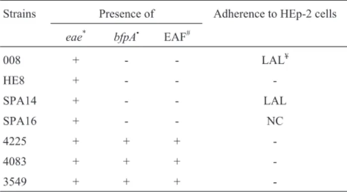
Seven EPEC strains were used, four atypical (eae+)
and three typical (eae+, bfpA+) strains (Beaudry et al.,
1996; Nakazato,et al., 2004), as shown in Table 1. All
sam-ples were negative for the Shiga toxin gene (stx) (Blancoet al., 1997). Adherence characteristics of the strains used in
the present study are shown in Table 1.
Animals
Twenty-five male and female Boxer dogs aged 60 days were used. The animals were selected from five lit-ters of four sislit-ters aged one and a half year. All animals were from the same breeder.
The parents were vaccinated (Vaccine Duramune® Max (Fort Dodge)) when they were pups and regularly dewormed (Vermifuge Endal Plus® (10 mg/kg)) at every four months. Pups were dewormed with Praziquantel and Pyrantel Pamoate when aged 20 days and vaccinated against distemper, infectious hepatitis, adenovirus type 2, parainfluenza, parvovirus and coronavirose when aged 45 days.
Clinical examination and complete blood count were performed for the parents of experimental animals in order to evaluate their health status during mating. All pups were subjected to clinical evaluation at birth, at 20 days and 45 days after birth and before the beginning of the experi-mental period.
Facilities
After mating, females were kept in individual ma-sonry kennels with covered area and solarium. From birth to weaning, at 30 days after birth, pups remained close to their mother in the same kennel. During the whole preg-nancy and breastfeeding period, the mothers received ani-mal food (AGR®Royal Canin) and clean and fresh water
ad libitum. During weaning, the pups of each litter were
housed in kennels, where 3 pups of one same litter were kept in one masonry kennel which also had covered area and solarium; pups received the same diet provided to their mothers.
Experimental infection
The spread ofE. colistrains was performed in BHI for
24 h at 37 °C without agitation. This bacterial growth was centrifuged and resuspended in saline (0.85% NaCl) until reaching Mcfarland scale 8 (2.4 x10⁹ bacteria/mL). The quantity of bacteria was verified by counting the colony forming units (cfu) in McConkey agar (Sigma). The ani-mals were experimentally infected with 1 mL of resuspend-ed bacteria by the oral route using gelatin capsules of intestinal release at a single dose. After 48 h of experimen-tal infection, all animals presented liquid-to-pasty diarrhea without vomit. Feces were collected by using a sterile swab and inoculated into GN broth incubated at 37 °C overnight for 24 h, which was then sown on McConkey agar (Sigma) incubated at 37 °C for 24 h; one sample per animal was
Table 1- Characteristics of dog EPEC strains used in the experimental in-fectionin vivo(1,20).
Strains Presence of Adherence to HEp-2 cells
eae* bfpA• EAF#
008 + - - LAL¥
HE8 + - -
-SPA14 + - - LAL
SPA16 + - - NC
4225 + + +
-4083 + + +
-3549 + + +
-*eae: gene that codes for intimin.
•bfpA: one of the genes responsible for codifying bundle-forming pilus. #EAF: EAF plasmid detection.
¥LAL: localized adherence-like pattern.
NC:non-characteristic adherencepattern. +: positive.
used. One colony from each growth was selected, inocu-lated into Brain Heart 1nfusion (BHI – Oxoid) broth and in-cubated at 37 °C overnight for genomic DNA extraction, as described by Gouveiaet al.(2011).
PCR
For PCR of theeaegene, 50 ng DNA template were
added to 2.5 UTaqDNA polymerase (Invitrogen), 50 pmol
of each primer, 200mM deoxynucleoside triphosphate
(Invi-trogen), 1.5 mM MgCl2 (Invitrogen) and 1X PCR buffer (Invitrogen) at 25mL final volume (Nakazatoet al., 2001).
After an initial denaturation at 94 °C for three min-utes, samples was subjected to 35 thermal cycles at 94 °C (denaturation) for one minute, 56 °C (annealing) for one min and 72 °C for 40s. Reactions were performed in a thermocycler BioRad Laboratories, USA. A 5mL volume
of each reaction was subjected to 0.8% agarose gel electro-phoresis, stained with ethidium bromide or SYBR Gold (Invitrogen) and visualized in a transilluminator (Ultra Vi-olet Products). Amplification of an 815 bp fragment was expected (Nakazatoet al., 2001).
For amplification of theeaegene, the following
prim-ers were used (Gannonet al., 1993):
EAE1: 5’ACGTTGCAGCATGGGTAACTC3’ and EAE2: 5’GATCGGCAACAGTTTCACCTG3’
Experimental design
Five experimental groups were defined: Group A - no diarrhea induction and no MOS supply; Group B - diarrhea induction by the strain 4083 and no MOS supply; Group C -diarrhea induction by the strain SPA14 and no MOS sup-ply; Group D - diarrhea induction by the strain 4083 and MOS supply; Group E - diarrhea induction by the strain SPA14 and MOS supply
MOS was orally administered at 2 g/kg live weight diluted in 2 mL water once a day during 20 days from 24 h after experimental infection. The inoculation day was con-sidered the Day zero of the experiment.
Experimental design and statistical analyses followed the instructions of Kaps and Lamberson (2004). Experi-mental design was completely randomized with five repli-cates, and the effect of groups was analyzed at each moment. To evaluate the effect of moments for each experi-mental group, a randomized complete block design was adopted, considering the animal a blocked factor.
Response variables
All animals were clinically evaluated according to Jones (2003) at every six hours after experimental infection in the first five days. After this period, examinations were performed at every 24 h.
Clinical evaluation
In all five moments of the experimental period (days 0, 5, 10, 15 and 20), the animals were also clinically
evalu-ated through measurement of body temperature and assess-ment of dehydration degree (mild, moderate and intense), mucosal color (pale, pink and congested), vitality (apa-thetic, plays when stimulated and agitated), presence of blood in the feces (absent, little and much blood) and fecal consistency (normal, pasty and liquid).
Laboratory evaluation
Soon after clinical evaluation, a 5 mL blood sample was collected from each animal by jugular puncture. These samples were separated into two aliquots. The first aliquot was conserved in a sterile flask with anticoagulants (EDTA) at -2 to 8 °C for subsequent hemogram using an ABC Vet automated analyzer. The second aliquot was cen-trifuged and, after separation, the serum was kept at -2 to 8 °C for subsequent determination of total IgA and IgG lev-els by ELISA (using the commercial kits IgA ELISA quan-titation kit and Dog IgG ELISA quanquan-titation set, Bethyl Laboratories). Of the samples obtained with the second aliquot, only those of days 0, 10 and 20 after inoculation were used. After collection of each blood sample, the ani-mals were individually weighed.
Statistical analysis
The results of hemogram, immunoglobulin dosages and fecal consistency of groups were compared at each mo-ment by the Kruskal-Wallis test, while these responses at the different moments were compared for each group by the Friedman test, according to Kaps and Lamberson (2004). For all comparisons, significance level was set at 5% (a= 0.05).
Ethics committee
This study was duly approved by theAnimal Experi-mentation Ethics Committee/Federal University of Mato
Grosso do Sul (UFMS), protocol number: 116/2006. After the experiment, all animals had good health status.
Results
PCR
At the beginning of the experiment, seven EPEC strains were evaluated, 3549, 4083, 4025, 008, HE8, SPA14 and SPA16. All strains were tested by PCR and showed amplification of an 815 bp fragment, correspond-ing to theeaegene.
Hemogram
all animals were within the normal parameters for blood cells and had negative results for hemoparasite survey.
Immunoglobulins IgG and IgA
The medians of IgA and IgG levels at different dpi for animals of all experimental groups were also calculated. There was no significant differences (p > 0.05) among groups at the several dpi and among dpi for the several groups.
Clinical evaluation
The body temperature of all animals remained within the normal limits, ranging from 38.5 °C to 39 °C. No animal vomited or had dehydration symptoms, indicating absence of parenteral hydration over the experiment. Mucosal color remained normal and the animals were alert (not apathetic). Blood was not detected in the feces at any moment. Ampli-fication of theeaegene (815 bp) by PCR led to
identifica-tion of EPEC SPA14 and 4083 strains in the feces of all experimentally infected animals.
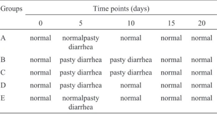
Results of fecal consistency for the different experi-mental groups and at different dpi are shown in Table 2.
All animals inoculated with EPECs had pasty diar-rhea for 12-24 h, including the treated groups D and E. No control animal had symptomatology of intestinal infection, whereas animals of the remaining groups had intense diar-rhea, presenting liquid feces at a certain moment over the experimental period. Bio-Mos®-treated animals (Groups D and E) showed a significantly (p < 0.05) faster recovery rel-ative to the animals that did not receive the prebiotic (Groups B and C). At fifteen days after treatments, all ani-mals had already recovered.
Discussion
The action of MOS is to bind to type-1 mannose site in the bacterium, preventing the latter from binding to glycoproteins of intestinal cells. According to Ferket
(2004), mannanoligosaccharides may stimulate the im-mune response against specific pathogens, preventing their colonization and promoting their presentation to the im-mune system as attenuated antigens. On account of such binding capacity with enteric pathogens (Spring et al.,
2000), the process of activation by antigens of blood cells (neutrophils, lymphocytes and monocytes) would be im-paired since the bacterium would leave the intestine with-out causing damage to the host. This may be the reason why cell immunity was not activated to a sufficient level. It must be emphasized that the type-I fimbria receptor (most com-mon acom-mongE. coli) has D-mannose as receptor (the same
as that of MOS); thus, the most probable mechanism of EPEC inhibition would be by competition of adhesion re-ceptors. However, the hypothesis of immunoglobulin stim-ulation should not be ruled out.
There are few studies about the effect of prebiotics on the blood parameters of the several species, and those for the canine species are even scarcer. A situation similar to that of the present study was reported by Budiño et al.
(2004) who carried out an experiment to test the addition of prebiotics for weaned piglets and did not find differences between treatments for haematological variables.
Laue and Tucker (2006) stated that pathogens and toxins associated with MOS form a great blend easily iden-tified by the immune system. In our study, there was no change in serum IgA and IgG levels. Thus, future studies should quantify IgA from samples of the intestinal mucosa, which can be more easily stimulated by the blend formed by the association between MOS and pathogens/toxins.
The mucosa surfaces of the intestine develop active defense mechanisms mediated by cells and chemical fac-tors, both related to innate (nonspecific) and acquired (spe-cific) immunity. These systems show differences that, although not marked, are due to the different external fac-tors to which they are subjected (Silva, 2006). For pups, these external factors, such as stress, cold and heat, are dif-ficult to control, which influences the levels of immuno-globulins. This could be minimized if the number of animals were larger, as well as the number of replicates. In the present study, samples were from pups from sister mothers and the difficulty in carrying out experiments with a large number of specimens limited the number of samples per treatment.
Based on virulence markers associated with diarrhea in humans and animals, five diarrheagenic groups ofE. coli
were defined (Beutin, 1999), including the enteropatho-genic group (EPECs), expressing colonization factors such as intimin (codified by theeaegene) and bundle-forming
pili (BFP).
In the present study, typical (strain 4083) and atypical (strain SPA14) EPEC strains caused diarrhea in the animals at 24, 48 and 72 h after induction, suggesting that both strains were pathogenic for the tested dogs. There was no difference between the results of typical and atypical
sam-Table 2- Fecal consistency at five days of the experimental period for
dogs subjected to different treatments*.
Groups Time points (days)
0 5 10 15 20
A normal normalpasty diarrhea
normal normal normal
B normal pasty diarrhea pasty diarrhea normal normal C normal pasty diarrhea pasty diarrhea normal normal D normal pasty diarrhea normal normal normal E normal normalpasty
diarrhea
normal normal normal
ples, which leads us to believe that the BFP fimbria was not essential for diarrhea manifestation; thus, BFP was not deeply investigated in the present study.
EPECs have virulence factors associated with several intestinal diseases in humans (Levine, 1984) and other ani-mals (Pestana de Castroet al., 1984; Franciset al., 1991;
Blancoet al., 1993). Since the dog is a domestic animal
which lives closely with humans, the symptoms of natural infection caused by the studied strains indicate that animals could potentially contaminate humans and vice versa.
Nakazatoet al.(2004) reported that the EPEC
sero-types found in humans were also identified in other animals including dogs, demonstrating the zoonotic risk of EPEC from dogs. AllE. colisamples isolated from the feces of
in-oculated animals had theeaegene,i.e.100% of the isolated
colony-forming units were EPECs, evidencing intense bac-terial colonization of the intestinal epithelium, which indi-cates that dogs can represent an important source of EPEC infection for humans.
MOS had an effect on fecal consistency, and MOS-treated animals recovered more rapidly from diarrhea than infected animals not treated with MOS. By means of com-petitive inhibition by mannose receptors, MOS decreases the effect of bacterial adherence by type-I fimbria in the testinal epithelium of animals, an important process in-volved in EPEC pathogenicity. Since type-I fimbria is found in mostEscherichia coli samples (Law, 1994), the
presence of MOS may potentially have reduced the coloni-zation or the perpetuation of colonicoloni-zation. Furthermore, the evaluation of fecal consistency (presence of diarrhea) showed that the process of bacterial adherence is extremely important in the emergence of diarrhea in EPEC-infected dogs.
In the present study, the EPEC infection model in dogs was established to determine the pathogenicity symp-toms of these samples. Dogs infected with EPEC receiving MOS demonstrated faster remission of diarrhea.
Acknowledments
This study was supported by “Coordenação de Aper-feiçoamento de Pessoal de Nível Superior (CAPES)” and by “Embrapa Gado de Corte”.
References
Beaudry M, Zhu C, Fairbrother M, Harel J (1996) Genotypic and Phenotypic Characterization of Escherichia coli isolates
from dogs manifesting attaching and effacing lesions. J Clin Microbiol 34:144-148.
Beutin L (1999)Escherichia colias a pathogen in dogs and cats. Vet Res 30:285-298.
Blanco M, Blanco JE, Ramos J (1993) Enterotoxigenic, veroto-xigenic and necrotoveroto-xigenicEscherichia coli isolatedfrom cattle in Spain. Am J Vet Res 54:1446-1451.
Blanco M, Blanco JE, Gonzalez EA, Mora A, Jansen W, Gomes TA, Zerbine LF, Yano T, Pestana de Castro AF,Blanco J
(1997) Genes coding for enterotoxins and verotoxins in
por-cineEscherichia colistrains belonging to different O:K:H serotypes: relationship with toxic phenotypes.J Clin Micro-biol 35:2958-2963.
Budiño FEL, Thomaz MC, Kronka RN, Pizauro Jr, JM, Santana AE, Tucci FM, Fraga AL, Scandolera AJ, Huaynate RAR (2004) Influencia da adição de probiótico e/ou prebiótico em dietas de leitoes desmamados sobre as atividades das enzi-mas digestivas e parametros sanguineos. Acta Sci Anim Sci 26:529-536.
Collet S (2000) Nutrição, Imunidade e Produtividade. In: 10ª Ronda Latino-Americana. Alltech - O Futuro da Alimen-tação. Alltech, Nicholasville, pp 20-30.
Cray WC, Moon HW (1995) Experimental infection of calves and adult cattle with Escherichia coliO157:h7. Appl Environ
Microbiol 61:1586-1590.
Drasar BS, Hill MJ (1974) Human intestinal flora. Academic
Press Ltd., London.
Ferket PR (2004) Raising drug-free poultry - What are the alterna-tives? Proc. 29th Annual Poultry Service Industry Work-shop. Banff, Alberta Canada, October 5-7, pp 95-104.
Francis CL, Jerse AE, Kaper JB, Falkow S (1991)
Characteriza-tion of interacCharacteriza-tions of enteropathogenic Escherichia coli
O127:H6 with mammalian cells in vitro. J Infect Dis 164:693-703.
Gannon VP, Rashed M, King RK, Thomas EJ(1993)Detection and characterization of theeaegene of shiga-like toxin
pro-ducingEscherichia coliusing polymerase chain reaction. J
Clin Microbiol 31:1268-1274.
Gouffaux F, China B, Janssen L, Mainil J (2000) Genotypic char-acterization of enteropathogenic Escherichia coli(EPEC) isolated in Belgium from dogs and cats. Res Microbiol 151:865-871.
Gouveia EMF, Silva IS, Nakazato G, Araújo FR, Chang MR (2011) Experimental infection with enteropathogenic Esch-erichia coli(EPEC) identified by PCR using enteric-coated
capsules in boxer pups. Acta Cir Bras 26:144-148. Jones D, Anamnese e exame físico (2003) In: Birchard, S.J,
Sherding, R. G (eds). Manual Saunders: Clínica de Peque-nos Animais. 2 ed. Rocca, São Paulo, pp 5-6.
Kaper JB (1996) Defining EPEC. Vet Microbiol 27:130-133. Kaps AM, Lamberson WR (2004) Biostatistics for Animal
Sci-ence. CABI Publishing, London.
Laue D, Tucker LA (2006) Recent Advances in Pet Nutrition. Nottingham University Press, UK.
Law D (1994) Adhesion and its role in the virulence of entero-pathogenicEscherichia coli. Clin Microbiol Rev 7:152-173. Levine MM (1984)Escherichia coliinfections.In: Germanier R. (ed). Bacterial Vaccines. Academic Press, London, pp 187-235.
Nakazato G, Gyles C, Ziebell K, Keller R, Trabulsi LR, Gomes TAT, Irino K, Silveira WD, Pestana de Castro AF (2004) Attaching and effancingEscherichia coliisolated from dogs
in Brazil: characteristics and serotypic relationship to human enteropathogenicE. coli(EPEC). Vet. Microbiol 101:169-277.
Nataro JP, Kaper JB (1998) DiarrheiogenicEscherichia coli.Clin
Microbiol Rev 11:142-201.
Pestana de Castro AF, Serafim MB, Brito JRF, Barcellos DSEN, Colli IAG (1984) Virulence factors present in cultures of
Escherichia coliisolated from pigs in the region of Con-cordia, SC, Brasil. Pesq Vet Bras 4:109-114.
Silva VK (2006) Extrato de levedura (Saccharomyces cerevisiae) e prebiótico na dieta pre-inicial para frangos de corte criados em diferentes temperaturas. Jaboticabal, Brasil, 151 pp.
(M.Sc. Dissertation. Faculdade de Ciencias Agrárias e Vete-rinárias, UNESP).
Spring P, Wenk C, Dawson A, Newman KE (2000) The effects of dietary mannaoligosaccharides on cecal parameters and the concentrations of enteric bacteria in the ceca of salmo-nella-challenged broiler chicks. Poult Sci 79:205-211.