online | memorias.ioc.fiocruz.br
Do Archaea and bacteria co-infection have a role in the pathogenesis
of chronic chagasic cardiopathy?
Maria de Lourdes Higuchi/+, Joyce Kawakami, Renata Ikegami, Maysa Beatriz Mandetta Clementino,
Flavio M Kawamoto, Marcia M Reis, Edimar Bocchi
Laboratório de Anatomia Patológica, Instituto do Coração, Faculdade de Medicina, Universidade de São Paulo, Av. Dr. Enéas de Carvalho Aguiar 44, 05403-000 São Paulo, SP, Brasil
Chronic cardiopathy (CC) in Chagas disease is a fibrotic myocarditis with C5b-9 complement deposition. My-coplasmaand Chlamydia may interfere with the complement response. Proteolytic enzymes and archaeal genes that have been described in Trypanosoma cruzi may increase its virulence. Here we tested the hypothesis that different ratios of Mycoplasma, Chlamydia and archaeal organisms, which are frequent symbionts, may be associated with chagasic clinical forms. Materials and methods: eight indeterminate form (IF) and 20 CC chagasic endomyocardial biopsies were submitted to in situ hybridization, electron and immunoelectron microscopy and PCR techniques for detection of Mycoplasma pneumoniae (MP), Chlamydia pneumoniae(CP), C5b-9 and archaeal-like bodies. Results: MP and CP-DNA were always present at lower levels in CC than in IF (p < 0.001) and were correlated with each other only in CC. Electron microscopy revealed Mycoplasma, Chlamydia and two types of archaeal-like bodies. One had electron dense lipid content (EDL) and was mainly present in IF. The other had electron lucent content (ELC) and was mainly present in CC. In this group, ELC correlated negatively with the other microbes and EDL and positively with C5b-9. The CC group was positive for Archaea andT. cruzi DNA. In conclusion, different amounts of Mycoplasma, Chlamydia and archaeal organisms may be implicated in complement activation and may have a role in Chagas disease outcome.
Key words: Chlamydia pneumoniae - Mycoplasma pneumoniae - Archaea - Chagas disease - complement C5b-9
Financial support:FAPESP, CNPq, Fundação Zerbini + Corresponding author: [email protected] Received 23 March 2009
Accepted 1 June 2009
Chagas heart disease is described as a cardiomyopa-thy secondary to Trypanosoma cruzi infection. Antigens (Higuchi et al. 1993a, Bellotti et al. 1996, 1998) and DNA (Jones et al. 1993) from T. cruzi are found within the inflammatory infiltrate of the myocardium. The scarce amount of parasite product present is associated with an intense inflammatory infiltrate suggesting alterna-tive mechanisms of pathogenesis (Higuchi et al. 2003a, Marin-Neto et al. 2007). One proposed theory is that the parasite releases polyclonal activators, which would favour the development of autoimmune injury (Gao et al. 2003). Alternatively, the presence of the parasite may alter myosin in such away that it would be recognized as non-self myosin by lymphocytes (Cunha-Neto et al. 1995). One theory not yet explored is the possible con-comitance of additional infectious agents with T. cruzi.
Interactions between agents and co-infections have been reported in experimental studies with T. cruzi and in clinical reports of myocarditis (Andreoli et al. 2006, Cortez et al. 2006, Chimenti et al. 2007, Walder et al. 2007) and non-myocardial diseases. Mycoplasma and
Chlamydia are frequent symbionts (Horn & Wagner 2004) and Mycoplasma in the cytoplasm of Trichomo-nas causes chronic resistant genital infection (Dessi et al. 2005). Biofilms may be involved in development of plaque vulnerability (Katz & Shannon 2006). We have frequently found Mycoplasma pneumoniae and Chla-mydia pneumoniae antigens in the coronary arteries and valves of normal hearts and in increased amounts in hearts affected by chronic inflammatory disorders involving stenotic aortic valves (Higuchi et al. 2002) and ruptured atherosclerotic plaques (Higuchi et al. 2000, 2003b). We observed that the intimal association between Mycoplasma and Chlamydia occurred in the presence of archaeal-like forms associated with myxoid matrix in vulnerable plaques (Higuchi et al. 2006b).
Archaea, one of the three domains of life, is a highly diverse and abundant group of prokaryotes and includes a number of “extremophiles” that thrive in such environ-ments as hot springs, salt lakes and submarine volcanic habitats (Madigan et al. 2000). Recent molecular studies have also revealed that archaea, like bacteria, are com-monly mesophilic (DeLong 1992).
of archaeal cellular processes. Some archaeal charac-teristics, such as M32 metallo carboxypeptidase (MCP) and two types of proteasome similar to those found in a common archaeal-eubacterial ancestor, were described in T. cruzi (Niemirowicz et al. 2007, Gille et al. 2003). A microbial consortium between a bacterium and a phylogenetically, distantly related archaeon has been suggested to promote the growth of both microbes in freshwater and marine sediments (Boetius et al. 2000, Raghoebarsing et al. 2006).
However, over half of the archaeal genes encode unique proteins of unknown function and no defini-tive virulence genes or factors have been described in archaea to date. Archaea share access to their host and are capable of long-term colonization and coexistence with endogenous flora in the host. The detection of an-aerobic archaea among the human colonic (Miller et al. 1982, Miller & Wolin 1983), vaginal (Belay et al. 1990) and oral microbial flora (Belay et al. 1988) demonstrates their ability to colonize the human host. Details regard-ing their survival in such human niches, includregard-ing their ability to evade the human immune system and compete
with normal human flora, however, are practically un -available (Eckburg et al. 2003).
Lepp et al. (2004) established correlations between the presence of disease and the presence of archaeal DNA, the severity of periodontal disease and the relative abundance of archaeal DNA in subgingival plaque, and between disease resolution and diminished archaeal DNA abundance. Archaeal genomes contain partial tad loci,
which may encode Tad-like proteins involved in fibril
formation and surface adherence (Lepp et al. 2004). The complement system is a major component of in-nate immunity and provides an effective host defence mechanism against pathogens (Muller-Eberhard 1988). Mycoplasma may cause chronic disease in part by evading the immune response through antigenic varia-tion and resistance to complement lysis (Simmons et al. 2004). The terminal complement complex C5b-9 was detected in the myocardium of chronic cardiac (CC) chagasic patients (Aiello et al. 2002). C5b-9 participates in several inflammatory and proliferative processes by releasing pro-inflammatory cytokines and growth fac-tors from target cells. Sublytical doses of C5b-9 may promote cell proliferation (Niculescu et al. 1999, Hal- perin 1993) and participate in the development of severe fibrosis in chagasic cardiomyopathy.
We hypothesized that a consortium involving bac-teria, archaea and T. cruzi might be related to different chronic chagasic disease outcomes, including CC and indeterminate asymptomatic form (IF). CC is associ-ated with increased complement activation, fibrosis and lymphocytic myocarditis. IF is associated with the presence of Mycoplasma and Chlamydia, which leads to decreased complement activation and degradation of myosin, preventing an “auto-immune” reaction.
In the present work we investigated whether en-domyocardial biopsies of CC or IF chagasic patients present forms and antigens or DNA from M. pneumo-niae, C. pneumoniae or archaeal organisms, and whether they are associated with each other and with C5b-9.
MATERIALS AND METHODS
This work was approved by the Ethical and Scientific Committee of Heart Institute, University of São Paulo, Shool of Medicine, São Paulo, Brazil. We reviewed endomyocardial biopsies from chagasic patients with indeterminate or chronic cardiac forms, performed between 1980-2001 at the Heart Institute of São Paulo. We compared the amount of M. pneumoniae DNA (MP-DNA), M. pneumoniae DNA (CP-DNA) and archaeal-like bodies in myocardial fragments of chagasic patients with the IF versus CC form using in situ hybridization (ISH), immunoelectron microscopy, electron microscopy and PCR. As the fragments were too small, we could not use the same case for multiple techniques. Therefore, correlations between the results obtained with different techniques were not determined in this study. Archaea-like bodies were quantified according to their morphol-ogy by electron microscopy since we do not have spe-cific antibodies or probes. DNA from CC fragments was amplified with specific primers for the Archaea domain.
ISH analysis - in situ myocardial detection of MP-DNA and CP-MP-DNA by light microscopy - Serial sections of paraffin embedded endomyocardial biopsy fragments from 24 patients with Chagas disease were submitted to ISH for identification of MP-DNA and CP-DNA. Of the 24 fragments, seven were of indeterminate form (men age 34 ± 11 years, 3 males), and 17 were associated with heart failure (37 ± 12 years, 11 males).
Cell permeabilization was achieved using a 0.01 M citrate buffered solution, pH 6.0 ± 0.1, in the microwave oven following the protocol for heat-induced target re-trieval described for IHC. Endogenous peroxidase was blocked with H2O2 at 3% for 20 min at 37o C. Tissue
pro-teins were blocked with serum free protein block (Dako, Carpinteria, CA, USA) for 15 min at 37oC. Next, 20 µL
of hybridization mixture containing the probe, deionized formamide, 50% dextran sulfate, 20X SSC, Denhardt so-lution, salmon sperm DNA, yeast tRNA, poly A and poly C, and diethyl pyrocarbonate pure water, was applied to the sections and overlaid with a coverslip. The M. pneu-moniae probe was prepared from a highly specific M. pneumoniae clone (Enzo Diagnosis, Farmingdale, NY, USA). The biotin-labelled M. pneumoniae oligonucle-otide probe was synthesized by GIBCO/BRL (Rock-ville, MD, USA). The target DNA double-strands were denatured at 95 ± 5oC for 6 min. DNA hybridization was
performed for 18 h in a humidity chamber at 37oC. After
hybridization, the coverslip was removed with 0.0 5M Tris/HCl and 0.003 mM NaCl, pH 7.6, containing 0.1% Tween 20. Non-specific hybrids were excluded with post hybridization washes in 0.2x SSC for 10 min at 50oC
and 2x SSC for 10 min at 37oC. The signal was
all reactions, we used human tissue sections known to be positive for M. pneumoniae and C. pneumoniae. In ad-dition, as a positive control for ISH, we used a repetitive Alu sequence probe (alu1/alu2) by Genetic Research (Al, USA) and for negative control we used a plasmid DNA labelled with biotin (Dako, Carpinteria, CA, USA).
The positive brown dots that represent DNA of M. pneumoniae and C. pneumoniae were detected by an im-age analysis system (Quantimet-500 Leica) and the mean percentage area for each DNA in all EMB was obtained.
Electron microscopy quantification of archaea by ultrastructural morphology and M. pneumoniae and C. pneumoniae by immune staining - Araldite embed-Araldite embed-ded endomyocardial fragments collected between 1980-1986 from five patients with the IF and five with the CC form were submitted to semi-thin and thin sectioning and were examined by electron microscopy.
Morphological analysis - The material was photo-graphed at a magnification of 2600X, allowing clear vi-sualization of the myocardial fibre alterations and count-ing of the archaeal-like bodies.
Immunoelectron microscopy-Immunogold labelling was performed for detection of M. pneumoniae, C. pneu-moniae and C5b-9 from nine biopsy fragments. Of these, five were from patients with indeterminate form (2 males and 3 females, mean age 33 ± 13 years) and four were from patients with chronic Chagas disease with heart failure (3 males and 1 female, mean age 38 ± 22 years).
Ultrathin sections were submitted to immunogold electron microscopy after etching with 12.5% ultrafil-tered sodium metaperiodate. The slices were incubated with primary antibodies for 20 h. The primary antibod-ies use were monoclonal M. pneumoniae antibody (clone M2110182, diluted 1:300) from Fitzgerald Industries International Inc, Concord MA, USA, C. pneumoniae antibody (clone RR-402, anti-protein from the outer membrane; non diluted) from Dako, Carpinteria, CA, USA, and anti-Human C5b-9-Terminal Complement Complex (rabbit polyclonal antibody; diluted 1:75) from Calbiochem-Novabiochem Corporation, San Diego, CA, USA). The detection was performed with anti-rabbit IgG (whole molecule) gold conjugate, 10 nm (1:20) from Sig-ma Immunochemicals, Saint Louis, MO, USA.
Morphometric analysis - Five photos at 5.500X original magnification were used for morphometry. We counted the mean numbers of dots (colloidal gold particles of 10nm)/ photo, which correspond to an area of 23 µm2. We also counted two types of rounded
structures enclosed by double membranes. These were not clearly stained, suggestive of archaeal bodies. One had mild electron dense lipidic (EDL) content and the other had an apparently clear empty content. We named these structures respectively: EDL and electron lucent content (ELC) bodies.
The sections were observed by electron transmission microscope Philips EM-301 (Eindhoven, Holland).
PCR technique for detection of Archaea and T. cruzi DNA - Five paraffin myocardial fragment blocks from pa-tients with CC were submitted to the PCR technique for
detection of archaeal and T. cruzi DNA. DNA from the fragments was obtained using the PureLink Viral RNA/ DNA Mini Kit (Invitrogen) following the manufacturer’s instructions. Five paraffin myocardial fragment blocks from patients with CC and three normal adult myocar-dium samples was submitted to the PCR technique for detection of archaeal and T. cruzi DNA. For the isolation and purification of genomic DNA we used the PureLink Viral RNA/DNA Mini Kit (Invitrogen) following the manufacturer’s instructions. Archaeal DNA amplifica-tion was carried out in 25 µL of reacamplifica-tion mixture un-der the following conditions: 0.2 mM deoxynucleoside triphosphates, 2.5 mM MgCl2, 2.5 U of Platinum Taq DNA Polymerase (Invitrogen), 50 pmol of each primer, 1x PCR buffer and 100 ng of DNA, using universal archaeal primers 1100F (5’ -AGTCAGGTAACGAGCGAG-3’) and 1400R (5’ -GTGCAAGGAGCAGGGAC-3’) (Kudo et al. 1997). PCR was performed in a BioRad DNA Engine thermocycler using the following program: denaturation of DNA at 94°C for 3 min, followed by 35 cycles at 94°C for 1 min, 54°C for 1.5 min and 72°C for 1 min, with 10 min at 72°C for extension at the end. Genomic DNA of Halobacterium salinarum INCQS A2 (DZMZ 668) (50 ng) was used as a positive control. It was kindly provided by Prof. Maysa Mandetta Clementino from the Bacteria and Archaea Reference Laboratory of the National Insti-tute of Quality Control in Health, Fiocruz, Rio de Janeiro, Brazil. A negative control without DNA was included and the PCR reactions were repeated three times to confirm the reproducibility of the amplification.
T. cruzi DNA amplifications were carried out with 25 µL of reaction mixture under the following conditions: 100 ng of DNA, 50 pmol each primer S34F (5’ -TAT-ATTACACCAACCCCAATCGAACC-3’) and S67R (5´ - TGGTTTTGGGAGGGGSSKTCAAMTTT-3’) (Oli-vares-Villagómez et al. 1998) 0.2 mM of each deoxynu-cleoside triphosphate, 1x PCR buffer (pH 9.0), 2,5mM MgCl2 and 2 U of Platinum Taq DNA Polymerase (In-vitrogen). Reactions were run in a BioRad DNA Engine thermocycler using the following program: denatu- ration of DNA at 94°C for 6 min, followed by 45 cycles at 94°C for 40 sec, 57°C for 1 min and 72°C for 45 sec, with 10 min at 72°C for extension. As a positive con-trol, we used genomic DNA from T. cruzi. The mixture was run on a 1% electrophoresis agarose gel in 1x TAE buffer (40 mM Tris base, 20 mM sodium acetate, 1.0 mM EDTA, pH 8.0) with a 100-bp DNA ladder (Invi- trogen Carlsbad, CA, USA). The gels were stained with ethidium bromide (0.5 mg/mL) to visualize the ampli-fied PCR products under UV illumination.
Statistical analysis - The comparison of DNA quan-tities and archaeal-like bodies between the two clini-cal groups was performed using the Student t-test. The Pearson correlation test was used to determine the rela-tionships between different microbe DNAs or antigens and complement.
RESULTS
groups of patients are shown in Table I. MP-DNA and CP-DNA were significantly higher in the IF group than in the CC group (p < 0.001). There was a significant pos-itive correlation between MP-DNA and CP-DNA values in the CC group of patients (r = 0.64; p = 0.005) but not in the IF group of patients (r = -0.28; p = 0.547).
Qualitative and quantitative analysis - Endomyocar-dial biopsies from the IF group revealed myocarEndomyocar-dial fi-bres with well preserved morphology, large amounts of glycogen-like granules in the sarcoplasm and moderate dilatation of T tubules, sarcoplasmic reticulum and inter-calated discs, which frequently contained membranous elements in the lumen. Fibrosis in the interstitium usually exhibited mycoplasmal-like forms characterized by only one outer membrane with a rounded or cylindrical shape. Chlamydial bodies characterized by a double membrane and dark homogeneous chromatin were rarely seen in myocardial fibres, macrophages or the extracellular ma-trix (Fig. 1A, B). Many EDL balls were present in the myocytes either at the subsarcolemal region or more in-ternally. These were similar to lysosomes but enclosed by a barely visibly double external membrane with a darker aspect at the periphery. EDLs were always located near mitochondria and glycogen-like granules (Fig. 2A, B).
A second type of round organelle enclosed by a double membrane was also present. These had ELC with irregu-lar thin flat tubules, an irreguirregu-lar folded external mem-brane and sometimes contained a large, clear periplasmic space. These EDL and ELC bodies are compatible with archaeal morphology and were sometimes fused to each other (Fig. 2A). EDL and ELC bodies presented variable diameters ranging from around 0.10-1.5 µm.
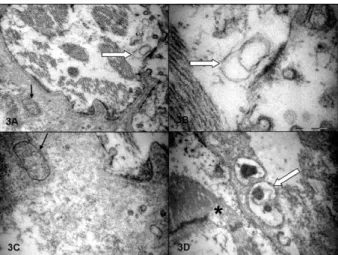
In CC cases, the myocardial fibres presented more intense alterations represented by mitochondrial crystol-ysis, a clear matrix and dilated reticulum sarcoplasmic organelles frequently presenting irregular membranes in their lumens. There were many foci of myocytolysis usually associated with ELC bodies and a lack of gly-cogen granules (Fig. 3A, B). There was a severe fibrosis in the interstitium, with mycoplasmal forms among the collagen fibres. There were many forms of ELC among the mononuclear inflammatory cells and in the myocar-dial fibres. These were associated with mycoplasmal forms and round dark structures that may have been derived from C. pneumoniae elements (Fig. 3C, D). ELC structures were sometimes very large (Fig. 4A, B). Glycogen-like granules were usually absent.
Immunogold electron microscopy counting - Immu-noelectron microscopy demonstrated that the monoclo-nal antibodies anti M. pneumoniae and C. pneumoniae stained surface antigens and free nanolipidic particles suggestive of lipoprotein or lipopolysaccharide from these bacteria, as had been shown in a previous study (Higuchi et al. 2006c). All chagasic EMBs presented high amounts of M. pneumoniae and C. pneumoniae as extra-cellular antigens, as well as in the extraextra-cellular matrix and myocardial fibres. M. pneumoniae and C. pneumo-niae were apparently associated with lipid nanovesicles. Table II shows the mean numbers and standard devi-ation of gold particles/100 µm2 detected by
immunoelec-tron microscopy and the number of EDL and ELC bod-ies detected by transmission electron microscopy in both the IF group and the CC group. The unicaudal Student t test showed lower levels of C. pneumoniae and higher levels of C5b-9, as well as non-significantly lower lev-els of M. pneumoniae and EDL values, in the CC group than in the IF group. ELC values were higher in the CC group, without statistical significance. The lack of statis-tical significance may be linked to the small number of samples analyzed.
Table III shows the correlations between the vari-ables in Table II. The CC group had higher R correlation values than the IF group, with significance only between C5b-9 and C. pneumoniae antigens (r = -0.96, p = 0.04) and an almost significant negative correlation between EDL and ELC values (r = -0.97; p = 0.06).
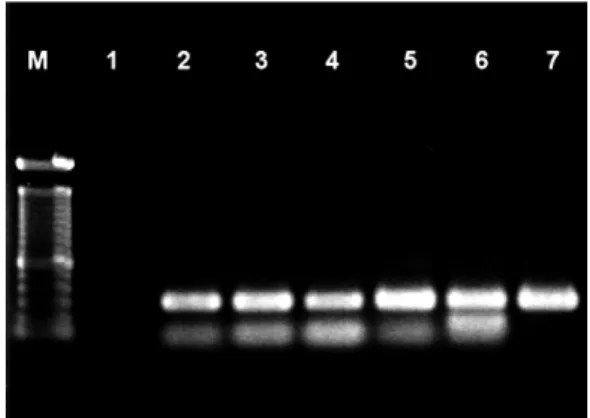
PCR technique for detection of archaeal and T. cruzi DNA - All five of the CC paraffin myocardial fragment blocks yielded a 300-bp fragment, including the H. sali-narum reference strain, after PCR with the universal archaeal primers (Fig. 5). The same samples described above yielded a 122-bp fragment using T. cruzi specific primers (Olivares-Villagómez et al. 1998).
DISCUSSION
The exact mechanism for the development of chronic dilated cardiopathy in some individuals infected with T. cruzi is a still matter of debate. Our results provide evi-dence that association of mycoplasmal, chlamydial and/or archaeal elements might be related to this phenomenon.
Optical, electron and immunoelectron microscopy showed that high levels of DNA from C. pneumoniae and M. pneumoniae and the presence of archaeal-like bodies of the EDL type were associated with IF. Since these values did not correlate with each other and be-cause the morphology was of isolated forms, we con-clude that these microbes are not closely associated with IF. Alternatively, fusion of ELC archaeal-like bodies with the microbes described in the IF group was present in the CC group. An intimal interaction is suggested by the following: (i) the high correlation between CP-DNA and MP-DNA, which correlated negatively with ELC values; (ii) the negative correla-tion between the numbers of ELC with the numbers of EDL and (iii) the positive correlation between ELC and C5b-9 values.
The five samples submitted to PCR analysis yielded fragments compatible with the specific primers. Since we did not have enough material to perform PCR in the IF cases, we could not compare this item. Further studies are needed, including a better characterization of EDL and ELC organelles to determine if they present differ-ent archaeal genome sequences.
Mycoplasma can either grow symbiotically with eu-karyotic cells without producing profound cytotoxicity or it may provoke a marked host response, acting as a super antigen by triggering apoptosis of the CD4+ T
My-coplasma may increase the virulence of other microbes (Dessi et al. 2005) and may induce pro-inflammatory
cytokines such as TNFα, IL-6 and TGFβ (Blanchard
& Bebear 2002, Yang et al. 2004), which are related to heart failure (Sharma et al. 2001, Sugamori et al. 2002). Chlamydia is also a frequent symbiont of microbes and has the ability to prevent apoptosis of the host cell (Fan et al. 1998, Sharma & Rudel 2009). Archaea have anti-oxidative enzymes, such as superoxidismutase, that pro-mote microbe survival (Cannio et al. 2000) and pepti-dases and proteasomes that contribute to degradation of aggregation-prone proteins and reduce cellular toxicity in mammalian cells (Yamada et al. 2006).
The presence of M32 family MCP, which is known to be specific to the Archaea and bacteria kingdoms, was described in T. cruzi (Niemirowicz et al. 2007). The authors suggest that the protozoan has acquired these genes by horizontal transfer of an ancestral archaea gene. Consistent with this hypothesis, the T. cruzi MCP has high biochemical similarity with Archaeon P. fu-riosus (Pfu). Pfu M32 MCP seems to be involved in the utilization of peptides and proteins for the metabolism of the organism (Schul et al. 2003).
Sequences of both the 20S proteasomes and hsIV were found in T. cruzi, suggesting the presence of
en-TABLE I
Percentage of area positive for Mycoplasma pneumoniae DNA (MP-DNA) and Chlamydia pneumoniae DNA (CP-DNA) in endomyocardial biopsies from chagasic patients in indetermi-nate (IF) or chronic cardiac (CC) forms by in situ hybridization
technique in light microscopy and Pearson correlation test
Group % MP-DNA mean (± SD)
% CP-DNA mean (± SD)
Correlation coefficient MP vs. CP IF (n = 7) 8.75 (4.00) 9.66 (4.22) - 0.28 (p = 0.547) CC (n = 17) 2.65 (1.64) 3.43 (2.65) 0.64 (p = 0.005) p (t test) 0.001 < 0.001
Fig. 1: ultrastructural aspects of an indeterminate asymptomatic form endomyocardial biopsy. A: a group of microbes, suggestive of myco-plasmal and chlamydial bodies, well individualized each other, amidst the fibrosis; B: intercalated disc (ID) with well preserved desmosomes (thin arrows) and dilated fascia adherens containing a membranous element in the lumen (white arrow). CP: Chlamydia pneumoniae; FA:
Fascia Adherens MP: Mycoplasma pneumoniae.
Fig. 3: ultrastructural aspects of a heart failure group endomyocar-dial biopsy. A: severe myocytolysis of the myocarendomyocar-dial fiber. The white arrow indicates an electron lucent content (ELC) structure formed by two compartments, which is in close view in B. The black arrow indicates a mycoplasmal body; C: close view; D: a myocardial fiber with myocytolysis (asterisk), with some apparently symbiotic forms of mycoplasma, ELC and Chlamydia pneumoniae.
Fig. 2: ultrastructural aspects of an indeterminate asymptomatic form endomyocardial biopsy. A: a panoramic view of a myocardial fiber contain mainly many electron dense lipidic (EDL) bodies (thin ar-electron dense lipidic (EDL) bodies (thin ar- bodies (thin ar-rows) and few electron lucent content bodies, one fused with one EDL body (empty arrow); B: higher magnification of an EDL body divided in two lobes (arrow), exhibiting doubled external membrane, electron dense lipidic internal material darker at the periphery.
dosymbionts, for example, an α-proteobacterial pro -genitor of mitochondria. Alternatively, horizontal gene transfer by temporal association with bacteria within the intestines of insects could account for the presence of these sequences (Gille et al. 2003). T. cruzi undergoes profound morphological changes using proteins from the host cell during its life cycle in the myocardial fibres. An essential role for proteasomes in the degradation of cytoplasmic proteins has been demonstrated (Gonzalez et al. 1996, De Diego et al. 2001).
In CC EMBs we observed ELC archaeal-like bodies. Their morphology is similar to the morphology of those present in the myxoid matrix of vulnerable atheroma plaques that are associated with vessel dilatation and adventitial inflammation in correlation with C. pneumo-niae and M. pneumopneumo-niae elements (Higuchi et al. 2003b, 2006b). The ELCs were in the interstitium among the chronic inflammatory infiltrate, sometimes with a huge periplasmic space containing nanoarchaeal-like bodies. This morphology is similar to that of Ignicoccus hospi-talis (Junglas et al. 2008).
The unique ether glycerolipids of Archaea can form vesicles (archaeosomes or liposomes from Archaea) with strong adjuvant activity for MHC class II presentation. The archaeosomes are superior adjuvants that induce a long-term CD8+ cytotoxic T cell response to entrapped
soluble protein in the absence of help from CD4+ T cells
(Krishnan et al. 2000). It has been shown that chronic chagasic active myocarditis in CC patients presents in-flammatory infiltrate composed mainly of CD8+ T cells
with increased numbers in the presence of T. cruzi
an-tigens, and a lack of CD4+ T cells (Higuchi et al. 1993b,
1997, Reis et al. 1997). We argue that T. cruzi antigens in archaeosomes may not be the cause of this increased inflammatory infiltrate.
Based on the literature, we may speculate that symbi-otic forms involving Mycoplasma, Chlamydia and ELC organelles in the human chronic infection by T. cruzi causes increased immune activation by complement de-position and lymphocyte activation leading to the devel-opment of myocarditis. However, the presence of non-at-tached mycoplasmal, chlamydial and EDL archaeal-like bodies leads to reduced myocardial inflammation.
The current view of microbiologists is that prokary-otes’ predominant state of being is inside microbial com-munities. Direct interaction between archaea has been proved to mediate exchange of chromosomal DNA, me-tabolites or substrates from the cytoplasm of one cell to another (Junglas et al. 2008). The association of dif-ferent species of microorganisms leads to the develop-ment of more resistant and productive new associated forms (Hansen et al. 2007). Also, the presence of one pathogen may inhibit the growth of others. Mycoplasma inhibited the growth of Chlamydia in in vitro studies
TABLE II
Mean numbers and standard deviation of gold particles/100µm2
Mycoplasma pneumoniae (MP), Chlamydia pneumoniae (CP) and membrane attack complex (MAC) antigens in immuno-eletron microscopy and archaeal-like bodies/100µm2 in
elec-tron microscopy, in indeterminate (IF) and chronic cardiac (CC) forms
Groups MP CP MAC EDL ELC IF (n = 5) 48 ± 22 48 ± 17 56 ± 13 0.13 ± 0.04 0.04 ± 0.04 CC (n = 4) 39 ± 17 17 ± 8.7 100 ± 43 0.09 ± 0.09 0.06 ± 0.04 p (t test) 0.30 0.005 0.04 0.19 0.31 EDL: electron dense lipidic bodies; ELC: electron lucent con-tent bodies.
TABLE III
Correlation coefficient and p value between mean numbers of Mycoplasma pneumoniae (MP), Chlamydia pneumoniae
(CP) and membrane attack complex (MAC) antigens at the immunoelectron microscopy and electron dense lipidic (EDL) bodies and electron lucent content (ELC) bodies at the
elec-tron microscopy
CP MAC EDL ELC
MP IF - 0.51 (0.38) 0.33 (0.59) - 0.21 (0.73) - 0.04 (0.95) CC - 0.71 (0.29) - 0.81 (0.19) 0.77 (0.23) - 0.85 (0.14) CP IF 0.007 (0.99) - 0.31 (0.61) - 0.34 (0.57) CC - 0.96 (0.04) 0.34 (0.66) - 0.64 (0.36) MAC IF 0.37 (0.53) - 0.60 (0.28) CC - 0.33 (0.67) 0.60 (0.40)
EDL IF - 0.53 (0.34)
CC - 0.97 (06)
Fig. 4: details of a foci of inflammatory infiltrate in a chronic cardiac case. A: among the mononuclear inflammatory cells, there are some suggestive of Archaeal large bodies characterized by rounded struc-tures surrounded by two membranes, large periplasmic space contain-ing amorphous and granules content and an internal vacuole (small ar-rows); B: a close view of the periplasmic space with many clear double membrane round structures suggestive of nanoarchaea (large arrow).
Fig. 5: PCR amplifications of Archaea (primers 1100/1400) in tissues samples of chagasic patients. Lane M: molecular size marker (100-bp DNA ladder); Lane 1: negative control with no added template DNA; Lanes 2-6: tissue samples of chagasic patients; Lane 7: positive control
(Van Nerom et al. 2000). Escherichia coli inhibited the growth of T. cruzi (Cortez et al. 2006). High ratios of M. pneumoniae/C. pneumoniae were related to an increased amount of growth factors and fibrosis in atheroma plaques (Higuchi et al. 2006a).
Limitations and clinical implications - The present work is a retrospective study with a small number of cases that cannot result in definite conclusions. However, our results may suggest a new frontier of investigation in Chagas cardiopathy pathogenesis, specifically the symbi-otic interaction among prokaryotes and a trypanosome.
In conclusion, different amounts of Mycoplasma, Chlamydia and archaeal elements may be implicated in complement activation and may have a role in Chagas disease outcome. IF was associated with isolated forms of these microbes and with the EDL type of archaeal-like bodies. CC was associated with fusion of these microbes and ELC archaeal-like bodies in association with increased myocardial complement deposition. These findings open a new direction for pathogenetic studies and the possibility of new prognostic biomark-ers in Chagas disease.
REFERENCES
Aiello VD, Reis MM, Benvenuti LA, Higuchi ML, Ramires JA, Halp-erin JA 2002. A possible role for complement in the pathogenesis of chronic chagasic cardiomyopathy. J Pathol197: 224-229.
Andreoli WK, Taniwaki NN, Mortara RA 2006. Survival of
Trypano-soma cruzi metacyclic trypomastigotes within Coxiella burnetii
vacuoles: differentiation and replication within an acidic milieu.
Microbes Infect8: 172-182.
Belay N, Johnson R, Rajagopal BS, de Macario EC, Daniels L 1988. Methanogenic bacteria from human dental plaque. Appl Environ
Microbiol54: 600-603.
Belay N, Mukhopadhyay B, Conway de Macario E, Galask R, Daniels L 1990. Methanogenic bacteria in human vaginal samples. J Clin
Microbiol28: 1666-1668.
Bellotti G, Bocchi EA, de Moraes AV, Higuchi ML, Barbero-Marcial M, Sosa E, Esteves-Filho A, Kalil R, Weiss R, Jatene A, Pileg-gi F 1996. In vivo detection of Trypanosoma cruzi antigens in hearts of patients with chronic Chagas’ heart disease. Am Heart J 131: 301-307.
Bellotti G, Bocchi EA, de Moraes AV, Higuchi ML, Barbero-Marcial M, Sosa E, Esteves-Filho A, Kalil R, Weiss R, Jatene A, Pileg-gi F 1998. In vivo detection of Trypanosoma cruzi antigens in hearts of patients with chronic Chagas’ heart disease. Am Heart J 135: 550.
Blanchard A, Bebear CM 2002. Mycoplasmas of humans. In S Razin, R Herrmann (eds.), Molecular biology and pathogenicity of
my-coplasmas, Kluwer Academic/Plenum Publisheres, New York,
p. 45-71.
Boetius A, Ravenschlag K, Schubert CJ, Rickert D, Widdel F, Gieseke A, Amann R, Jørgensen BB, Witte U, Pfannkuche O 2000. A ma-rine microbial consortium apparently mediating anaerobic oxida-tion of methane. Nature407: 623-636.
Cannio R, Florentino G, Morana A, Ossi M, Bartolucci S 2000. Oxy-gen: friend or foe? Archaeal superoxide dismutases in the pro-tection of intra- and extracellular oxidative stress. Front Biosci 5: 768-779.
Chimenti C, Del Nonno F, Topino S, Abbate I, Licci S, Paglia MG, Capobianchi MR, Petrosillo N, Frustaci A 2007. Fatal myocardial
co-infection by Toxoplasma gondii and Parvovirus B19 in an HIV patient. AIDS21: 1386-1388.
Cortez M, Atayde V, Yoshida N 2006. Host cell invasion mediated by
Trypanosoma cruzi surface molecule gp82 is associated with
F-actin disassembly and is inhibited by enteroinvasive Escherichia coli. Microbes Infect8: 1502-1512.
Cunha-Neto E, Duranti M, Gruber A, Zingales B, De Messias I, Stolf N, Bellotti G, Patarroyo ME, Pilleggi F, Kalil J 1995. Autoim-munity in Chagas disease cardiopathy: biological relevance of a cardiac myosin-specific epitope crossreactive to an immu-nodominant Trypanosoma cruzi antigen. Proc Natl Acad Sci USA92: 3541-3545.
De Diego JL, Katz JM, Marshall P, Bessy G, Manning J, Nussen-zweig V, Gonzalez J 2001. The ubiquitina-proteasome pathway plays an essential role in proteolysis during Trypanosoma cruzi remodeling. Biochem40: 1053-1062.
DeLong EF 1992. Archaea in coastal marine environments. Proc Natl
Acad Sci USA89: 5685-5689.
Dessì D, Delogu G, Emonte E, Catania MR, Fiori PL, Rappelli P 2005. Long-term survival and intracellular replication of
Myco-plasma hominis in Trichomonas vaginalis cells: potential role of
the protozoon in transmitting bacterial infection. Infect Immun 73: 1180-1186.
Eckburg PB, Lepp PW, Relman DA 2003. Archaea and their potential role in human disease. Infect Immun71: 591-596.
Fan T, Lu H, Hu H, Shi L, Mcclarty GA, Nance DM, Greenberg AH, Zhong G 1998. Inhibition of apoptosis in Chlamydia infected cells: blockade of mitochondrial cytochrome c release and cas-pase activation. J Exp Med 187: 487-496.
Gao W, Luquetti AO, Pereira MA 2003. Immunological tolerance and its breakdown in Chagas’ heart disease: role of parasitokines.
Front Biosci8: E218-227.
Gille C, Goede A, Schlöetelburg C, Preiβner R, Kloetzel PM, Göbel
UB, Frömmel C 2003. A comprehensive view on proteasomal se-quences: implications for the evolution of the proteasomo. J Mol Biol326: 1437-1448.
Gonzalez J, Ramalho-Pinto EJ, Frevert U, Ghiso J, Tomlinson S, Scharfstein J, Corey EJ, Nussenzweig V 1996. Proteasome activ-ity is requires for the stage-specific transformation of a protozoan parasite. J Exp Med184: 1909-1918.
Halperin JA, Taratuska A, Nicholson-Weller A 1993. Terminal com-plement complex C5b-9 stimulates mitogenesis in 3T3 cell. J Clin
Invest91: 1974-1978.
Hansen SK, Rainey PB, Haagensen JA, Molin S 2007. Evolution of spe-cies interactions in a biofilm community. Nature445: 533-536.
Higuchi ML, Benvenuti LA, Reis MM, Metzger M 2003a. Pathophys- Pathophys-iology of the heart in Chagas’ disease: current status and new developments. Cardiovasc Res60: 96-107.
Higuchi ML, Britto T, Reis MM, Barbosa A, Bellotti G, Pereira Bar-reto AC, Pileggi F 1993a. Correlation between Trypanosoma cruzi parasitism and myocardial inflammatory infiltrate in human chronic chagasic myocarditis: light microscopy and immunohis-tochemical findings. Cardiovasc Pathol2: 101-106.
Higuchi ML, Góis JM, Reis MM, Higuchi-Dos-Santos MH, Diament J, Sousa JM, Ramires JA, Oliveira SA 2006a. Co-infection ratios versus inflammation, growth factors and progression of early atheromas. APMIS114: 338-344.
comparison with myocardial rejection process. Virchows Arch A
Pathol Anat Histopathol423: 157-160.
Higuchi ML, Higuchi-dos-Santos MH, Pierri H, Palomino S, Sambi-ase NV, Ramires JAF, Wajngarten M 2002. Mycoplasma
pneu-moniae and Chlamydia pneumoniae in calcified nodules of aortic
stenotic valves. Rev Inst Med Trop Sao Paulo44: 209-212.
Higuchi ML, Higuchi-dos-Santos MH, Rogério A, Kawakami JT, Bezerra HG, Canzian M 2006b. A role for archaeal organisms in development of atherosclerotic vulnerable plaques and myxoid matrices Clinics 61:473-478.
Higuchi ML, Pierri H, Sesso A, Santos MHH, Timenetski J, Strunz CMC, Fukasawa S, Ramires JAF, Wajngarten M 2006c. C-reactive protein and Mycoplasma pneumoniae antigen mor-phological particles are positively correlated and increased in the serum of elderly atherosclerotic patients. InLV Clark (ed.),
New resesarch on atherosclerosis, Nova Science Publisher Inc,
New York, p. 1-17.
Higuchi ML, Reis MM, Aiello VD, Benvenutti LA Gutierrez PS, Bel-lotti G, Pileggi F 1997. Asssociation of an increase in CD8+ T
cells with the presence of Trypanosoma cruzi antigens in chronic, human, chagasic myocarditis. Am J Trop Med Hyg56: 485-489.
Higuchi ML, Reis MM, Sambiase NV, Palomino SAP, Castelli JB, Gutierrez PS, Aiello VD, Ramires JA 2003b. Co-infection with
Mycoplasma pneumoniae and Chlamydia pneumoniae in
rup-tured plaques associated with acute myocardial infarction. Arq
Bras Cardiol 81: 12-22.
Higuchi ML, Sambiase N, Palomino S, Gutierrez P, Demarchi LM, Aiello V, Ramires JÁ 2000. Detection of Mycoplasma
pneu-moniae and Chlamydia pneumoniae in ruptured atherosclerotic
plaques. Braz J Med Biol Res33: 1023-1026.
Horn M, Wagner M 2004. Bacterial endosymbionts of free-living amoebae. J Eukaryot Microbiol51: 509-514.
Jones EM, Colley DG, Tostes S, Lopes ER, Vnencak-Jones CL, Mc-Curley TL 1993. Amplification of a Trypanosoma cruzi DNA sequence from inflammatory lesions in human chagasic cardio-myopathy 1993. Am J Trop Med Hyg48: 348-357.
Junglas B, Briegel A, Burghardt T, Walther P, Wirth R, Huber H, Ra-chel R 2008. Ignicoccus hospitalis and Nanoarchaeum equitans: ultrastructure cell-cell interaction, and 3D reconstruction from serial section of freeze-substituted cells and by electron cryoto-mography. Arch Microbiol190: 395-408.
Katz JT, Shannon RP 2006. Bacteria and coronary atheroma: more fingerprints but no smoking gun. Circulation113: 920-922.
Krishnan L, Sad S, Patel GB, Sprott GD 2000. Archaeosomes induce long-term CD8+ cytotoxic T cell response to entrapped soluble
protein by the exogenous cytosolic pathway, in the absence of CD4+ T cell help. J Immun165: 5177-5185.
Kudo Y, Nakajima T, Miyaki T, Oyaizu H 1997. Methanogen flora of paddy soils in Japan. FEMS Microbiol Ecol22: 39-42.
Lepp PW, Brinig MM, Ouverney CC, Palm K, Armitage GC, Rel-man DA 2004. Methanogenic Archaea and huRel-man periodontal disease. Proc Natl Acad Sci USA101: 6176-6181.
Lo S-C 1992. Mycoplasmas and AIDS. In J Maniloff, RN Mc El-haney, LR Finch, JB Baseman (eds.), Mycoplasmas, molecular
biology and pathogenesis, Am Soc Microbiol, Washington DC,
p. 525-545.
Madigan MT, JM Martinko, J Parker 2000. Prokaryotic diversity: the Archaea. In MT Madigan, JM Martinko, J Parker (eds.), Brock
bi-ology of microorganisms, Prentice-Hall Inc, Upper Saddle River,
p. 546-572.
Marin-Neto JA, Cunha-Neto E, Maciel BC, Simões MV 2007. Pathogenesis of chronic Chagas heart disease. Circulation115: 1109-1123.
Miller TL, Wolin MJ 1983. Stability of Methanobrevibacter smithii populations in the microbial flora excreted from the human large bowel. Appl Environ Microbiol45: 317-318.
Miller TL, Wolin MJ, de Macario EC, Macario AJ 1982. Isolation
of Methanobrevibacter smithii from human feces. Appl Environ
Microbiol 43: 227-232.
Muller-Eberhard HJ 1988. Molecular organization and function of the complement system. Ann Rev Biochem57: 321-347.
Niculescu F, Badea T, Rus H 1999. Sublytic C5b-9 induces prolifera-tion of human aortic smooth mucles cells: role of mitogen acti-vated protein kinase and phosphatidylinositol 3-kinase. Athero-sclerosis142: 47-56.
Niemirowicz G, Parussini F, Aguero F, Cazzulo JJ 2007. Two met-allocarboxypeptidases from the protozoan Trypanosoma cruzi belong to the M32 family, found so far only in prokaryotes.
Bio-chem J401: 399-410.
Olivares-Villagómez D, McCurley TL, Vnencak-Jones CL, Correa-Oliveira R, Colley DG, Carter CE 1998. Polymerase chain re-action amplification of three different Trypanosoma cruzi DNA sequences from human chagasic cardiac tissue. Am J Trop Med Hyg59: 563-570.
Raghoebarsing AA, Pol A, van de Pas-Schoonen KT, Smolders AJ, Ettwig KF, Rijpstra WI, Schouten S, Damsté JS, Op den Camp HJ, Jetten MS, Strous M 2006. A microbial consortium couples anaerobic methane oxidation to denitrification. Na-ture440: 918-921.
Reis MM, Higuchi ML, Benvenuti LA, Aiello VD, Gutierrez PS, Belotti G, Pileggi F 1997. An in situ quantitative immunohis-tochemical study of cytokines and IL-2R+ in chronic human
chagasic myocarditis: correlation with the presence of myocar-dial Trypanosoma cruzi antigens. Clin Immunol Immunopathol 83: 165-172.
Sant’Anna C, Parussini F, Lourenço D, de Souza W, Cazzulo JJ, Cunha-e-Silva NL 2008. All Trypanosoma cruzi developmental forms present lysosome-related organelles. Histochem Cell Biol 130: 1187-1198.
Schul G, Brehm SD, Dallas S, Adams MW 2003. Whole-genome DNA microarray analysis of a hyperthermophile and na
archae-on: Pyrococcus fusiosus grown on carbohidrates or peptides.
J Bacteriol185: 3935-3947.
Sharma M, Rudel T 2009. Apoptosis resistance in Chlamydia -infect-ed cells: a fate worse than death? FEMS. Immunol Med Microbiol 55: 154-161.
Sharma R, Al Nasser FO, Anker SD 2001. The importance of tumor necrosis factor and lipoproteins in the pathogenesis of chronic heart failure. Heart Fail Monit2: 42-47.
Simmons WL, Denison AM, Dybvig K 2004. Resistance of
My-coplasma pulmonis to complement lysis is dependent on the
number of Vsa tandem repeats: shield hypothesis. Infect Immun 72: 6846-6851.
Srinivasan V, Morowitz HJ 2006. Ancient genes in contemporary per-sistent microbial pathogens. Biol Bull210: 1-9.
factor-alphawith systemic and local production of nitric oxide.
Circ J66: 627-632.
Van Nerom A, Ducatelle R, Charlier G, Haeseebrouck F 2000. Inter-action between turkey monocytes and avian - Chlamydiapsittaci - in the presence of Mycoplasma sp.: the importance of nitric oxide. Dev Comp Immunol24: 417-432.
Walder G, Gritsch W, Wiedermann CJ, Pölzl G, Laufer G, Hotzel H, Berndt A, Pankuweit S, Theegarten D, Anhenn O, Oehme A,
Dier-ich MP, Würzner RC 2007. Co-infection with two Chlamydophila species in a case of fulminant myocarditis. Care Med35: 623-626.
Yamada S, Niwa J, Ishigaki S, Takahashi M, Ito T, Sone J, Doyu M, Sobue G 2006. Archaeal proteasomos effectively degrade aggre-gation-prone proteins and reduce cellular toxicities in mamma-lian cells. J Biol Chem281: 23842-2351.
Yang J, Hooper WC, Phillips DJ, Talkington DF 2004. Cytokines in
Mycoplasma pneumoniae infections. Cytokine Growth Factor