Expression of
mdr
isoforms in mice during estrous cycle and under hormone
stimulation
Marion Schiengold
1, Lavínia Schwantes
1, Maria F. Ribeiro
2, Nívia Lothhammer
3, Tatiana P. Gonzalez
1,
Jose Artur Bogo Chies
1and Nance B. Nardi
11
Departamento de Genética, Universidade Federal do Rio Grande do Sul, Porto Alegre, RS, Brazil.
2Departamento de Fisiologia, Universidade Federal do Rio Grande do Sul, Porto Alegre, RS, Brazil.
3Departamento de Ciências Morfológicas, Universidade Federal do Rio Grande do Sul, Porto Alegre,
RS, Brazil.
Abstract
The multidrug resistance (MDR) phenotype is associated with the expression of P-glycoprotein (Pgp), coded by the multigenicmdr family. Mice present the isoforms mdr1 and mdr3, which are responsible for multidrug resistance, and mdr2, that is involved in the transport of phospholipids. mdr1 expression has more recently been associated also with the secretion of steroid hormones. This work presents an RT-PCR analysis of the expression ofmdr isoforms, in sev-eral organs of mice during different phases of the estrous cycle. Additionally, females were ovariectomized, submit-ted to different hormone treatments, and their uterus was analyzed for the expression of mdr isoforms. The results show that in the adrenal gland and ovaries mdr1 is the main isoform during proestrus, and that progesterone or a combination of progesterone and estrogen induce the expression of allmdr isoforms in the uterus of ovariectomized females. We suggest that the functions of mdr1 and mdr3 are overlapping, that mdr3 may be the more efficient isoform in the detoxification function, and that mdr1 may be more closely related to the secretion of steroid hor-mones.
Key words:adrenal gland, multidrug resistance, ovaries, steroids, uterus.
Received: September 15, 2005; Accepted: April 4, 2006.
Introduction
The multidrug resistance (MDR) phenotype is associ-ated with the expression of P-glycoprotein (Pgp), coded by the multigenicmdr family. Pgp acts as an energy-depen-dent efflux pump, preventing accumulation of cytotoxic drugs within the cell. Themdr gene family presents two members in humans (MDR1/ABCB1 andMDR3/ABCB4) and three in rodents (mdr1,mdr2, andmdr3). MDR1, mdr1 and mdr3 are responsible for the MDR phenotype, whereas MDR3 and mdr2 seem to be involved only with the trans-port of phospholipids (Gottesman and Pastan, 1993; Sha-piro and Ling, 1998).
The tissue distribution of Pgp has been investigated in humans and rodents through different approaches. In hu-mans, Paulyet al. (1992) detected low levels of Pgp mRNA in the brain, bone marrow, esophagus, ovary and stomach, intermediate levels in the colon, liver and lung, and high
levels in the adrenal gland, kidney and pancreas. In mice,
mdr1mRNA was observed mainly in the pregnant uterus, adrenal gland, kidney and heart; high levels of mdr2 were expressed in the liver, spleen and muscles, and mdr3 was found in the intestine, brain, kidney, liver and spleen (Cro-opet al., 1989). It has been demonstrated that the placental
mdr3Pgp is present in fetus-derived epithelial cells, and can greatly limit the passage of various toxic or beneficial Pgp substrate drugs into the fetus (Lankaset al., 1998; Smit
et al., 1999). Ushigome et al. (2000) observed that Pgp (MDR1) is expressed on the brush-border membrane (ma-ternal side) of human placental trophoblasts.
Borstet al. (1993) reviewed the possible physiologi-cal roles of Pgp isoforms as a function of their tissue distri-bution. However, the review was based mainly on the results published by Croopet al. (1989), who did not ob-serve the expression of mdr3 in the adrenal gland, ovaries and uterus, or of mdr1 in the ovaries. The presence of Pgp in virtually all kinds of tumors, however, suggests that low levels of the protein are normally present in all tissues but could not be detected with the techniques available at that time. Alternatively, it was proposed that the MDR
pheno-www.sbg.org.br
Send correspondence to Marion Schiengold. Departamento de Genética, Universidade Federal do Rio Grande do Sul, Av. Bento Gonçalves 9500, Caixa Postal 15053, 91501-970 Porto Alegre, RS, Brazil. E-mail: [email protected].
type could have been acquired as a consequence of chemo-therapy (Herweijer et al., 1990; Goldstein et al., 1990; Schneideret al., 1989). It is possible that few pre-existing cells expressing Pgp are selected by chemotherapy (No-onanet al., 1990).
The role of Pgp expression in the secretion of steroid hormones has been studied in mice. Arceciet al. (1988) re-ported the predominant location of Pgp on the luminal sur-face of secretory epithelial cells of the endometrium. Yang
et al. (1989) observed that progesterone interacts with Pgp in the endometrium of the pregnant uterus, and that the ef-fect of steroids on the accumulation of other drugs is related to their hydrophobicity. The expression ofmdrgenes in the secretory epithelium of the endometrium was shown to be increased by the combination of estrogen and progesterone, although the level of expression of mdr mRNA did not seem to increase during the normal estrous cycle (Arceciet al., 1990). Pierkarzet al. (1993) reported that progesterone, whose level is increased during pregnancy, regulates the activity of themdr1promoter, which has an element re-sponsive to that hormone on its first untranslated exon.
The importance of mdr1 on the secretion of steroid hormones by the adrenal gland was also pointed out by Altuviaet al. (1993). According to Raoet al. (1994), the product ofMDR1in humans is observed at high levels in tissues synthesizing steroid hormones. The authors detec-ted ATPasic activity of Pgp with different steroids and con-cluded that progesterone was the most effective inducing agent for that activity, although â-estradiol could also be ef-fective, but in higher concentrations.
The relationship of progesterone with Pgp remains unclear. Hamiltonet al. (2001) showed that steroids vary in their ability to modulate Pgp. According to Lewinet al. (2002), although Pgp transports many steroids, it does not transport progesterone, unless it is treated with compounds that modify Pgp phosphorylation. On the other hand, Uhret al. (2002) suggested that the endogenous steroid hormones corticosterone, cortisol, aldosterone and, to a lesser extent, progesterone are physiological substrates for Pgp. In
mdr1/3(-/-) mice, the uptake of the four hormones into the brain is significantly increased compared to wild-type mice. In addition, the four endogenous steroid hormones were found to significantly accumulate in the testes of
mdr1/3 (-/-) mice. These organs need to be protected against the entry of a wide range of potentially toxic xenobiotics and drugs.
Kuoet al. (1995) observed that the rate ofmdr1 tran-scription in ovariectomized mice treated with estradiol and progesterone was only slightly higher than that of non-treated mice. However, the experimental conditions did not allow the distinction between the highly similar sequences ofmdr1andmdr3. Moraleset al. (2000) demonstrated that in renal tubular cells the mdr1 isoform activity can be mod-ulated by aldosterone.
In a previous work (Schiengoldet al., 2001), we in-vestigated the expression of the mdr isoforms during mu-rine ontogeny. The three isoforms were observed in all eight organs analyzed (spleen, brain, liver, adrenal gland, intestine, kidney, testes and ovaries), although with differ-ent frequencies. In adult mice, mdr3 was found to be the most frequently expressed isoform in the ovary and adrenal gland. The similarity observed among females was mainly due to absence of mdr1 expression. Interestingly, in all 20 females analyzed for the eight different organs (with the sole exception of the adrenal gland of a 45-day old animal),
mdr1expression was always observed in coexpression with
mdr3.
In this work, we investigated the expression of mdr isoforms during the different phases of the estrous cycle in normal mice, as well as in the uterus of ovariectomized ani-mals submitted to different hormonal treatments.
Material and Methods
Mice
We used female BALB/c mice aged 3 to 6 months, raised in our animal house under standard conditions.
Identification of the estrous cycle phases
The females were analyzed twice daily, during one week, for the determination of the different phases of the estrous cycle (proestrus, estrus and diestrus). Vaginal se-cretion was collected by scraping the vaginal opening or by aspirating the suspension fluid with a Pasteur pipette. Indi-vidual slides from each sample were prepared and stained with Harris hematoxylin. Identification of the estrous cycle phases depends on the cell types present in the samples (Knobil and Neil, 1994), and was considered positive whenever both collection methods gave identical results.
Five females in each of the cycle phases were studied. Immediately after identification of the phase, the mice were sacrificed and organs were collected, under sterile condi-tions, for RNA extraction and analysis of mdr expression. The organs studied included the brain, spleen, liver, adrenal gland, intestinal tract, kidney and ovary.
Hormonal treatment
alone, respectively, during 5 days. The control group was constituted of six females (three of them ovariectomized) that received saline injections. The mice were sacrificed 8 h after the last injection, the uteruses were collected, and total RNA was extracted for analysis of mdr expression.
RNA extraction
Total RNA was extracted with TRIZOL (Life Tech-nologies, Gaithersburg, MD), according to the manufac-turer’s instructions. From each organ, 0.5 g was used as source for RNA extraction. The organs were minced, TRI-ZOL was added, the suspension was homogenized, and chloroform was added. The mixture was centrifuged, and isopropanol was added to the collected aqueous phase, for RNA precipitation. Precipitated RNA was washed with 75% ethanol, suspended in diethylpyrocarbonate (DEPC, Sigma, St Louis, MO)-treated water and stored at -20 °C until used. RNA quality and integrity were tested by elec-trophoresis in 1.4% agarose gel containing ethidium bro-mide, by verifying the presence of ribosomal RNA.
cDNA synthesis
cDNA was synthesized from total RNA using the Bulk First Strand cDNA Reaction Mix kit (Amersham-Pharmacia Biotech, Buckinghamshire, UK). Briefly, 5mL Bulk First Strand cDNA Reaction Mix, 1mL DTT, 1mL pd(N)6 at 0.2mg/mL, and 8mL total RNA were added to-gether in a microcentrifuge tube. After 90 min of incubation at 37 °C and 5 min of incubation at 90 °C, the material was stored on ice until used.
PCR
Primers specific for the three mdr genes in mice (mdr1, mdr2 and mdr3) were synthesized according to Vollrathet al. (1994); as a control for cDNA integrity, we used primers specific for 28S ribosome RNA (Mulleret al., 1995). The primer sequences were 5’TGCTTATGGATCC CAGAGTGAC3’ and 5’TTGGTGAGGATCTCTCCGG CT3’ formdr1; 5’CTCGTTAACATGCAGACAGCAG3’ and 5’GACCAGGGAGAACATGTTACAC3’ for mdr2; 5’AGCTATCACGGACAACATCTCC3’ and 5’TGTCC GCTCTTCACCTTCAGAT3’ formdr3; and 5’GAAAGA TGGTGAACTATGCC3’ and 5’TTACCAAAAGTGGCC CACTA3’ for rRNA, as previously described by us in Schiengoldet al. (2001).
The PCR reactions for eachmdrgene were performed in individual tubes. When no amplification was detected for a given mdr gene, the PCR’s were repeated using both primers, for 28S ribosome RNA and for the specificmdr
gene, in the same tube, in order to rule out possible false-negatives. This multiplex system was tested, and in these cases a positive sample for the expression of both genes was also amplified as a control. Each PCR included all isoforms from all organs from different animals in different cycle phases or under different hormone treatments.
All PCR reactions consisted of: an initial denaturation cycle at 94 °C for 2 min, followed by 40 cycles at 95 °C for 30 s, 61 °C for 45 s, and 72 °C for 1 min, and a final exten-sion step at 72 °C for 7 min. Each reaction contained 10 mM of each dNTP, 3 mM MgCl2, 1 unit Taq polymerase (CENBIOT, Porto Alegre, Brazil), and 0.2 mM of each spe-cific primer. The final volume was 25mL, of which 10mL were applied onto a 2% agarose gel for electrophoresis with 0.5 x TBE containing ethidium bromide.
Data analysis
Data were grouped according to positive or negative amplification (presence or absence, in the gel, of the spe-cific band that indicates gene expression) in each organ of each individual studied. The results were analyzed for the different phases of the estrous cycle, according to two simi-larity coefficients (Sneath and Sokal, 1973) which can be expressed as fractions or as percentages. The Simple Matching Coefficient (SSM) is based on both positive and negative concordances. The Jaccard Similarity Coefficient (SJ), on the other hand, does not consider the negative con-cordances, measuring similarity based only on the expres-sion of the gene under analysis. The result (presence or absence of expression) for each individual organ was com-pared with those obtained for the same organ in each of the other four animals in the same phase of the estrous cycle. Thus, for each individual organ, an average index of associ-ation with the same organ of the other matched animals was calculated. These indexes were then used for the calcula-tion of the average SSMand SJfor each organ in each phase. The Kolmogorov-Smirnov one-sample test (Sokal and Rohlf, 1995) and the chi-square test using the Yates correc-tion for continuity (Sneath and Sokal, 1973) were em-ployed for testing the significance levels observed. Differences were considered significant when p < 0.05.
Results
Expression of mdr isoforms during the estrous cycle
The estrous cycle phase influenced the expression of
mdrgenes only in the adrenal gland and in the ovary. In the other organs, the patterns of expression were uniform throughout all phases (not shown). As detailed elsewhere (Schiengoldet al., 2001), all isoforms are seen in all organs, with considerable individual variation.
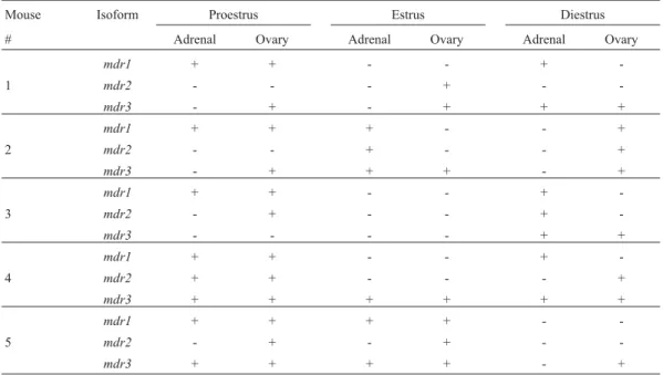
Table 1 shows the results obtained for the presence or absence of mdr isoforms in the adrenal gland and ovary, for each estrous phase analyzed. The average similarity coeffi-cients (SSMe SJ) for the five females in the different estrous cycle phases are shown in Table 2.
In the adrenal gland it can be observed that, whereas
al-though the detection of mdr2 and mdr3 was not variable (considering presence x absence) among the cycle phases (p > 0.2,a= 0.05), mdr1 was significantly more detectable in proestrus (p < 0.01,a= 0.05).
In the ovaries, mdr3 was the most frequently ob-served isoform, and an analysis of the different phases showed that the similarity coefficient due to expression of
mdr1was 1.00 for females in proestrus, and 0.00 in estrus and diestrus (Kolmogorov-Smirnov one-sample test, p < 0.01,a= 0.05).Mdr2expression was rarely seen, and expression ofmdr1was observed in the absence ofmdr3
expression.
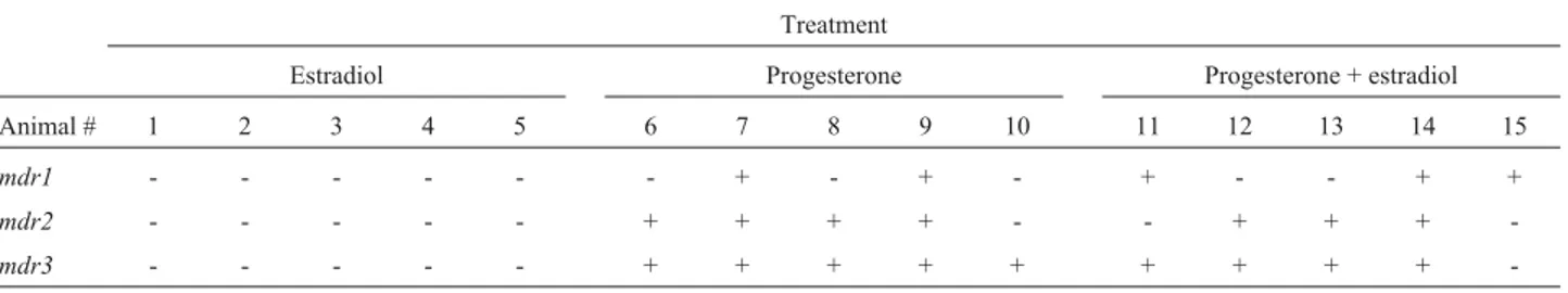
Expression of mdr isoforms in the uterus of females stimulated with steroid hormones
The expression of mdr genes was analyzed in the uterus of the six females in the control group, three of which were ovariectomized. The only isoform observed
was mdr3, in two of the ovariectomized females (not shown).
Results for the mice treated with hormones are pre-sented in Table 3. Nomdrexpression was detected in any of the five ovariectomized animals treated with estradiol. All mdr isoforms were frequently observed in ovariectomized females treated with progesterone. Estrogen plus progester-one also appears to induce the expression of the different mdr isoforms in the uterus of ovariectomized females. In the last case,mdr1expression was observed independently of mdr3, contrasting with the progesterone treatment, wheremdr1expression, when detected, was concomitant withmdr3expression. Both progesterone and estrogen plus progesterone treatments inducedmdrexpression as com-pared to the control group (c2y= 5.689, p = 0.017,a= 0.05)
Discussion
In rodents, the estrous cycle averages 4 to 5 days. The first phase is known as proestrus (proliferation) and is cyto-- Presence (+) or absence (cyto--) ofmdrisoforms in the adrenal gland and ovary of females in different phases of the estrous cycle.
Mouse Isoform Proestrus Estrus Diestrus
# Adrenal Ovary Adrenal Ovary Adrenal Ovary
mdr1 + + - - +
-1 mdr2 - - - + -
-mdr3 - + - + + +
mdr1 + + + - - +
2 mdr2 - - + - - +
mdr3 - + + + - +
mdr1 + + - - +
-3 mdr2 - + - - +
-mdr3 - - - - + +
mdr1 + + - - +
-4 mdr2 + + - - - +
mdr3 + + + + + +
mdr1 + + + + -
-5 mdr2 - + - + -
-mdr3 + + + + - +
Table 2- Average Jaccard Similarity Coefficient for themdrisoforms in the adrenal gland and ovary in proestrus, estrus and diestrus.
Isoform Coeff. Proestrus Estrus Diestrus
Adrenal Ovary Adrenal Ovary Adrenal Ovary
mdr1 SSM 1.00 1.00 0.40 0.60 0.40 0.60
SJ 1.00 1.00 0.20 0.00 0.30 0.00
mdr2 SSM 0.60 0.40 0.60 0.40 0.60 0.40
SJ 0.00 0.30 0.00 0.10 0.00 0.10
mdr3 SSM 0.40 0.60 0.40 0.60 0.40 1.00
SJ 0.20 0.60 0.30 0.60 0.30 1.00
S SSM 0.67 0.67 0.47 0.53 0.47 0.67
logically characterized by the predominance of nucleated epithelial cells, which are round, bear an easily visible nu-cleus and may appear in clusters or individually. Peaks of estradiol and progesterone secretion occur in this phase, which lasts about 12 h. The following phase is the estrus (sexual receptivity), which lasts 26 h and is cytologically characterized by large numbers of cornified squamous ir-regularly shaped epithelial cells, occurring in clusters. The next phase is the diestrus (sometimes divided into diestrus I and diestrus II), of relative sexual rest and in which the ovarian secretions prepare the reproductive tract for receiv-ing the fertilized egg. If fertilization does not occur, the ani-mal returns to proestrus. The diestrus lasts from two and a half to three days and is cytologically characterized by the predominance of small leukocytes interspersed by a few nucleated epithelial or cornified squamous epithelial cells (Knobil and Neil, 1994). Our results concerning the differ-ential expression of themdrgenes along the various phases of the estrous cycle in ovaries and adrenal gland suggest an involvement of the P-glycoprotein in the secretion of ste-roid hormones. Special attention is given tomdr1, which presents a progesterone-responsive element on its first un-translated exon (Pierkarzet al., 1993).
High expression levels of themdr1gene in the adre-nal gland have been reported in mice (Croopet al., 1989), although Bradleyet al. (1990) did not detectmdr1 expres-sion in the adrenal gland of Chinese hamster females. As there are no reports in the literature aboutmdrexpression during the estrous phases of animals, our results may ex-plain these contrasting results, showing that in females the adrenal gland does not display detectable expression of the
mdr genes during most of the estrous cycle (estrus and diestrus), whereas in proestrus, possibly related to steroid synthesis, the expression ofmdr1appears to be increased. This hypothesis is supported by the report of Altuviaet al. (1993), who observed that an increase in the steroid bio-synthesis, induced by ACTH, resulted in an increase in the level of expression ofmdr1in a murine adrenal gland cell line. In line with this finding, Séréeet al. (1998) observed that the inhibitory effect of dexamethasone on adreno-corticotropin hormone (ACTH) production can explain the decreasedmdr1expression. The rare expression of mdr1
during estrus and diestrus would be mostly related to its role as an adjuvant to mdr3 in detoxification rather than in hormone secretion.
mdr3 was the most frequently observed isoform in the ovaries, irrespective of the estrous cycle. Formdr1, how-ever, expression was much higher in proestrus (SJ= 1.00), indicating its phase-related regulation. In the proestrus ova-ries, it was also possible to observe the expression ofmdr1
in the absence ofmdr3expression.Although this data con-cerns only one animal, this was never observed in ovaries before, as discussed in our previous study, where we stated thatmdr1andmdr3expression were always concomitant (Schiengoldet al., 2001). Noteworthy, considering the ho-mologous genes to mousemdr1andmdr3in humans, is that the activation of theMDR3gene seems to be independent of the activation of the closely linkedMDR1gene (van der Blieket al., 1988; Raymondet al., 1990; Chinet al., 1992).
The uterus is poor inmdrexpression, and mdr3 is the main isoform present. Estradiol-treated females did not ex-pressmdrgenes (Table 3), a result similar to that reported by Arceciet al. (1990), who employedin situhybridization and the same experimental conditions used in the present study. As opposed to other results in that same report, how-ever, in this studymdrexpression was observed in all fe-males treated with progesterone, with the expression of
mdr3in all animals.
A similar situation was observed for the ovariec-tomized females treated with progesterone and estradiol. Moreover, in the present study, in one uterus,mdr1 expres-sion was observed in the absence of mdr3 expression. Croop et al. (1989), using Northern blot, detected only mdr1 in the pregnant uterus and, although Arceci et al. (1990) believed that mdr1 was the isoform detected in their experiments, they also stated that the probes employed were not able to discriminate between the differentmdr
genes. Bello-Reuss et al. (2000) determined the role of MDR1 in the secretion of aldosterone by a human adrenal cell line. It is noteworthy that, whereas in humans MDR1 (equivalent to the murine mdr3 isoform) functions in detox-ification and in the transport of steroids, mice present two isoforms to which different function have been ascribed. mdr3 is referred to as the most effective isoform in detoxifi-cation, and mdr1 as the isoform preferably associated with the transport of steroid hormones (Yanget al., 1989; Got-tesman and Pastan, 1993). Interestingly, Taylor et al.
(1999) found no significant differences between the mdr3 and mdr1 isoforms in the nature of drug-binding sites and suggested that the presence of multiple isoforms of Pgp
al-Table 3- Presence (+) or absence (-) ofmdrisoforms in the uterus of 15 ovariectomized females submitted to different hormone treatments.
Treatment
Estradiol Progesterone Progesterone + estradiol
Animal # 1 2 3 4 5 6 7 8 9 10 11 12 13 14 15
mdr1 - - - + - + - + - - + +
mdr2 - - - + + + + - - + + +
-lows subtle quantitative and qualitative regulations of their respective cellular activity.
Mice which are homozygous for a disruption ofmdr1
ormdr3are apparently healthy (Borstet al., 1993). In 1997, Schinkel et al. obtained mice which, although homozy-gously deficient for themdr1andmdr3genes combined, were healthy and fertile. These results suggest that no strict functions ofmdr1ormdr3are essential to survival, or that the mdr2 isoform can compensate for the absence of the other isoforms (Smithet al., 2000, demonstrated that the protein encoded byMDR3, although not concerned with the MDR phenotype, can transport drugs). Also, other proteins associated to the MDR phenotype can compensate for the absence of Pgp.
Our results suggest that the functions of mdr1 and mdr3 in mice are not restricted. mdr3 is probably more effi-cient in the detoxification function. The detection ofmdr1
expression independently ofmdr3under hormonal stimula-tion and during proestrus is very surprising (according to Smitet al., 1999, themdr1andmdr3genes are linked, and hence behave essentially as one locus) and indicates that its function is closely related to the secretion of steroid hor-mones. It is also interesting to observe that in mice the three
mdrgenes are located in tandem on chromosome 5 (mdr3,
mdr1, mdr2), which suggests that transcription of more than one isoform due to an initial transcription ofmdr3may be a common event. According to Lee and Ling (2003), while little is known about the molecular mechanism gov-erning the changes in Pgp expression at the tissue level, ac-cumulated evidence suggests that post-transcriptional control at the RNA stability level plays a key role.
In conclusion, we investigated the expression of the mdr isoforms during the phases of the estrous cycle in dif-ferent organs of normal mice. We observed that only in the adrenal gland and the ovary the estrous cycle influenced the expression ofmdrgenes. In these organs we observed that
mdr2expression is rare, irrespective of the phase. All fe-males in proestrus expressed mdr1 in the adrenal gland. In the ovaries, mdr3 was the most frequently observed iso-form. mdr1 expression in the absence of mdr3 was ob-served in the ovaries and in the adrenal gland in proestrus. These results could be related to peaks of secretion of estradiol and progesterone that are seen in proestrus. In the uterus, the only isoform observed was mdr3. Estradiol does not seem to inducemdrexpression. Progesterone and estro-gen plus progesterone induced the expression of all mdr isoforms in ovariectomized females, and this last treatment may also have inducedmdr1expression alone in one ani-mal. Our results suggest that the mdr1 and mdr3 functions are overlapping. While mdr3 may be the more efficient isoform in the detoxification function, the detection of
mdr1expression independently of mdr3under hormonal stimulation indicates that its function is closely related to the secretion of steroid hormones.
Acknowledgments
We are grateful to Dr. Carmen C. R. Saavedra and Dr. Karen L. Haag for valuable comments during the prepara-tion of this work and for critically reviewing the manu-script. The present work was supported by grants from Conselho Nacional de Desenvolvimento Científico e Tec-nológico (CNPq).
References
Altuvia S, Stein WD, Goldenberg S, Kane SE, Pastan I and Gottesman MM (1993) Targeted disruption of the mouse
mdr1b gene reveals that steroid hormones enhance mdr gene expression. J Biol Chem 268:27127-27132.
Arceci RJ, Croop JM, Horwitz SB and Housman D (1988) The gene encoding multidrug resistance is induced and expres-sed at high levels during pregnancy in the secretory epithe-lium. Proc Natl Acad Sci USA 85:4350-4354.
Arceci RJ, Baas F, Raponi R, Horwitz SB, Housman D and Croop JM (1990) Multidrug resistance gene expression is con-trolled by steroid hormones in the secretory epithelium of the uterus. Mol Reprod Dev 25:101-109.
Bello-Reuss E, Ernest S, Holland OB and Hellmich M (2000) Role of multidrug resistance P-glycoprotein in the secretion of aldosterone by human adrenal NCI-H295 cells. Am J Physiol Cell Physiol 278:C1256-C1265.
Borst P, Schinkel AH, Smit JJM, Wagenaar E, Van Deemter L, Smith AJ, Eijdems EWHM, Baas and Zaman GJR (1993) Classical and novel forms of multidrug resistance and the physiological functions of P-glycoproteins in mammals. Pharmacol Ther 60:289-299.
Bradley G, Georges E and Ling V (1990) Sex-dependent and in-dependent expression of the P-glycoprotein isoforms in Chi-nese hamster. J Cell Physiol 145:398-408.
Chin KV, Chauhan SS, Abraham I, Sampson KE, Krolczyk AJ, Wong M, Schimmer B, Pastan I and Gottesman MM (1992) Reduced mRNA levels for the multidrug-resistance genes in cAMP-dependent protein kinase mutant cell lines. J Cell Physiol 152:87-94.
Croop JM, Raymond M, Haber D, Devault A, Arceci RT, Gros P and Housman DE (1989) The three mouse multidrug resis-tance (mdr) Genes are expressed in a tissue-specific manner in normal mouse tissues. Mol Cell Biol 9:1346-1350. Goldstein LJ, Fojo AT, Ueda K, Crist W, Green A, Brodeur G,
Pastan I and Gottesman MM (1990) Expression of the multi-drug resistanceMDR1gene in neuroblastomas. J Clin Oncol 8:128-136.
Gottesman MM and Pastan I (1993) Biochemistry of multidrug resistance mediated by multidrug transporter. Annu Rev Biochem 62:385-427.
Hamilton KO, Yazdanian MA and Audus KL (2001) Modulation of a P-glycoprotein activity in Calu-3 cells using steroids andb-ligands. Int J Pharm 228:171-179.
Herweijer H, Sonneveld P, Baas F and Nooter K (1990) Expres-sion ofMDR1andMDR3multidrug resistance genes in hu-man acute and chronic leukemias and association with stimulation of drug accumulation by cyclosporine. JNCI 82:1133-1140.
Kuo MT, Julian J, Husain F, Song R and Carson DD (1995) Regu-lation of multidrug resistance genemdr1b/mdr1expression in isolated mouse uterine epithelial cells. J Cell Physiol 164:132-141.
Lankas GR, Wise LD, Cartwright ME, Pippert T and Umbenhauer DR (1998) Placental P-glycoprotein deficiency enhances susceptibility to chemical induced birth defects in mice. Reprod Toxicol 12:457-463.
Lee CH and Ling V (2003) Superinduction of P-glycoprotein messenger RNAin vivoin the presence of transcriptional in-hibitors. J ExpTher Oncol 3:14-26.
Lewin J, Cooper A and Birch B (2002) Progesterone: A novel ad-junct intravesical chemotherapy. BJU International 90:736-741.
Morales MM, Capella MAM, Sanches MV, Lopes AG and Gug-gino WB (2000) Modulation of themdr-1bgene in the kid-ney of rats subjected to dehydration or a high-salt diet. Eur J Physiol 439:256-362.
Muller C, Goubin F, Ferrandis E, Cornil-Schwartz I, Bailly JD, Bordier C, Benard J, Sikic BI and Laurent G (1995) Evi-dence for transcriptional control of humanMDR1gene ex-pression by verapamil in multidrug resistance leukemic cells. Mol Pharmacol 47:51-56.
Noonan KE, Beck C, Holzmayer TA, Chin JE, Wunder JS, An-drulis IL, Gazdar AF, Willman CL, Griffith B, Von Hof DD and Roninson IB (1990) Quantitative analysis of MDR1
(Multidrug resistance ) gene expression in human tumors by polymerase chain reaction. Proc Natl Acad Sc USA 87:7160-7164.
Pauly M, Ries F and Dicato M (1992) The genetic basis of multidrug resistance. Pathol Res Pract 188:804-807. Pierkarz RL, Cohen D and Horwitz SB (1993) Progesterone
regu-lates the murine multidrug resistance mdr1b gene. J Biol Chem 268:7613-7616.
Rao US, Fine RL and Scarborough GA (1994) Antiestrogens and steroid hormones: Substrates of the human P-glycoprotein. Biochem Pharmacol 48:287-292.
Raymond M, Rose E, Housman DE and Gros P (1990) Physical mapping, amplification, and overexpression of the mouse mdr gene family in multidrug-resistant cells. Mol Cell Biol 10:1642-1651.
Schiengold M, Schwantes L, Schwartsmann G, Chies JAB and Nardi NB (2001) Multidrug resistance gene expression dur-ing the murine ontogeny. Mech Agedur-ing Dev 122:255-270. Schinkel AH, Mayer U, Wagenaar E, Mol CAAM, Van Deemter
L, Smit JJM, Van Der Valk MA, Voordouw AC, Spits H, Van Tellingen O, Zijlmans JM, Fibbe WE and Borst P (1997) Normal viability and altered pharmacokinetics in mice lacking mdr1-type (drug-transporting) P-glycopro-teins. Proc Natl Acad Sci USA 94:4028-4033.
Schneider J, Bak M, Efferth TH, Kauffmann M, Mattern J and Volm M (1989) P-glycoprotein expression in treated and un-treated human breast cancer. Br J Cancer 50:815-818. Sérée E, Villar PH, Hevér A, Guidal N, Puyoou F, Charvet B,
Point-Scomma H, Lechevalier E, Lacarelle B and Barra Y (1998) Modulation of MDR1 and CYP3A expression by dexamethasone: Evidence for an inverse regulation in adre-nals. Biochem Biophys Res Comm 252:392-395.
Shapiro AB and Ling V (1998) The mechanisms of ATP-depen-dent multidrug transport by P-glycoprotein. Acta Physiol Scand Suppl 643:227-234.
Smit JW, Huisman MT, van Tellingen O, Wiltshire HR and Schinkel AH (1999) Absence or pharmacological blocking of placental P-glycoprotein profoundly increases fetal drug exposure. J Clin Invest 104:1441-1447.
Smith AJ, van Helvoort A, van Meer G, Szabó K, Welker E, Szakács G, Váradi A, Sarkadi B and Borst P (2000) MDR3 P-glycoprotein, a phosphatidylcholine translocase, trans-ports several cytotoxic drugs and directly interacts with drugs as judged by interference with nucleotide trapping. J Biol Chem 275:23530-23539.
Sneath PHA and Sokal RR (1973) Numerical Taxonomy: The Principles and Practice of Numerical Classification. W.H. Freeman and Company, San Francisco, 513 pp.
Sokal RR and Rohlf FJ (1995) Biometry. W.H. Freeman and Company, New York, 887 pp.
Taylor JC, Ferry DR, Higgins CF and Callaghan R (1999) The equilibrium and kinetic drug binding properties of the mouse O-go1a and Pgp1b P-glycoproteins are similar. Br J Cancer 81:783-789.
Uhr M, Holsboer F and Müller MB (2002) Penetration of endoge-nous steroid hormones corticosterone, cortisol, aldosterone and progesterone into the brains is enhanced in mice defi-cient for bothmdr1aandmdr1bP-glycoproteins. J Neuro-endocrinol 14:753-759.
Ushigome F, Takanaga H, Matsuo H, Yanai S, Tsukimori K, Nakano H, Uchiumi T, Nakamura T, Kuwano M, Ohtani H and Sawada Y (2000) Human placental transport of vinblas-tine, vincrisvinblas-tine, digoxin and progesterone: Contribution of P-glycoprotein. Eur J Pharmacol 408:1-10.
van der Bliek AM, Baas F, van der Velde-Koertz T, Biedler JL, Meyers MB, Ozols RF, Hamilton TC, Joenje H and Borst P (1988) Genes amplified and overexpressed in human multi-drug-resistant cell lines. Cancer Res 48:5927-5932. Vollrath V, Wielandt AM, Acuna C, Duarte I, Andrade L and
Chianale J (1994) Effect of colchicine and heat shock on multidrug resistance gene and P-glycoprotein expression in rat liver. J Hepatol 21:754-763.
Yang CPH, Cohen D, Greenberger LM, Hsu SIH and Horwitz SB (1989) Differential transport properties of two mdr gene products are distinguished by progesterone. J Biol Chem 265:10282-10288.