Expression of S-100, EMA, CD34 and presence
of mast cells in eight oral neuroibromas,
and a review of 127 cases of the literature.
Expressão de S-100, EMA, CD34 e presença de mastócitos em
oito neurofibromas orais e revisão de 127 casos da literatura
Francisco Artur Forte Oliveira1; Clarissa Pessoa Fernandes1; Mário Rogério Lima Mota2; Fabrício Bitu Sousa3; Malena Regina de Freitas e Silva4; Carolina Rodrigues Teóilo1; Régia Maria do S. V. Patrocínio5; Fábio Wildson Gurgel Costa6; Ana Paula Negreiros Nunes Alves7
First submission on 16/05/13; last submission on 17/07/13; accepted for publication on 17/07/13; published on 20/10/13
1. Master’s degree in Clinical Dentistry from Universidade Federal do Ceará (UFC); doctoral student of the Postgraduate Program in Dental Medicine at Faculdade de Farmácia, Odontologia e Enfermagem (FFOE)-UFC.
2. Doctorate in Pharmacology from UFC; associate professor of Stomatology and Pathology in the Dentistry Course at FFOE-UFC.
3. Post-doctoral degree in Dermatology from Faculdade de Medicina da Universidade de São Paulo (FMUSP); associate professor of Stomatology and Pathology of the Dentistry Course at FFOE-UFC.
4. Master’s degree in Clinical Dentistry from UFC; doctoral student of Oncology, Fundação Antônio Prudente, Instituto do Câncer do Ceará. 5. Master’s degree in Pathology from UFC; pathologist at UFC.
6. Doctorate in Clinical Dentistry from UFC; associate professor of Radiology in the Dentistry Course at FFOE-UFC. 7. Doctorate in Pharmacology from UFC; associate professor of Pathology in the Dentistry Course at FFOE-UFC.
ABSTRACT
Introduction: The rarity of oral neuroibromas (ONs) generates problems regarding their epidemiological and immunohistochemical characterization. Objectives:The aim of this study was to evaluate the expression of different markers in ONs and review epidemiologic data reported in the literature. Material and methods: Clinicopathologic, immunohistochemical (markers S-100, epithelial membrane antigen [EMA], CD34) and histochemical (modiied-Ziehl-Neelsen-method) studies were performed in eight cases of ON diagnosed in the Department of Pathology and Legal Medicine (DPML), Universidade Federal do Ceará (UFC), Ceará, Brazil, between 1994 and 2010.
Results: Oral neuroibromas represented 0.2% of the oral lesions diagnosed by our service in 16 years, and the buccal mucosa was the most frequent oral site (71.4%). Seven (87.5%) and 8 (100.0%) cases were positive for S-100 and CD34, respectively, and none for EMA. Mast cells were identiied in seven cases (87.5%). The literature search indicated that solitary ONs are more common and occur preferentially in females, affecting patients between 30 and 40 years old. The alveolar ridge is the most commonly involved site. Conclusion: S-100- and CD34 markers proved to be of great value as a diagnostic tool, unlike EMA staining. Identiication of mast cells in most cases suggests their involvement in this tumor pathogenesis. The clinicopathologic data retrieved from the literature enabled the establishment of a more consistent epidemiological proile.
Key words:neuroibroma; immunohistochemistry; oral cavity; mast cells.
INTRODUCTION
Neuroibroma is a slow-growing, relatively circumscribed, benign tumor. Along with schwannoma, it is one of the most common benign neoplasms of the neural sheath(2, 13, 28, 36).
Neuroibromas can occur as solitary lesions or in association with multiple other neuroibromas of the skin, café-au-lait macules, freckling in the axillary and inguinal regions, Lisch nodules,
and abnormalities of bone development as part of a syndrome known as neuroibromatosis type 1 (NF1) or von Recklinghausen
disease(12, 18, 36).
NF1 is a disorder of unknown etiology that is triggered by a mutation in the NF1 tumor suppressor gene, located on the
pathways. In the absence of neuroibromin, the RAS cascade is constitutively activated, resulting in increased cellular proliferation
and survival(14, 33, 35).
Neuroibromas are rare tumors that typically affect the skin and subcutaneous tissue. In cases of isolated neuroibromas, patients are usually diagnosed between the ages of 20 and 40 years, although patients with tumors associated with the NF1 syndrome
are usually diagnosed at younger ages(4, 18, 36). The prevalence of
isolated oral neuroibromas (ONs) is uncertain; however, in patients with the syndrome, oral manifestations of the condition occur in up to 17% of patients(23). Some studies indicate a
slight female predominance for both the syndrome and isolated cases(5, 7, 23, 28), whereas others observe no distinction between the
sexes(8). Approximately 25% of neuroibromas are found in the
head and neck region, and rare involvement of the oral cavity has been reported in sites such as the lips, tongue, buccal mucosa and alveolar ridge. Neuroibromas usually present as a supericial, painless nodule with nonspeciic clinical symptoms and resemble ibrous hyperplasia, ibromas, or lipomas(7, 23, 24, 28).
Ultrastructurally, neuroibromas exhibit signiicant
heterogeneity, largely due to the involvement of a variety of cellular constituents: Schwann cells, perineural-like cells, endoneurial ibroblasts, intermediate cells, and mast cells(15, 35). Immunohistochemistry (IHC) has been instrumental in
conirming this heterogeneity and represents an auxiliary method in the diagnosis and identiication of this neural tumor(6, 15, 29). The
S-100 protein marker has been widely used in the identiication of Schwann cells and is important for the diagnosis of neural sheath
tumors(30). However, the differential diagnosis of neuroibromas
from other lesions (schwannomas, leiomyomas, and myoibromas) requires the use of additional immunohistochemical stains and histochemical reagents to identify the several components of the
tumor(1, 11, 24, 26).
The rarity of oral cavity involvement in neuroibroma is relected by the paucity of studies that exclusively consider this tumor and its intra-oral location, what generates problems regarding the epidemiological and immunohistochemical characterization of ONs(7, 11, 28, 29).
The aim of this paper was to immunohistochemically characterize eight cases of neuroibromas located in the oral cavity using S-100, epithelial membrane antigen (EMA) and CD34 markers in addition to histochemical analyses using the modiied Ziehl-Neelsen method. A literature review highlighting the epidemiological characteristics of ONs was performed to contribute to the identiication and/or understanding of the behavior of neuroibromas.
MATERIAL AND METHODS
This study was approved by the Research Ethics Committee of Universidade Federal do Ceará (Protocol 280/05). We reviewed all cases of oral cavity neuroibromas that had been recorded between 1994 and 2010 in the iles of the Department of Pathology and Legal Medicine (DPML), UFC, Ceará, Brazil. Qualitative analyses of the microscopic characteristics of the tumors were performed by an oral pathologist (A.P.N.N.A) after the tissue was sectioned, stained with hematoxylin and eosin (HE), and prepared for histochemical (modiied Ziehl-Neelsen stain) and immunohistochemical study. Clinical data related to the patients, such as sex, age, and the tumor characteristics and anatomical site, were retrieved from the biopsy records (Table 1).
TABLE 1 – Clinical data of oral neuroibromas diagnosed between
1994 and 2010 in the DPML, Universidade Federal do Ceará, Brazil
Case nº Age (years) Sex Location
1 49 F Buccal mucosa
2 69 M Buccal mucosa
3 76 M Buccal mucosa
4 69 M Buccal mucosa
5 39 F Buccal mucosa
6 NI F Hard palate
7 41 M NI
8 NI M Inferior lip
DPML: Department of Pathology and Legal Medicine; NI: non-informed.
Immunohistochemical analyses were performed using the standard streptavidin-biotin-peroxidase method in 5-μm-thick tissue sections that had been obtained from parafin-embedded blocks and mounted on silanized microscopic slides. The steps from deparafinization to heat-induced epitope retrieval were performed with an EZ Prep Solution (Ventana Tucson, AZ, USA) for 30 minutes. The primary antibodies used in our study included S-100 protein (1:2000, DakoCytomation® Carpinteria, CA, USA), EMA (1:200, DakoCytomation® Carpinteria, CA, USA) and CD34 (1:100, DakoCytomation® Carpinteria, CA, USA). TheBenchMarkTM XT IHC/in situ hybridization (ISH) (Ventana
The positive results were expressed as the intensity (+ mild, ++ moderate or +++ intense) and extent (focal/diffuse) of staining. In addition, the term rare cells was applied to the samples in which the number of immunoreactive cells was considered very low.
To identify mast cells, a modiied Ziehl-Neelsen stain was performed. Histological slides were prepared from the 5-μm-thick sections of the parafin-embedded blocks. To deparafinize the sections, a 60ºC solution of liquid parafin and turpentine was dripped onto the histological sections and left for 20 minutes. Subsequently, the sections were hydrated with absolute ethanol and washed in running water. The sections were then stained with Ziehl Fuchsin for 50 minutes.After a second wash, the material was decolorized with 5% sulfuric acid until the sections were pale pink. Finally, the slides were counterstained, washed rapidly in running water, dried and mounted.
The histochemical reaction of the modiied Ziehl-Neelsen stain was evaluated with an Olympus CX 41 optic microscope. The letter P (positive reaction) was used to identify positive cases, and the letter A (absence of expression), to identify negative cases
(Table 2).
TABLE 2 – Immunohistochemical and histochemical
proile of oral neuroibromas diagnosed between 1994 and 2010 in the DPML, Universidade Federal do Ceará, Brazil
Case nº S-100 EMA CD34
Ziehl-Neelsen
1 +++, D A +++, D P
2 ++, F A ++, D P
3 +++, D A ++, D, RC A
4 A A +, D, RC P
5 +++, D A +++, D, RC P
6 ++, F, RC A +, F P
7 ++, D A ++, F, RC P
8 +++, D A +++, F P
EMA: epithelial membrane antigen; +++: intense immunoexpression; ++: moderate immunoexpression; +: mild immunoexpression; D: diffuse extent of staining; F: focal extent of staining; A: absence of expression; RC: rare cells; P: positive reaction.
In addition, we searched the literature (PubMed Database) for case series of ONs that included epidemiological data. A manual search and the key words "neuroibroma", "oral cavity", and "immunohistochemistry" were used. Only the articles published in indexed journals were considered. On the basis of this search, seven articles within the period 1971 to 2011 were selected without date restrictions and after critical analyses (Table 3).
RESULTS
ONs represented eight (0.2%) of the 3,730 oral lesions collected between 1994 and 2010 from the DPML of UFC.
Analyses of the eight cases of ONs revealed a male predominance (5:3), with patient ages varying from 39 to 76 years (mean = 57.1 ± 5.65 years). The most frequently involved intraoral site was the buccal mucosa (71.4%), and none of the lesions was associated with NF1 (Table 1).
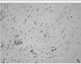
Positive immunohistochemical staining for S-100 protein
(Figure 1) was observed in seven specimens, four of which
exhibited moderate staining, and three of which stained intensely. Of the seven S-100 positive specimens, ive were diffusely stained, and two were focally stained (with one case exhibiting rare cells). EMA immunoexpression was not observed in any of the evaluated cases. All of the samples were immunopositive for CD34 (Figure 2),
and the degree of staining intensity varied from mild (two cases) or moderate (three cases) to intense (three cases). The extent of staining was diffuse in ive cases (with three cases exhibiting rare cells), and focal in three cases (with one case exhibiting rare cells). In all cases, vessels were positive for CD34 (Table 2). Histochemical analysis using a modiied Ziehl-Neelsen stain revealed a small number of mast cells that were largely localized to the perivascular region and the periphery of the lesion (Table 2).
DISCUSSION
A literature search for case studies of ONs identiied a total of 127 cases(3, 9-13, 22). ONs were observed in female patients in 64.3% of
cases, with a male to female ratio of 1.00:1.80. The mean patient
FIGURE 1 – Positive immunohistochemical expression of S-100 protein in
TABLE 3 – Clinicopathological data of 127 intraoral neuroibromas reported in the literature
within the period 1971-2011 (PubMed database). Case studies and case reports
Authors Number of cases Cases associated with NF1 N (%) Mean age (variation) years Gender (M/F) Location (n)
Cherrik and Eversole, 1971 19 4 (21) NI 7/12 Buccal mucosa (7)
Tongue (6) Palate (3) Lip (1) Alveolar ridge (1) Others♦ (1)
Chen and Miller, 1979 49 NI 39.5 (9 to 72) 21/28 Alveolar ridge* (24)
Palate (9) Tongue (6) Buccal mucosa (3) Lip (3)
Mandible (2) NI (2)
Crysomali et al., 1997 16 NI 45.1 (-) 5/10 Alveolar ridge (7)
Palate (5) Lip (2)
Buccal mucosa (1) NI (1)
Marocchio et al., 2007 11 2 (18.1) NI NI Alveolar ridge (5)
Tongue (2) Buccal mucosa (1) Lip (1)
Mandible (1) Others♦ (1)
Salla et al., 2009 12 2 (16.6) 31.2 (9 to 50) 2/10 Palate (4)
Alveolar ridge (4) Tongue (2) Lip (1) Others♦ (1)
El-Gehani et al., 2009 8 NI NI 4/4 Mandible (3)
Tongue (3) Maxilla (1) Others♦(1)
do Nacimento et al., 2009 12 3 (25) 35.7 (6 to 78) 2/10 Alveolar ridge (3)
Lip (2) Tongue (1) Maxilla (1) Buccal mucosa (1) Others♦ (4)
Total 127 11 (20.1) 37.8 (6 to 78) 41/74 Alveolar ridge (44)
Palate (21) Tongue (20) Lip (10) Mandible (6) Buccal mucosa (13) Maxilla (2) Others♦ (8)
NI (3)
NF1: neuroibromatosis type 1; M: male; F: female; NI: non-informed; *: gingival cases were included in this group; others♦: retromolar area, mental region, loor of
age was 37.8 years and varied from 6 to 78 years. The alveolar ridge was the most frequently involved intraoral site (35.4%). Solitary neuroibromas accounted for approximately 80% of cases, and 20% of cases were associated with the NF1 syndrome (Table 3).
The involvement of the oral cavity, as reported by Salla
et al.(28) and Chrysomali et al.(5), is rare. These reports are
consistent with the indings of the present study, in which only 0.2% of the oral lesions reported over a period of 16 years were neuroibromas. None of the cases was associated with the NF1 syndrome, which corroborates literature reports indicating that solitary neuroibromas are more common in the oral cavity than neuroibromas associated with NF1 (Table 3).
The literature also indicates that the tumors occur preferentially in the oral cavities of females and affect patients between the 1st and 8th decades of life, with the mean patient age
between 30and 40 years. Soft tissue was more commonly affected by neuroibromas, with the alveolar ridge, palate and tongue reported as the most commonly involved sites. The mandible was the preferred site for intraosseous involvement in the oral cavity (Table 3). In our case series, neuroibromas were more frequently diagnosed in males, but the sample was too small to suggest a predilection. The mean age of patients in our sample was 57 years, which can be explained by the predominantly adult population supported by our service. Soft tissue was exclusively affected, and the buccal mucosa was the most commonly involved site, consistent with this site being a typical location for the neoplasm.
Ultrastructural and immunohistochemical studies have been of great importance in recognizing the cellular components of neuroibromas. Schwann cells with mutations in both alleles of the NF1 gene (Nf1-/-) seem to be the initiators of neuroibroma
formation, promoting neoangiogenesis and secretion of the stimulation factor Kit ligand, which interacts with the c-kit receptor expressed by mast cells with mutation in one of the NF1 alleles
(Nf1+/-). The consequent mast cell migration causes the secretion
of the growth factors and cytokines necessary for the initiation and progression of the tumor, as well as the factors necessary for neoangiogenesis, including nerve growth factor (NGF), vascular endothelial growth factor (VEGF), ibroblast growth factor, transforming growth factor beta (TGF-β), and others.Fibroblasts, Schwann cells, perineural-like cells and endothelial cells with a mutation in one of the NF1 alleles (Nf1+/-) seem to be responsive
to the neoplastic environment(14, 33, 35).
We performed a modiied Ziehl-Neelsen stain as an effective and low-cost alternative for the identiication of mast cells(16, 22).
Mast cells were identiied in most of the cases, frequently localized to the perivascular region and the periphery of the lesions, as reported in other studies(9, 23), thereby suggesting their migration
from blood vessels and/or peripheral tissues. Mast cells can also be identiied by other histochemical techniques: for example, by the use of dyes such as toluidine blue solution(31), and by
immunohistochemistry using tryptase(27), CD 117 (c-kit) or
calretinin(11) as markers. Few studies have identiied mast cells
in neuroibromas by immunohistochemical and histochemical methods, despite the important role attributed to these cells in the pathogenesis of this lesion(14, 33, 35). The identiication of mast
cells, even in small numbers, can aid in the diagnosis of this neoplasm, particularly if the identiication is associated with other immunomarkers of neuroibromas.
Immunohistochemical markers have commonly been used in the identiication of this tumor, especially when a differential diagnosis is necessary. Lesions such as schwannomas, leiomyomas, leiomyosarcomas, myoibromas, ibrosarcomas, ibrohistiosarcomas and malignant peripheral nerve sheath tumors also exhibit spindle-shaped cells(1, 7, 20, 26).
S-100 protein is found predominantly, but not exclusively, in the nervous system. This protein is expressed by Schwann cells(5)
and is very useful for the identiication of tumors composed of these cells(15). Neuroibromas present a variable number of
S-100 positive cells that may localize to different areas of the
same tumor(5), corroborating our indings of the variable extent
of staining (diffuse or focal) for these cells. In addition, the cell nuclei and cytoplasm in the present study exhibited frequent variations in the staining intensity, which corroborates the indings of Nascimento et al.(7). Neuroibromas are immunopositive for the
S-100 protein in 85% to 100% of the occasions(19, 32), although rare
indings of negative S-100 immunostaining can occur(6), as seen
in one case in the present study.
FIGURE 2 – Positive immunohistochemical expression of CD34 in neuroibromas (LSAB 200×)
In our series, none of the cases exhibited spindle cells whose nuclei or cytoplasm stained positive for EMA. This marker is expressed in perineural cells, and normal nerves are characterized by EMA-positive cells and S-100 protein-negative cells(15). Despite
the existence, as demonstrated by ultrastructural studies, of perineural-like cells in neuroibromas, immunohistochemical studies using EMA as a marker for these cells have produced variable expression(15, 25, 38).
The CD34 antigen, expressed by hematopoietic progenitor cells of myeloid and lymphoid lineages, has also been recently identiied in peripheral nerve sheath tumors(21). CD34-positive
cells in neuroibromas are distinct from Schwann cells, perineural cells and "conventional" ibroblasts, suggesting that they may correspond to the endoneurial ibroblasts that play a supportive role for Schwann cells(15, 21). The variable expression of CD34
immunostaining in the present study corroborates other reports(15, 21, 37), and those differences in expression levels have been
attributed to the type of stroma, ibrous or myxoid(37), and the age
of the lesion(21). The CD34 staining pattern in neuroibromas is
different from the pattern seen in schwannomas. In the latter, only Antoni B areas, located near the capsule of the neoplasm, stain positive for CD34(37). Malignant peripheral nerve sheath tumors,
neurothekeomas and perineuriomas express very low levels of the CD34 marker or none at all(15, 17, 34, 37).
The antibodies used in the present study were selected from a literature review. In addition to S-100 protein, other markers, such as EMA and CD34, have been used in ultrastructural studies to highlight the heterogeneity of neuroibromas(15). However, the
shortage of immunohistochemical studies using these markers to identify intraoral tumors is relected in the relative dearth of knowledge of these immunostains and their use in the daily practice of neuroibroma diagnosis. This lack of knowledge is a complicating factor when the identiication of other components of the tumor is necessary for the differential diagnosis from other lesions.
With the rare incidence of solitary ONs and their unique histological and clinical characteristics, the diagnosis of this neoplasm is a challenge for many clinicians and oral pathologists. Therefore, we hope that the epidemiological and immunohistochemical data provided by this study will contribute to the understanding of the pathogenesis of these lesions and guide their correct diagnosis and treatment.
CONCLUSION
In the present study, the prevalence of ONs was rare, and no cases were associated with NF1 lesions. The analyzed lesions exhibited positive S-100 and CD34 expression as well as positive mast cells, which were identiied through a modiied histochemical method, and negative EMA expression. The association of these data with the clinicopathologic data of 127 cases retrieved from the literature enabled the establishment of a more consistent epidemiological proile. In addition, the immunohistochemical and histochemical aspects addressed here proved to be important to the determination of markers useful for the identiication of tumorous cells, thereby aiding in correct diagnoses.
ACKNOWLEDGMENTS
The authors are grateful to the Laboratory of Anatomopathology BIOPSE and João Carlos Silva (Department of Pathology and Legal Medicine, Universidade Federal do Ceará) for their immunohistochemical and histochemical support, respectively. In addition, the authors thank Fundação Cearense de Apoio ao Desenvolvimento Cientíico e Tecnológico (FUNCAP) and Coordenação de Aperfeiçoamento de Pessoal de Nível Superior (CAPES) for inancial support with the scholarships of postgraduate students.
RESUMO
Introdução: A rara ocorrência de neuroibromas orais (NO) gera problemas com relação à caracterização epidemiológica e imuno-histoquímica dessas lesões. Objetivos: Avaliar a expressão celular de diferentes marcadores em NO e revisar dados epidemiológicos reportados na literatura. Material e métodos: Estudos clinicopatológico, imuno-histoquímico (marcadores S-100,
epithelial membrane antigen [EMA] e CD-34) e histoquímico (método Ziehl-Neelsen modiicado) foram realizados em oito casos
de literatura indicou que NO solitários são mais comuns e ocorrem preferencialmente em mulheres, afetando pacientes entre 30 e 40 anos. O rebordo alveolar é a localização intraoral mais comum. Conclusão: Os marcadores S-100 e CD34 provaram ser de grande valor como ferramentas diagnósticas, diferente da coloração EMA. A identiicação de mastócitos na maioria dos casos sugere seu envolvimento na patogênese desse tumor. Os dados clinicopatológicos da revisão de literatura ajudaram no estabelecimento de um peril epidemiológico mais consistente.
Unitermos: neuroibroma; imuno-histoquímica; boca; mastócitos.
REFERENCES
1. AJURA, A. J.; LAU, S. H. Gingival myoibroma in children: Report of 4 cases with immunohistochemical indings. Malays J Pathol, v. 29, p. 53-6, 2007.
2. ARRIBAS-GARCÍA, I. et al. Traumatic neuroma of the inferior alveolar nerve: a case report. Med Oral Patol Oral Cir Bucal, v. 13, p. E186-8, 2008.
3. CHEN, S. Y.; MILLER, A. S. Neuroibroma and schwannoma of the oral cavity. A clinical and ultrastructural study. Oral Surg Oral Med Oral Pathol, v. 47, p. 522-8, 1979.
4. CHERRICK, H. M.; EVERSOLE, L. R. Benign neural sheath neoplasm of the oral cavity. Report of thirty-seven cases. Oral Surg Oral Med Oral Pathol, v. 32, p. 900-9, 1971.
5. CHRYSOMALI, E. et al. Benign neural tumors of the oral cavity: a comparative immunohistochemical study. Oral Surg Oral Med Oral Pathol Oral Radiol Endod, v. 84, p. 381-90, 1997.
6. DEICHLER, J. et al. Solitary intraosseous neuroibroma of the mandible. Apropos of a case. Med Oral Patol Oral Cir Bucal, v. 16, p. e704-7, 2011. 7. Do NASCIMENTO, G. J. et al. A 38-year review of oral schwannomas
and neuroibromas in a Brazilian population: clinical, histopathological and immunohistochemical study. Clin Oral Investig, v. 15, p. 329-35, 2011.
8. EL-GEHANI, R. et al. Benign tumours of orofacial region at Benghazi, Libya: a study of 405 cases. J Craniomaxillofac Surg, v. 37, p. 370-5, 2009.
9. ERLANDSON, R. A.; WOODRUFF, J. M. Peripheral nerve sheath tumors: an electron microscopic study of 43 cases. Cancer, v. 49, p. 273-87, 1982. 10. FERNER, R. E.; O’DOHERTY, M. J. Neuroibroma and schwannoma.
Curr Opin Neurol, v. 15, p. 679-84, 2002.
11. FINE, S. W.; MCCLAIN, S. A.; LI, M. Immunohistochemical staining for calretinin is useful for differentiating schwannomas from neuroibromas.
Am J Clin Pathol, v. 122, p. 552-9, 2004.
12. FRIEDMAN, J. M. Neuroibromatosis 1: clinical manifestations and diagnostic criteria. J Child Neurol, v. 17, p. 548-54, 2002.
13. GHOSH, A.; TALWAR, O. P.; PRADHAN, S. V. Tumour and tumour-like conditions of peripheral nerve origin: ten years’ experience. Kathmandu Univ Med J, v. 8, p. 97-101, 2010.
14. GOTTFRIED, O. N. et al. Molecular, genetic, and cellular pathogenesis
of neuroibromas and surgical implications. Neurosurgery, v. 58, p. 1-16, 2006.
15. HIROSE, T. et al. Immunohistochemical demonstration of EMA/ Glut1-positive perineurial cells and CD34-positive ibroblastic cells in peripheral nerve sheath tumors. Mod Pathol, v. 16, p. 293-8, 2003. 16. HONG, S. P.; AHN, S. K. Abundant eosinophil iniltration in a neuroibroma. Am J Dermatopathol, v. 29, p. 187-9, 2007.
17. IDE, F. et al. Comparative ultrastructural and immunohistochemical study of perineurioma and neuroibroma of the oral mucosa. Oral Oncol,
v. 40, p. 948-53, 2004.
18. JETT, K.; FRIEDMAN, J. M. Clinical and genetic aspects of neuroibromatosis 1. Genet Med, v. 12, p. 1-11, 2010.
19. JOHNSON, M. D.; GLICK, A. D.; DAVIS, B. W. Immunohistochemical evaluation of Leu-7, myelin basic-protein, S100-protein, glial-ibrillary acidic-protein, and LN3 immunoreactivity in nerve sheath tumors and sarcomas. Arch Pathol Lab Med, v. 112, p. 155-60, 1988.
20. JORDAN, R. C.; REGEZI, J. A. Oral spindle cell neoplasms: a review of 307 cases. Oral Surg Oral Med Oral Pathol Oral Radiol Endod, v. 95,
p. 717-24, 2003.
21. KHALIFA, M. A. et al. What are the CD34+ cells in benign peripheral nerve sheath tumors? Double immunostaining study of CD34 and S-100 protein. Am J Clin Pathol, v. 114, p. 123-6, 2000.
22. KIM, Y. D. et al. Giant ibroepithelial polyp of the glans penis. Korean J Urol, v. 50, p. 619-21, 2009.
23. MAROCCHIO, L. S. et al. Sporadic and multiple neuroibromas in the head and neck region: A retrospective study of 33 years. Clin Oral Investig, v. 11, p. 165-9, 2007.
24. OHNO, J. et al. Solitary neuroibroma of the gingiva with prominent differentiation of Meissner bodies: a case report. Diagn Pathol, v. 5, p. 61, 2010.
25. PERENTES, E. et al. Expression of epithelial membrane antigen in perineurial cells and their derivatives. An immunohistochemical study with multiple markers. Acta Neuropathol, v. 75, p. 160-5, 1987.
26. REDDY, B. et al. Leiomyoma of the mandible in a child. J Oral Maxillofac Pathol, v. 15, p. 101-4, 2011.
27. RIBATTI, D. et al. Tryptase-positive mast cells and CD8-positive T cells
in human endometrial cancer. Pathol Int, v. 61, p. 442-4, 2011. 28. SALLA, J. T. et al. Retrospective analysis of oral peripheral nerve sheath tumors in Brazilians. Braz Oral Res, v. 23, p. 43-8, 2009.
29. SALLA, J. T. et al. Immunohistochemical study of GLUT-1 in oral peripheral nerve sheath tumors. Oral Dis, v. 14, p. 510-3, 2008. 30. SHARMA, S. et al. Benign nerve sheath tumors: a light microscopic,
electron microscopic and immunohistochemical study of 102 cases.
MAILING ADDRESS
Francisco Artur Forte Oliveira
Rua Padre Mororó, 1.813, casa 32; Farias Brito; CEP: 60015-221; Fortaleza-CE, Brazil; Tel.: (85) 9656-6343/3281-0801; e-mail: [email protected]. 31. SHIBATA, N. et al. Solitary neuroibroma without neuroibromatosis
in the superior tarsal plate simulating a chalazion. Graefes Arch Clin Exp Ophthalmol, v. 250, p. 309-10, 2011.
32. SOUZA, L. B. et al. Neuroibroma paciniano: relato de um caso raro de localização intra-oral. Rev Bras Otorrinolaringol, v. 69, p. 851-4, 2003.
33. STASER, K.; YANG, F. C.; CLAPP, D. W. Mast cells and the neuroibroma microenvironment. Blood, v. 116, p. 157-64, 2010.
34. VERED, M. et al. Classic neurothekeoma (nerve sheath myxoma) and cellular neurothekeoma of the oral mucosa: immunohistochemical proiles. J Oral Pathol Med, v. 40, p. 174-80, 2011.
35. VISKOCHIL, D. H. It takes two to tango: mast cell and Schwann cell interactions in neuroibromas. J Clin Invest, v. 112, p. 1791-3, 2003.
36. WANG, B. Y.; ZAGZAG, D.; NONAKA, D. Tumors of the nervous system. In:Barnes, L. Surgical pathology of the head and neck. New York: 2009. p. 669-772.
37. WEISS, S. W.; NICKOLOFF, B. J. CD-34 is expressed by a distinctive cell population in peripheral nerve, nerve sheath tumors, and related lesions.
Am J Surg Pathol, v. 17, p. 1039-45, 1993.