Chediak-Higashi syndrome:
case report in afro-descendant individual
Síndrome de Chédiak-Higashi: relato de caso em indivíduo afrodescendente
Judde Lacerda Andrade Carlos1; Márcio Vasconcelos Oliveira2; Claudio Lima Souza2
First submission on 23/03/14; last submission on 09/04/14; accepted for publication on 11/04/14; published on 20/06/14
1. Pharmacist.
2. PhD in Public Health and Epidemiology at Universidade Federal de Minas Gerais (UFMG); assistant professor II at Instituto Multidisciplinar em Saúde at Universidade Federal da Bahia (IMS-UFBA).
ABSTRACT
This is a Chediak-Higashi Syndrome (CHS) case report in afro-descendant individual, male, 3 months old, born from consanguineous union. On admission he had fever for a month, unresolved pneumonia, and hepatosplenomegaly. He evolved to bacterial sepsis, septic shock, and death. CHS presents quantitative and morphological and hematological changes. Abnormal leukocyte inclusions are the pathognomonic inding of the disease; its recognition and differentiation from other leukocyte inclusions is essential for diagnosis and institution of therapy. Early diagnosis of CHS increases the life expectancy of the individual and provides appropriate therapeutic approach for patients affected by the disease.
Key words: Chediak-Higashi syndrome; immunodeiciency; afro-descendant; diagnosis.
INTRODUCTION
Chediak-Higashi Syndrome (CHS) is a primary immunodeiciency with autosomal recessive heritage, more common in the presence of inbreeding, and very rare in black(5). CHS is the result of a series of genetic changes, and the
main one is the mutation in the LYST gene(3, 7), which provides
irregular clustering of lysosomes, affecting hematopoietic cells, renal tubular cells, neurons, Schwann cells, melanocytes, and ibroblasts(25). This condition leads to several complications, such
as prolonged and recurrent infections, due to phagocytes primary dysfunction, tendency to bleeding, progressive neurological involvement, lymphoproliferative syndrome, and oculocutaneous albinism(7, 8, 10, 11, 24). In leukocytes, there is a formation of giants
azurophic granules, that are organized in a linear fashion, similar to rosary beads, and are pathognomonic feature of the disease(5, 12, 18, 24).
This study was performed with the family consent, by signing the Informed Consent form and approved by the Research Ethics Comittee on Human Beings from the Instituto Multidisciplinar
em Saúde, campus Anísio Teixeira-Universidade Federal da Bahia
(IMS/CAT-UFBA) under number 330.657 on June 25th, 2013.
CASE REPORT
Male, 3 months old, afro-descendant, irst-born of consanguineous marriage, was admitted to Emergency Room at General Hospital, come from smaller city agreed by Sistema Único de Saúde ([SUS] – Brazilian National Health System). The main complain was fever for one month. Physical examination revealed hepatomegaly and splenomegaly, 8 to 10 cm from the respective costal margins.
On admission, he was diagnosed with pulmonary infection, although he was in antimicrobial treatment, as prior medical prescription. During hospitalization, with antibiotics and supportive care, the patient had recovered infection, but remains hospitalized due to persistent neutropenia and need of investigation for diagnosis of the underlying condition. Throughout the
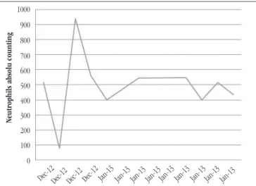
hospitalization period, patient presented neutropenia (mean
494/mm3) (Figure 1), thrombocytopenia (mean 13,477/mm3), and
elevated C-reactive protein (CRP) (mean 54.79 mg/l), besides low prothrombin activity (international normalized ratio [INR] = 1.64),
and elevated aspartate aminotransferase (AST) (mean 130.4 U/l). Patient had increased serum levels of total bilirubin serum levels = 3.06 mg/dl and direct fraction = 2.1 mg/dl, due to infectious cholestasis and coagulopathy, and persistent
thrombocytopenia, mean = 13,477/mm3, and reduced prothrombin
activity, mean = 61.3%, (INR = 1.36), consistent with infectious cholestasis and worsening of the infectious process(3, 17, 25).
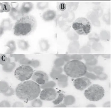
On the ifth day of hospitalization (DOH), the CBC showed abnormal leukocytes granules in patient’s peripheral blood smear, stained by Wrigth method (Figure 2). In this case report, inclusions were described as intracytoplasmic giant granules in all neutrophil-lineages, suggesting CHS investigation.
FIGURE 1 – Severe neutropenia in patient with CHS in the presence of infection
CHS: Chediak-Higashi syndrome.
FIGURE 2 – Photomicrograph of peripheral blood smear of CHS patient
Single giant azurophilic granules in lymphocyte (2A), multiple giant azurophilic granules in neutrophil (2B) and monocyte (2C) (Wright, 100×). CHS: Chediak-Higashi syndrome.
A
B
C
On the 34th day of hospitalization, he showed fever peek, facial
swelling, diarrhea, abdominal distension, and e tachypnea. On the 36th day of hospitalization, he was transferred to Intensive Care Unit
(ICU), where new antimicrobial therapeutic was implemented, including broad-spectrum drugs (meropenem) and measures of supportive therapy.
Two blood cultures and one catheter tip culture were performed during hospitalization. The result of irst blood culture
was positive, and Staphylococcus epidermidis was isolated,
which presents high potential to produce bioilm, especially in immunocompromised patients. After establishing the treatment, new blood and catheter tip cultures were performed, and both results were negative.
On the 41st day of hospitalization, he presented another
fever peek, abdominal distension, and diarrhea. And, despite the imposition of necessary support measures, there were worsening of general condition with severe tachypnea and hypotension,
which culminated in cardiac arrest and death in the 45th day of
hospitalization. The cause of death was reported as sepsis caused by CHS complication.
DISCUSSION
It is a rare disease, with 200 cases described in the literature(15),
and it is especially rare in black people(5).During our search in the
U.S. National Library of Medicine (PUBMED) baseline, in March 2014, we found seven cases of CHS diagnosis in black children. CHS diagnosis may be dificult, since characteristic clinical and laboratorial indings are not well deined, or are not properly reported by laboratorians or well interpreted by the prescribing physician.
Neutrophils absolu counting
Dec-12Dec-12 Dec-12 Dec-12 Jan-13 Jan-13 Jan-13 Jan-13Jan-13 Jan-13 Jan-13 Jan-13Jan-13
1000
900
800
700 600 500 400
300 200 100
In general, patients with CHS seek health service due to recurrent infections in early, and the disease is diagnosed when they are about 5 years old(1, 5). This period depends on disease
presentation and severity. In this case, the clinical presentation was early identiied, with pulmonary infection, not responsive to irst-line antibiotics, and persistent fever. Increased susceptibility to infections comes from phagocyte primary dysfunction, which keeps its ability to phagocytosis of pathogens; but digestion is not effective due to lysosomal deiciency(7, 8, 10, 11, 24).
Patients suffering from this disease have an average life expectancy of 10 years, if hematological symptoms of the disease are not treated(1, 5). In case reported, the patient progressed quickly
and unsatisfactorily, with recurrent infections caused by resistant microorganisms likely enough, due to his stay in the nosocomial environment. Although cultures kept negative results, patient presented persistently increased levels of CRP, indicative inding of inlammatory or infectious process, consistent with the case, considering the related immunodeiciency, that does not change the systemic inlammatory response against the pathogen, but it strongly affects phagocytic activity(4, 10, 11, 13).
Hematological dysfunction in CHS causes important changes in CBC, such as qualitative/morphological and quantitative abnormalities(5, 12, 18, 24). Neutropenia persisstende is observed by
increasing apoptosis, throughout hospitalization, evolving to febrile neutropenia. On the ifth day of hospitalization, it was observed giant azurophilic lysosomal granules in the neutrophils and other leukocytes cytoplasm(2, 4, 10, 11).
Establishing a diagnosis, goes through a suitable description of inclusions in CBC report, so that there is an accurate investigation and diagnostic differentiation from other hypotheses, such as Hermansky-Pudlak Syndrome (HPS), and Griscelli syndrome, which exhibit symptoms similar to CHS, differing only in regards to granular leukocytes(9, 14).
CHS lysosomal granules may be easily differentiated from typical toxic granulation of severe infectious processes (Figure 3), which represent benign response, helping neutrophil phagocytosis, and they are markers of infection severity(13). These granules are
present during the process, and cease in stimulus absence. CHS lysosomal granules are not restricted to neutrophils (Figure 2B), they may be present in lymphocytes (Figure 2A) and monocytes (Figure 2C), in addition, they are larger and rougher, and are present in all stages of disease, with or without symptomatology(12, 13, 16).
Other inclusions that may get confused with CHS are Alder Reilly inclusions, which are more common in individuals with Tay-Sachs disease and mucopolysaccharidosis, which present azurophils variable in size and shape from spherical to ellipsoid, that may
hide nucleus cell(16, 22). May-Hegglin Anomaly is another condition
that presents leukocytes inclusions, in which the spindle granules are similar to Döhle bodies, which are typically peripheral, bright blue basophils, and irregular contour(16).
FIGURE 3 – Photomicrograph of peripheral blood smear
Toxic granulation in neutrophils citoplasm (3A and 3B) and normal azurophilic granules in immature granulocytic precursors (3C) (Wright, 100×).
A
B
C
Platelets in CHS are also altered and have less ability to aggregate due to adenosine diphosphate and serotonin deiciency, resulting from Delta-storage pool deiciency, that inluences the quantity and content of dense-granules in megakaryocytes and platelets, and may result in thrombocytopenia(7, 23), exacerbated in
septic processes, consistent to patient outcome.
CHS course with periods of acceleration and chronicity of the disease. The accelerated phase is a complication with obscure and usually fatal prognosis, which must be avoided(1, 3, 11, 18).
Changes in hepatic markers may be directly related to the disease, or as the result of frequent infections. At this phase, activated T CD8 lymphocytes and macrophages iniltrate viscera in several organs, especially the highly perfused ones, leading to increase in transaminase and decrease in liver activity, also contributing to decrease in prothrombin activity(1, 3, 17).
The treatment considered curative for CHS is bone marrow transplantation (BNT)(6, 12, 19, 20), which should occur in the stable of
disease, although some authors assert that BNT is healing only for the hematological symptoms of disease(6, 21), showing no interference
CHS early and correctly diagnosis is especially important, due to complications during clinical course of disease, and its need for appropriate therapy. It is noteworthy that since CHS is a rare disorder, it may affect patients of different ages and ethnicities, including afro-descendant, as the case reported, and impose different degrees of immunodeiciency(3).
The accurate description of the CHS abnormal giant azurophilic granules, that are peroxidase-positive, is decisive for diagnosis. This fact requires clinical analyst to improve knowledge and skills necessary to differentiate leukocytes abnormalities, since
cytomorphological diagnosis is essential in the disease differential diagnosis process.
This report is of particular relevance since it presents CHS occurrence in an afro-descendant individual. As the occurrence is rare in this race, cytomorphologic differentiation with correct statement of characteristic leukocyte inclusions, and laboratory indings associated with the disease, may contribute decisively, together with clinical indings, to differential diagnosis and subsequent better clinical management.
RESUMO
Trata-se de relato de caso de síndrome de Chediak-Higashi (SCH) em indivíduo afrodescendente, sexo masculino, 3 meses de idade, filho de união consanguínea. Apresentava na admissão febre há um mês, pneumonia não resolvida e hepatoesplenomegalia. Evoluiu para sepse bacteriana, choque séptico e óbito. A SCH apresenta alterações hematológicas, morfológicas e quantitativas. As inclusões leucocitárias anormais constituem achado patognomônico da doença e seu reconhecimento e sua distinção de outras inclusões leucocitárias é fundamental para diagnóstico e instituição da terapêutica. O diagnóstico precoce da SCH aumenta a expectativa de vida do indivíduo e proporciona abordagem terapêutica adequada aos pacientes acometidos pela doença.
Unitermos: Chediak-Higashi; imunodeficiência; afrodescendente; diagnóstico.
REFERENCES
1. ABU SHARIF, M. A. et al. Chédiak-Higashi syndrome: an accelerated phase with hereditary elliptocytosis: case report and review of the
literature. Ann Saudi Med, v. 21, n. 3-4, p. 221-4, 2001.
2. BOXER, L. A. et al. Improvement of Chediak-Higashi leukocyte function by cyclic guanosine monophosphate. Blood, v. 49, n. 1, p. 9-17, 1977. 3. CERTAIN, S. et al. Protein truncation test of LYST reveals heterogenous mutations in patients with Chediak-Higashi syndrome. Blood, v. 95, n. 3, p. 979-83, 2000.
4. CLARK, R. A.; KIMBALL, H. A.; PADGETT, G. A. Granulocyte chemotaxis in the Chediak-Higashi syndrome of mink. Blood, v. 39, n. 1, p. 644-9, 1972.
5. COLLA, V. A. et al. Síndrome de Chediak-Higashi – relato de caso e revisão de literatura. Rev Bras Alerg Imunopatol, v. 21, n. 3, p. 83-90, 1998.
6. EAPEN, M. et al. Hematopoietic cell transplantation for
Chediak-Higashi syndrome. Bone Marrow Transplant, v. 39, n. 1, p. 411-5, 2007. 7. FAGUNDES, F. V. B. Aspectos clínicos dos pacientes com imunodeiciencia primária submetidos a transplante de células progenitoras hematopoiéticas em um centro de referência do Estado do Rio de Janeiro. Rio de Janeiro. 2010. Tese de mestrado. Instituto Fernandes Figueira/ Fundação Oswaldo Cruz.
8. FANTINATO, G.T. et al. Voce conhece esta síndrome? Síndrome de Chediak-Higashi. An Bras Dermatol, v. 86, n. 5, p. 1029-38, 2011. 9. GAHL, W. A. et al. Genetic defects and clinical characteristics of patients with a form of oculocutaneous albinism (Hermansky-Pudlak Syndrome).
N Engl J Med, v. 338, n. 18, p. 1258-64, 1998.
10. GRUMACH, A. S.; PIRES, R. B.; SAMPAIO, M. M. S. C. Distúrbios primários de fagócitos. Apresentação de 16 casos. Ped (São Paulo), v. 10, n. 3, p. 125-30, 1988.
11. JESSEN, B. et al. Subtle differences in CTL cytotoxicity determine susceptibility to hemophagocytic lymphohistiocytosis in mice and humans with Chediak-Higashi syndrome. Blood, v. 118, n. 17, 2011. 12. JÚNIOR, P. R. Imunodeiciências primárias: aspectos relevantes para o pneumologista. J Bras Pneumol, v. 35, n. 10, p. 1008-17, 2009. 13. MELLO, W. A.; SILVA, J. O. Sepse: importância do laboratório clínico no diagnóstico. Rev Multidisc Saúde, v. 1, n. 2, 2009.
14. MÉNASCHÉ, G.; FISCHER, A.; BASILE, G. S. Griscelli syndrome types 1 and 2. Am J Hum Genet, v. 71, n. 1, p. 1237-8, 2002.
15. ORPHANET REPORT SERIES. Rare Diseases collection. November 2013. Number 1: Listed in alphabetical order of disease or group of diseases. Availabte at: <http://www.orpha.net/orphacom/cahiers/docs/GB/Prevalence_of_rare_ diseases_by_alphabetical_list.pdf>. Accessed on: 15 jan. 2014.
MAILING ADDRESS
Claudio Lima Souza
Universidade Federal da Bahia; Instituto Multidisciplinar em Saúde; Campus Anísio Teixeira; Rua Rio de Contas, 58, Quadra 17, Lote 58; Candeias; CEP: 45.029-094; Vitória da Conquista-BA, Brazil; Phone: +55 (77) 3429-2709/9954-0509; e-mail: [email protected].
17. PAGE, A. R. et al. The Chediák-Higashi syndrome. Blood, v. 20, p. 330-43, 1962.
18. SEIXA, A. M. I. et al. Síndrome de Chediak-Higashi: relato de caso e revisão da literatura. An Bras Dermatol, v. 74, n. 6, p. 605-9, 1999. 19. ROSÁRIO FILHO, N. A. et al. Albinismo parcial com imunodeiciência.
Rev Bras Alerg Imunopatol, v. 21, n. 1, p. 28-31, 1998.
20. SPRITZ, R. A. Genetic defects in Chediak-Higashi syndrome and the
beige mouse. J Clin Immunol, v. 18, n. 2, 1998.
21. TARDIEU, M. et al. Progressive neurologic dysfunctions 20 years after allogeneic bone marrow transplantation for Chediak-Higashi syndrome.
Blood, v. 106, p. 40-2, 2005.
22. VIEIRA, T. A. et al. Granulações de alder-reilly em pacientes com mucopolissacaridose VI. Rev HCPA, v. 28, n. 2, p. 128-9, 2008.
23. VIZCARGÜÉNAGA, M. I. Síndrome de pool de depósito. Revisión. Presentación de estudios de laboratório. Acta Bioquím Clín Latinoam, v. 40, n. 3, p. 327-34, 2006.
24. WHITE, J. G. The Chediak-Higashi syndrome: a possible lysosomal
disease. Blood, v. 28, p. 143-56, 1966.
25. WINDHORST, D. B.; PADGETT, G. The Chediak-Higashi syndrome