417
ORIGINAL ARTICLEResearch on antimicrobial residues activity in urine
samples of hospitalized patients
Pesquisa da atividade de resíduos de antimicrobianos em
amostras de urina de pacientes hospitalizados
Daiane Cardozo1; Gislene Maria Botão Kussen2; Laura Lúcia Cogo3
First submission on 24/10/14; last submission on 24/11/14; accepted for publication on 24/11/14; published on 20/12/14
1. Degree in Generalist Pharmacy by Centro de Ensino Superior dos Campos Gerais; multidisciplinary resident in Hospital Care by Women’s Health Program at Hospital de Clínicas da Universidade Federal do Paraná (HC-UFPR).
2. MSc in Internal Medicine by UFPR; pharmaceutical biochemistry at Bacteriology Department of Diagnosis Support Unit-HC-UFPR. 3. PhD in Biotecnological Processes by UFPR; pharmaceutical biochemistry at Bacteriology Department of Diagnosis Support Unit-HC-UFPR.
ABSTRACT
Introduction: Urinary tract infection is quite frequent in a hospital environment, and the urine culture is the gold standard for diagnosis of this disease, because it allows bacterial identiication and performing antimicrobial susceptibility testing. Culture-negative urine samples result of patients with strong suspicion of infection may occur due to the activity of antimicrobial residues, which can interfere with bacterial growth in vitro and produce false-negative results. Objective: Verify the occurrence of
false-negative urine cultures due to the presence of antimicrobial residues in samples of patients admitted to the Clinical Hospital of Paraná Federal University. Material and methods: A total of 188 urine samples from hospitalized patients were randomly selected, during the period from July to December 2012. All samples were evaluated on the result of the urine culture, bacteriuria, and research on residues of antimicrobial activity by manual and automated techniques. Results: 44 (23.4%) presented positive urine culture, 121 (64.4%) negative urine culture, and 23 (12.2%) presented growth of many species. In 14 samples, negative urine cultures associated with the presence of bacteria and were positive for the research on antimicrobial residues activity (RARA), were observed. Conclusion:
Automated technique showed better performance when compared to manual technique, with sensitivity of 92.8% and 71.4%, respectively. The presence of antimicrobial residues may affect the recovery of bacteria in the urine, producing a false-negative result.
Key words: research on antimicrobial residues activity; urinary tract infection; urine culture.
J Bras Patol Med Lab, v. 50, n. 6, p. 417-420, dezembro 2014
INTRODUCTION
The urinary tract infection (UTI) is one of the most common infectious disease in humans, it occurs at high frequency in both inpatients and outpatients; it represents an important cause of morbidity and mortality(1, 7, 8).
The clinical course of the disease is related to the virulence of microorganism involved, host resistance, effectiveness of used clinical and antimicrobial therapy(2, 12). These factors make the
appropriate diagnosis an important tool for infection resolution(6).
The UTI diagnosis is performed based on the clinical condition of the patient and laboratory tests. The routine monitoring tests used for UTI diagnosis are: reactive strip,
analysis urine test
, smear microscopy, sediment analysis, and urine culture, which is considered the gold standard(5). The isolation and identiicationof bacteria, together with the antimicrobial susceptibility testing performance, are useful in the choice of drug therapy(1).
Samples of patients with strong suspicion of UTI that produced negative urine culture results may raise uncertainty about the reliability of the results released by the laboratory. Some studies suggest that the presence of inhibitory substances, such as antimicrobial residues in urine samples can interfere with the
in vitro growth of the
probable
bacteria causing the infection, producing a false-negative result(10).
OBJECTIVE
In order to verify the occurrence of false-negative urine cultures due to the presence of antimicrobial residues that interfere
418
with bacterial growth in vitro, this study aimed to research, using two methods, the activity of such substances in urine samples of patients admitted to the Clinical Hospital of the Paraná Federal University (HC-UFPR).
MATERIAL AND METHODS
Sampling
During the period from July to December 2012, a total of 188 midstream spontaneous urine specimen were randomly selected from patients admitted to HC-UFPR hospitalized. The samples sent to the Bacteriology and Urinalysis section of the Diagnostic Support Unit of the same Institution.
The study was approved by the Research Ethics Committee at HC-UFPR under CAAE record 06301912.5.0000.0096.
All urine samples were processed for bacteriuria detection, research on antimicrobial residues activity (RARA) by manual and automated techniques, besides culture in Petri dishes containing Cystine Lactose Electrolyte Deicient agar ([CLED] – MERCK, Darmstadt, Germany), which were incubated at 35-37ºC for 18 - 24 hours.
Bacteriuria detection
The direct presence of bacteria in the urine sample was veriied by three different methods: automated count performed in UF-1000i® equipment (SYSMEx - Kobe, Japan), direct
visualization, and Gram stain of a drop of uncentrifuged urine.
Manual technique for RARA
Sterile ilter paper discs (6-mm diameter) were soaked with 10 µl aliquots of the urine to be tested. They were placed in an incubator to dry at 56ºC for one hour. Standard strains of Escherichia coli
ATCC 25922 and Enterococcus faecalis ATCC 29212 were used in the
bacterial suspension preparation with standardized concentration according to 0.5 McFarland scale tube. The discs were placed on Petri dishes containing Mueller-Hinton Agar (MERCK, Darmstadt, Germany), previously seeded with the bacterial suspensions. After 18 to 24 hours of incubation at 35-37ºC the tests were read. The formation of an inhibition zone around the disc, i.e., the absence of growth around the disc was considered a positive result. The halo size was measured in millimeters using pachymeter(10).
For technical veriication, positive control (10 µg ampicillin disc) and negative control (disc soaked with sterile saline) were performed.
Automated technique for the research on
antimicrobial residues
The automated research was performed using Alfred 60®
equipment (ALIFAX - Padua, Italy) that offers the residual antimicrobial activity test (RAA). In this test, 500 µl of the urine sample were inoculated into a lask containing an enriched solution and a Staphylococcus epidermidis suspension, previously added.
The lasks were incubated under constant shaking and controlled temperature at 37ºC, for approximately 3 hours. Bacterial growth was monitored in real-time by turbidity compared to McFarland Scale. The same turbidity observed early in the reaction that does not change over time, indicates the absence of bacterial growth, resulting in positive RAA. The increase in suspension turbidity due to bacterial growth was reported as negative RAA test.
For method validation, we added 10 µg/µl Gentamicin Solution to positive control lask, and 500 µl of sterile saline to negative control lask.
RESULTS
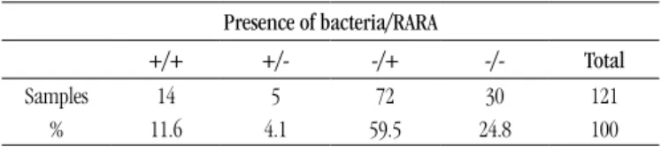
From the 188 studied samples, 44 (23.4%) showed positive urine culture, 121 (64.4%) showed negative urine culture, and 23 (12.2%) were considered contaminated because of the development of many bacterial species.
The negative cultures results associated with the presence of bacteriuria and RARA are inserted in Table 1.
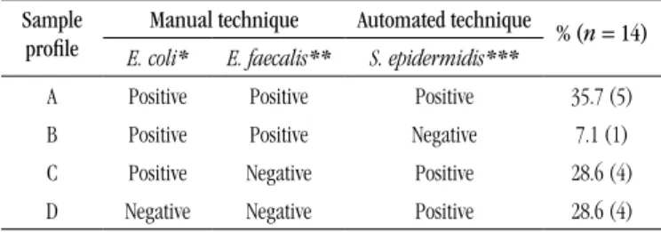
From the 14 samples that showed presence of bacteria and positive RARA, different results were obtained from the techniques evaluated, and were divided in four proiles. Table 2 shows the proile of results and the percentage found, according to the microorganism used.
RARA was detected by all the techniques in 64.3% of samples (proile A + C). When analyzing the different microorganisms tested, we observed positivity for the three strains in only 35.7% of samples (proile A). S. epidermidis strain was more sensitive to antimicrobial activity detection, because in 92.8% of samples (proile A + C + D) the test was positive, followed by E. coli ATCC 25922 with 71.4% of positivity (proile A + B + C), and E. faecalis
ATCC 29212 with 42.8% (proile A + B) of positivity. Daiane Cardozo; Gislene Maria Botão Kussen; Laura Lúcia Cogo
TABLE 1 – RARA and bacteriuria in negative urine culture samples
Presence of bacteria/RARA
+/+ +/- -/+ -/- Total
Samples 14 5 72 30 121
% 11.6 4.1 59.5 24.8 100
419
Research on antimicrobial residues activity in urine samples of hospitalized patients
DISCUSSION
Urine samples represent a large volume of specimens daily submitted to the microbiology laboratory for evaluation. Several factors such as use of antimicrobials, vaginal contamination and urethral colonization, may inluence culture result and should be carefully evaluated during the exam to ensure its quality.
The positivity rate found in this study (23.4%) for urine culture was similar to that reported by other authors(3, 14),
ranging from 15.9% to 22.8%. Most samples showed negative culture result, which is consistent with many studies showing that up to 80% of samples submitted for culture has no bacterial growth, or show negligible growth(9, 15). This may
be associated with excessive request iled by clinical material easily accessible, and due to UTI be one of the major infections acquired in hospitals environment(17). However, the negative
result must be carefully evaluated and correlated with other laboratory indings, such as the presence of bacteria and previous antimicrobial therapy(16, 18).
The presence of bacteria and RARA positive in 14 samples with negative culture suggest a false negative result, representing 7.45% of studied samples (n = 188). In the study carried out by Botão et al., cited by Kussen(10), the presence of antimicrobial residues was detected
in 1% of urine samples, this value is lower than we found in this study. However, their sample included urine of both inpatients and outpatients, whereas in this study, only samples of hospitalized patients were included, in whom antimicrobial therapy is more widely used.
The presence of bacteria associated to negative result for RARA was detected in 4.1% of samples (n = 5), amounting 2.66% of
samples (n = 188). This proile may occur due to an infection
caused by anaerobic microorganisms that are not recovered from routine culture, or by low-growth bacteria that require speciic diagnostic tests and appropriate isolation method(13).
Other researchers also report the potencial presence of non-viable bacterial cells in urine(15).
In the other samples evaluated, absence of bacteriuria
and positive RARA occurred in 59.5% (n = 72), and absence of
bacteriuria and negative RARA occurred in 24.8% (n = 30), which
characterized evidenced negative results, because bacteria were not detected in the three different methods used in the study.
The evaluation of the samples with positive RARA, negative
culture and bacteriuria (n = 14), the manual technique had a
sensitivity of 71.4% (n = 9), and the automated technique
had a sensitivity of 92.8% (n = 13). The low sensitivity of the
manual technique can be explained by the small sample volume used for agar diffusion method(4).
The use of E. faecalis strain did not increase the positivity of manual technique, demonstrating that this one, in subsequent studies, may be abolished or replaced. Some studies suggest the use of different microorganisms, such as Micrococcus luteus, Staphylococcus aureus, Bacillus stearothermophilus, among
others, to improve the sensitivity of the method(4, 11).
Recent reports also suggest that the results of RARA may be interfered
according to
the antimicrobial class used, due to the presence of inert metabolites excreted in urine, or due to the use of antibiotics to treat infections in other anatomical locations(16, 18). These studies reinforce the need for accurate information onantibiotic therapy at the time of exam request, since the use of antibiotics limits the sensitivity of the culture(18).
CONCLUSION
In this study, we demonstrated that in 7.45% of studied samples, the presence of antimicrobial residues compromised the recovery of uropathogens, producing a possible false negative result. The automated technique showed better performance when compared to the manual technique.
The research on antimicrobial residues activity can be considered a useful tool to rule out urinary infection, mostly in patients with bacteriuria which are under antimicrobial therapy, therefore, we suggest the implementation of the RARA as routine in microbiology laboratories.
AKNOWLEDGMENT
for the Diagnosis Support Unit of HC-UFPR for providing the structure for the accomplishment of the research, and to Alifax for materials and inputs supply.
TABLE 2 – Classiication of bacteriuria and culture-negative samples accord -ing to the proile found in RARA
Sample profile
Manual technique Automated technique % ( n = 14) E. coli* E. faecalis** S. epidermidis***
A Positive Positive Positive 35.7 (5)
B Positive Positive Negative 7.1 (1)
C Positive Negative Positive 28.6 (4)
D Negative Negative Positive 28.6 (4)
420
REFERENCES
1. BROEREN, M. A. C. et al. Screening for urinary tract infection with the Sysmex UF-1000i urine low cytometer. J Clin Microbiol, v. 49, n. 3, p. 1025-9, 2011.
2. CARVALHAL, G. F.; ROCHA, L. C. A.; MONTI, P. R. Urocultura e exame comum de urina: considerações sobre sua coleta e interpretação. Revista da AMRIGS, v. 50, n. 1, p. 59-62, 2006.
3. COSTA, L. C. et al. Infecções urinárias em pacientes ambulatoriais: prevalência e peril de resistência aos antimicrobianos. RBAC, v. 42, n. 3, 2010.
4. DRISCOLL, A. J. et al. Disk diffusion bioassays for the detection of antibiotic activity in body luids: applications for the pneumonia etiology research for child health project. Clin Infect Dis, v. 54, supl. 2, p. 159-64, 2012.
5. FOSTER, R. T. Uncomplicated urinary tract infections in women. Obstet Gynecol Clin North Am, v. 35, p. 235-48, 2008.
6. GRIEBLING, T. L. Urologic diseases in America project: trends in resource use for urinary tract infections in women. J Urol, v. 173, p. 1281-7, 2005.
7. HÖRNER, R. et al. Prevalência de microrganismos em infecções do trato urinário de pacientes atendidos no Hospital Universitário de Santa
Maria. RBAC, v. 38, n. 3, p. 147-50, 2006.
8. JOLKKONEN, S. et al. Screening of urine samples by low cytometry reduces the need for culture. J Clin Microbiol, v. 48, n. 9, p. 3117-121, 2010. 9. KELLOGG, J. A. et al. Clinical relevance of culture versus screens for the detection of microbial pathogens in urine specimens. Am J Med, v. 83, n. 4, p. 739-45, 1987.
RESUMO
Introdução: A infecção do trato urinário é bastante frequente em ambiente hospitalar, e a cultura de urina é padrão-ouro para o diagnóstico dessa doença, pois permite a identificação bacteriana e a realização do teste de suscetibilidade aos antimicrobianos. Amostras de urina de pacientes com forte suspeita de infecção que resultam em cultura negativa podem ocorrer devido à atividade de resíduos de antimicrobianos, os quais podem interferir no crescimento bacteriano in vitro e gerar resultados falso negativos. Objetivo:
Verificar a ocorrência de culturas de urina falso negativas devido à presença de resíduos de antimicrobianos em amostras de pacientes internados no Hospital de Clínicas da Universidade Federal do Paraná. Material e métodos: Um total de 188 amostras de urina de pacientes internados foi selecionado aleatoriamente, durante o período de julho a dezembro de 2012. Todas as amostras foram avaliadas quanto ao resultado da cultura de urina, da bacteriúria e da pesquisa da atividade de resíduos de antimicrobianos por meio das técnicas manual e automatizada. Resultados: Quarenta e quatro (23,4%) apresentaram cultura de urina positiva, 121 (64,4%), cultura negativa e 23 (12,2%), crescimento de várias espécies. Em 14 amostras foi observada cultura negativa associada à presença de bactérias e pesquisa da atividade de resíduos de antimicrobianos (PRA) positiva. Conclusão: A técnica automatizada apresentou melhor desempenho quando comparada com a técnica manual, apresentando sensibilidade de 92,8% e 71,4%, respectivamente. A presença de resíduos de antimicrobianos pode comprometer a recuperação de bactérias na urina, gerando resultado falso negativo.
Unitermos: pesquisa da atividade de resíduos de antimicrobianos; infecção do trato urinário; cultura de urina.
10. KUSSEN, G. M. B. Pesquisa de substâncias antimicrobianas em urinas destinas a cultura. In: ALBINI, C. A. A.; SOUZA, H. A. P. H. M.; SILVEIRA, A.
C.O. (Org.). Infecções urinárias: uma abordagem multidisciplinar. 1. ed.
Curitiba: Editora CRV, 2012. p. 579-83.
11. LIU, Y. C. et al. Detection of antimicrobial activity in urine for epidemiologic studies of antibiotic use. J Clin Epidemiol, v. 52, n. 6, p. 539-45, 1999.
12. LUCCHETTI, G. et al. Infecções do trato urinário: análise da frequência e do peril de sensibilidade dos agentes causadores de infecção do trato urinário em pacientes com cateterização vesical crônica. J Bras Patol Med Lab, v. 41, n. 6, p. 383-9, 2005.
13. McCARTER, Y. S. et al. Cumitech - laboratory diagnosis of urinary tract infections. American Society of Microbiology Press, 2009. 14. MULLER, E. V.; SANTOS, D. F.; CORRÊA, N. A. B. Prevalência de microrganismos em infecções do trato urinário de pacientes atendidos no laboratório de análises clínicas da Universidade Paranaense-Umuarama-PR. RBAC, v. 40, n. 1, 2008.
15. OKADA, H. et al. Enumeration of bacterial cell numbers and detection of signiicant bacteriuria by use of a new low cytometry-based device. J Clin Microbiol, v. 44, n. 10, p. 3596-9, 2006.
16. SRIRANGARAJ, S.; KALI, A.; CHARLES, M. V. P. Antibiotic screening of urine culture as a tool for internal quality audit. Australas Med J, v. 7, n. 2, p. 73-7, 2014.
17. VIEIRA, J. M. S. et al. Suscetibilidade antimicrobiana de bactérias isoladas de infecções do trato urinário de pacientes atendidos no Hospital Universitário Bettina Ferro de Souza, Belém PA. RBAC, v. 39, n. 2, 2007. 18. WILSON, G.; BADARUDEEN, S.; GODWIN, A. Antibiotic screening of urine culture as a useful quality audit. J Infect Dev Ctries, v. 5, n. 4, p. 299-302, 2011.
MAILING ADDRESS
Daiane Cardozo
Seção de Bacteriologia da Unidade de Apoio Diagnóstico do Hospital de Clínicas da UFPR; Rua Padre Camargo, 280; Alto da Glória; CEP 80060-240; Curitiba-PR, Brazil; e-mail: [email protected]