Comparison among three cold staining methods in the
primary diagnosis of tuberculosis: a pilot study*
,**
Comparação entre três métodos de coloração a frio no diagnóstico primário de tuberculose: um estudo piloto
Soham Gupta, Vishnu Prasad Shenoy, Indira Bairy, Sethumadhavan Muralidharan
Abstract
Objective: In developing countries, sputum smear microscopy is the main tool for pulmonary tuberculosis case finding. The objective of the present study was to evaluate the diagnostic efficacy of Gabbett’s staining (GS) and modified cold staining (MCS), both of which are two-step methods, in comparison with that of fluorescent staining
(FS), which is a three-step method, for the detection of AFB in sputum smears. Methods: Our sample comprised
260 sputum samples collected from individuals suspected of having pulmonary tuberculosis at Kasturba Hospital, in Manipal, India. Smears were prepared in triplicate: one each for FS, MCS, and GS. The smears were randomly
numbered so that the examiner was blinded to the sample identities. Results: Of the 260 samples, 16 (6.15%),
15 (5.77%), and 13 (5.00%) showed positive AFB results with FS, MCS, and GS, respectively. The sensitivity of GS and MCS, in comparison with that of FS, was 81.25% and 93.75%, respectively. The concordance of GS and MCS with FS was good (0.988 and 0.996, respectively), and no statistically significant differences were found.
Conclusions: Although MCS and GS were found to be less sensitive than was FS, which is evaluated under
fluorescence microscopy, the first two are promising methods for the diagnosis of tuberculosis.
Keywords: Tuberculosis, pulmonary; Diagnostic techniques and procedures; Microscopy, fluorescence; Sputum.
Resumo
Objetivo: Em países em desenvolvimento, a baciloscopia é a principal ferramenta para a identificação de casos de
tuberculose pulmonar. O objetivo do presente estudo foi avaliar a eficácia diagnóstica do método de coloração de Gabbett (MCG) e de um método modificado de coloração a frio (MMC), ambos em duas etapas, em comparação com a do método de coloração fluorescente (MCF), em três etapas, para a detecção de BAAR em esfregaços de
escarro. Métodos: Nossa amostra consistiu de 260 amostras de escarro coletadas de casos suspeitos de tuberculose
pulmonar no Kasturba Hospital, em Manipal, Índia. Os esfregaços foram preparados em triplicata, para cada um dos métodos: MCF, MMC e MCG. As lâminas foram numeradas aleatoriamente a fim de que o examinador fosse
cegado quanto à identidade das amostras. Resultados: Das 260 amostras, 16 (6,15%), 15 (5,77%) e 13 (5,00%)
foram positivas para BAAR com MCF, MMC e MCG, respectivamente. A sensibilidade de MCG e MMC em relação à de MCF foi de 81,25% e 93,75%, respectivamente. Houve boa concordância de MCG e MMC com MCF (0,988 e 0,996, respectivamente), e não houve diferenças estatísticas significativas. Conclusões: Embora MCG e MMC apresentaram menor sensibilidade que MCF, que é avaliado por microscopia de fluorescência, consideramos que os dois primeiros métodos sejam promissores no diagnóstico de tuberculose.
Descritores: Tuberculose pulmonar; Técnicas de diagnóstico e procedimentos; Microscopia de fluorescência;
Escarro.
*Study carried out in the Department of Microbiology, Kasturba Medical College, Manipal, India.
Correspondence to: Soham Gupta. Department of Microbiology, St. John’s Medical College, Bangalore, 560034, Karnataka, India. Tel 91 80 2206-5052. E-mail: [email protected]
Financial support: None.
Submitted: 23 January 2010. Accepted, after review: 10 May 2010.
with freshly filtered auramine-phenol (0.3 g of auramine and 3 mL of phenol in 97 mL of distilled water) for 7-10 min without heating. The smears were washed in running water and destained with a 3% acid/alcohol solution for 3-5 min. The slides were washed in running water and counterstained with a 0.1% potassium permanganate solution for 1 min, followed by washing and air-drying.(13)
For the preparation of smears to be submitted to GS, samples of each specimen were air-dried and heat-fixed. The slides were then flooded with 1% carbol-fuchsin stain (10 g of basic fuchsin, 100 mL of methylated spirit, and 50 g of phenol), and distilled water was added to make a final volume of 1,000 mL. This was allowed to stand at room temperature for 10 min. The smears were then washed in running water and counterstained with Gabbett’s methylene blue (1 g of methylene blue, 20 mL of sulfuric acid, 30 mL of absolute alcohol, and 50 mL of distilled water) for 2 min. The slides were then washed and air-dried.(1)
For the preparation of smears to be submitted to MCS, samples of each specimen were air-dried and heat-fixed. The slides were flooded with carbol-fuchsin stain, as for GS, for 5 min. The smears were then washed in running water and counterstained with a modified decolorizing counterstain (0.25 g of methylene blue, 25 mL of absolute alcohol, 10 mL of glycerol, 0.01 g of potassium hydroxide, 4.5 mL of glacial acetic acid, and 3 mL of hydrochloric acid), and distilled water was added to make a final volume of 100 mL. This was allowed to stand at room temperature for 3 min. The slides were then washed and air-dried.(6)
The triplicate smears from each specimen (FS, GS, and MCS smears) were randomly numbered so that the examiner was blinded to the sample identities, thereby ruling out selective bias. All of the smears were read by an experienced examiner, under oil immersion microscopy for GS and MCS and under fluorescence microscopy with a ×40 objective for FS (Leica, Wetzlar, Germany). The smears were read and classified as 3+, 2+, 1+, scanty, or negative, in accordance with the RNTCP guidelines.(7,13) After the smears had been read, the results were documented and, at the end of the study, the smear results were decoded and cross compared.
Introduction
Tuberculosis is a disease of great importance in developing countries, such as India, where it has caused considerable morbidity and mortality. The control of tuberculosis is defined as the reduction of its transmission, which reduces morbidity and mortality, and every case finding is quite important to maintaining this control. Sputum smear microscopy continues to be the main tool for case finding in developing countries.(1-6)
In India, the Ziehl-Neelsen (Z-N) method is the procedure recommended in the Revised National Tuberculosis Control Programme (RNTCP) guidelines.(7) However, the Z-N method is cumbersome and poses various operational problems, since it requires the application of heat during staining.(1,2,6)
For the proper implementation of a tuberculosis control program, as well as for overcoming the problems associated with the Z-N method, improvements and simplifications have long been sought. Various cold staining methods have been evaluated, and some have shown promising results.(1-6,8,9) Gabbett’s staining (GS) and, more recently, modified cold staining (MCS) have been advocated as alternative staining techniques, because they do not require a heat source during staining and they eliminate the decolorizing step.(1,2,6,8,10) The fluorescent staining method (FS, evaluated under fluorescence microscopy), which is also a cold staining method, is a rapid and reliable method for the detection of AFB and has been found to be more sensitive than is the Z-N method.(3-5,11,12)
The objective of the present study was to comparatively evaluate the diagnostic efficacy of these three cold staining methods in the detection of AFB.
Methods
Our sample comprised 260 sputum samples collected from individuals suspected of having pulmonary tuberculosis at Kasturba Hospital, located in the city of Manipal, India. The smears of each sample were prepared in triplicate; one each for FS, GS, and MCS.
were no significant differences between these methods and FS.
The smear results obtained with GS and MCS were compared. Two smears read as negative with GS were read as positive with MCS (scanty and 1+, respectively). The sensitivity, specificity, positive predictive value, and negative predictive value of GS in relation to MCS were, respectively, 86.66%, 100.00%, 100.00%, and 99.19%. There was good overall concordance between the two methods (0.992), and no statistically significant difference was found.
Discussion
Tuberculosis is a major public health problem, and its control has become a challenge in developing countries, such as India.(14) Case finding is quite important for the control of the disease and is chiefly achieved by sputum smear microscopy. Although culture is considered the gold standard, it needs a proper laboratory setting and takes longer, making its use impractical in resource-poor settings.
In India, in accordance with the RNTCP guidelines, the Z-N method is performed at primary health care facilities.(7) However, the Z-N method poses problems, such as the need for a regular supply of spirit/liquid petroleum gas, which is used for heating. It is also a
Results
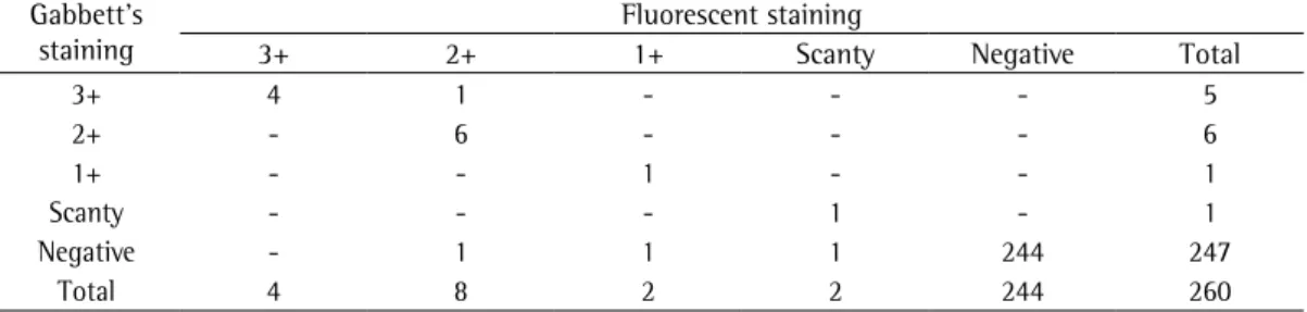
Among the 260 samples, 16 (6.15%), 15 (5.77%), and 13 (5.00%) showed positive AFB results with FS, MCS, and GS, respectively. Since FS showed the greatest number of positive results, it was considered the gold standard. Positive smears were confirmed by restaining all of the FS smears with the Z-N method. The smear results obtained with GS and MCS were compared with those obtained with FS, as shown in Tables 1 and 2.
All of the specimens that were AFB-positive with GS and MCS were also AFB-positive with FS, whereas the number of samples that were AFB-positive with FS but AFB-negative with GS and MCS was, respectively, 3 and 1. The 3 samples read as AFB-negative with GS were classified, respectively, as 2+, 1+, and scanty with FS, whereas the sample read as AFB-negative with MCS was classified as 1+ with FS.
In the cross-comparison of GS and MCS with FS, the sensitivity, specificity, positive predictive value, and negative predictive value of GS were, respectively, 81.25%, 100.00%, 100.00%, and 98.79%, whereas those of MCS were 93.75%, 100.00%, 100.00%, and 99.59%, respectively. The overall concordance of GS and MCS with FS was good (0.988 and 0.996, respectively). There
Table 1 - Comparison between Gabbett’s staining and fluorescent staining in terms of the AFB smear results.
Gabbett’s staining
Fluorescent staining
3+ 2+ 1+ Scanty Negative Total
3+ 4 1 - - - 5
2+ - 6 - - - 6
1+ - - 1 - - 1
Scanty - - - 1 - 1
Negative - 1 1 1 244 247
Total 4 8 2 2 244 260
Table 2 - Comparison between the modified cold staining method and the fluorescent staining method in
terms of the AFB smear results. Modified cold
staining
Fluorescent staining
3+ 2+ 1+ Scanty Negative Total
3+ 3 - - - - 3
2+ 2 5 - - - 7
1+ - 2 - - - 2
Scanty - - 1 2 - 3
Negative - - 1 - 244 245
be considered a promising tool for case findings in field research. Further studies should be conducted in order to establish and evaluate a reliable staining technique that is suitable for use in resource-poor settings in developing countries.
References
1. Gokhale S, Qadir S, Nagra JS, Chakraborty AK. Efficiency of cold staining method of AFB in sputum - a comparison with Ziehl Neelsen Method under field condition. Indian J Tuberc. 1990;37(3):135-7.
2. Selvakumar N, Gomathi M, Rehman F, Narayanan PR. Evaluation of a two-reagent cold staining method for detection of acid-fast bacilli. Int J Tuberc Lung Dis. 2002;6(8):728-31.
3. Ulukanligil M, Aslan G, Tasçi S. A comparative study on the different staining methods and number of specimens for the detection of acid fast bacilli. Mem Inst Oswaldo Cruz. 2000;95(6):855-8.
4. Ziaee M, Namaei M, Khazae M, Azarkar G. Comparison of the value of two different sputum staining for diagnosis of acid fast bacilli. Iranian J Clin Inf Dis. 2008;3(2):99-102.
5. Jain A, Bhargava A, Agarwal SK. A comparative study of two commonly used staining techniques for acid fast bacilli in clinical specimens. Indian J Tuberc. 2002;49(3):161-2.
6. Gupta S, Prasad V, Bairy I, Muralidharan S. Comparative evaluation of two cold staining methods with the Ziehl-Neelsen method for the diagnosis of tuberculosis. Southeast Asian J Trop Med Public Health. 2009;40(4):765-9.
7. TBC India [homepage on the Internet]. New Delhi: Ministry of Health and Family Welfare [cited 2008 Dec 3]. Manual for Laboratory Technicians. [Adobe Acrobat document, 76p.] Available from: http://www.tbcindia. org/pdfs/Module%20for%20Laboratory%20Technician. pdf
8. Tansuphasiri U, Kladphuang B. Evaluation of sputum staining by modified cold method and comparison with Ziehl-Neelsen and fluorochrome methods for the primary diagnosis of tuberculosis. Southeast Asian J Trop Med Public Health. 2002;33(1):128-35.
9. Rao KP, Naganathan N, Nair SS. A cold staining method for tubercle bacilli using chloroform. Indian J Tuberc. 1966;14:3-9.
10. Reuben J, inventor. Stain for acid-fast bacilli. United States patent US 4857459. 1989 Aug 15.
11. Kumar R, Agarwal M, Prasad M. Demonstration of acid fast bacilli in sputum. Indian J Tuberc. 1979;26(1):17-20.
12. Ba F, Rieder HL. A comparison of fluorescence microscopy with the Ziehl-Neelsen technique in the examination of sputum for acid-fast bacilli. Int J Tuberc Lung Dis. 1999;3(12):1101-5.
13. TBC India [homepage on the Internet]. New Delhi: Ministry of Health and Family Welfare [cited 2009 Sep 29]. Manual for Sputum Smear Fluorescence Microscopy. [Adobe Acrobat document, 16p.] Available from: http:// www.tbcindia.org/pdfs/Flourescence_Microscopy%20 Manual.pdf
14. Rao V G. Tuberculosis control: Current status and challenges. RMRCT Update. 2007;4(2):1-4.
cumbersome procedure and can be hazardous. Various attempts have been made to develop a cold staining technique in order to omit the step that involves the heating of carbol-fuchsin.(1,2,6)
At present, fluorescence microscopy appears to be an effective, rapid, and reliable tool for case finding and is more sensitive than is the Z-N method. It is also easier to perform,(3-5,11,12) but the cost-effectiveness of this technique needs to be considered, because it has high maintenance costs, including that of the ultra high pressure mercury lamp.
Various two-step cold staining methods, in which the heating step has been omitted and the deco lorizing and counters-taining steps have been combined, have been evalu a ted.(1,2,8,10) Among such techniques, GS has gained worldwide acceptance, and MCS has shown promising results in comparisons against Z-N.(6)
In the present study, we evaluated GS and MCS, comparing them with FS. With FS, AFB appear as slender bright yellow fluorescent rods, standing out clearly against a dark background. With the other two methods, AFB appear as delicate images that more closely depict their original morphology but are fainter, which might explain the false-negative results. However, we cannot ignore the possibility that FS also produced false-positive results, since culture was not performed. Therefore, to rule out false-positive FS results, all of the FS smears were restained with the Z-N method.
In our study, we obtained more positive results with FS. However, a study conducted in Thailand showed better results with GS than with FS.(8) Although MCS was less sensitive than FS, MCS was faster (< 10 min) and showed better sensitivity than did GS. In terms of cost, there is a negligible difference between GS and MCS, but FS is more expensive than are the other two methods, because of its maintenance and lamp costs. In addition, the preparation of smears with MCS was found to be easily performed; any referral laboratory can prepare the reagents and distribute them to the peripheral laboratories. Furthermore, MCS is considerably faster than is GS and FS, allowing more time to be spent on the examination of smears.
About the authors
Soham Gupta
Senior Research Fellow. Department of Microbiology, St. John’s Medical College, Bangalore, India.
Vishnu Prasad Shenoy
Selection Grade Lecturer. Department of Microbiology, Kasturba Medical College, Manipal, India.
Indira Bairy
Head. Department of Microbiology, Kasturba Medical College, Manipal, India
Sethumadhavan Muralidharan