Late complication of a renal calculus: fistulisation to
the psoas muscle, skin and bronchi
_______________________________________________
Ziga Snoj
1, Nenad Savic
1, Jaka Regvat
11 Radiology Institute, University Medical Centre, Zaloška 2, Ljubljana, Slovenia
ABSTRACT
_______________________________________________________________________________________
Kidney disease presenting with cutaneous fistula is a rare condition. We present a case of a 90-year-old woman with dementia who had no prior urological problems and had a cutaneous fistula in the left lumbar region. A fistulogram and computer tomography examination revealed a large staghorn calculus with signs of xanthogranulomatous pyelonephri-tis in the left kidney and renal fistulisation to the psoas muscle, skin and bronchi. To our knowledge this is the first report in the literature of coexisting renal fistulisation to the psoas major muscle, skin and bronchi. This report illustrates how computed tomography in combination with fistulography can resolve the diagnostic dilemma that pertains to the complex spread of the disease in cases involving nephrocutaneous fistula. Furthermore, the report shows how a renal calculus, even asymptomatic, can cause a serious medical condition, and highlights the importance of early medical intervention.
INTRODUCTION
Retroperitoneal and psoas muscle absces-ses are common complications in renal disease. Fistula formation between the kidney and its ad-jacent organs has been reported in the literatu-re due to a number of causes including surgical complications, infection, trauma and stone disease (1). However, nephrocutaneous fistula (NCF) and nephrobronchial fistula (NBF) are rare manifesta-tions in renal disease. To our knowledge this is the first report in the literature of coexisting renal fistulisation to the psoas muscle, skin and bronchi.
CASE REPORT
the patient’s good general condition no diagnostic procedures were conducted. In the last few days prior to admission the daughter noticed that the patient was getting weaker, had no appetite, had a dry cough and was unable to walk due to severe left-sided back pain and coxalgia. The patient had no history of urological problems. On admission the patient was subfebrile (37.7ºC) with no evident respiratory distress. Laboratory examination sho-wed anaemia (haemoglobin, 8.7g/L), leukocytosis (17.1x10⁹/L) and a raised CRP value (220mg/L). The results of the kidney function tests were normal.
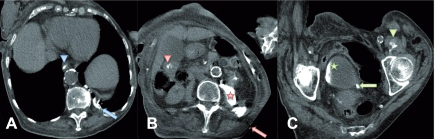
communicating with the renal pelvis and contrast was excreted into the bladder (Figure-2 C). NBF was communicating with the bronchi of the pos-terobasal segment of the left lower lobe through the lumbocostal trigone of the diaphragm (Figu-re-2 A). Contrast was observed in the left main bronchus and trachea. Furthermore, contrast was
also observed in the oesophagus, stomach and duodenum after expectoration from the bronchial tree (Figures 2 A, B and C). Caudal contrast leaka-ge was shown retroperitoneally along the whole length of the psoas and the iliacus muscle, all the way to the lesser trochanter (Figure-2 C). A mor-phologically deformed kidney was observed.
Figure 2 - The computer tomographic scan performed immediately after the fistulogram; the patient was positioned on her right flank. A) contrast in the oesophagus (blue arrowhead) and communication with the left lower lobe of the lung (blue arrow). B) cutaneous fistula (red arrow), contrast retroperitoneally (red star) and in the duodenum (red arrowhead). C) contrast in bladder (green star), ureter (green arrow) and in the psoas muscle (green arrowhead) just proximally to lesser trochanter.
Detailed examination of the deformed kid-ney and the caudal contrast leakage was perfor-med with a follow-up CT scan five days later with intravenous contrast application. A large staghorn calculus was observed within the collecting sys-tem of the enlarged left kidney (Figures 3 A and B). Non-enhancing areas of the dilated collecting system, surrounded by enhancing and extremely atrophic parenchyma, were observed in the left kidney–a finding known as the “bear paw” sign (Figure-3 A). These CT findings of the deformed left kidney are typical for xanthogranulomatous pyelonephritis. The caudal contrast leakage was found to be a retroperitoneal abscess fistulising along the psoas muscle and the iliacus muscle, forming a large pelvic abscess (Figure-3 C). The right kidney was normal.
E. coli grew from fistular smear and urine cultures. The QuantiFERON test was negative. The inflammatory markers were found to be depleting after the initial empirical therapy with fluclo-xacillin and ciprofloxacin that were changed to specific antibiotic coverage with piperacillin and tazobactam. Considering the general condition
DISCUSSION
We report on a unique case of triple renal fistulisation. To our knowledge this is the first re-port in the literature with fistulisation to the psoas muscle, skin and bronchi as a late complication of a renal calculus.
A psoas muscle abscess is a relatively rare condition with renal disease being the cause of abs-cess formation in 17.5% of cases (2, 3). Sponta-neous fistulisation to the adjacent organs due to in-fectious or inflammatory disease is not uncommon. However, fistulisation to the skin and bronchi wi-thout previous renal surgery is very rare. A Medline search revealed 16 cases of spontaneous NCF since 1983 and 24 cases of NBF since 1956. Furthermo-re, we found only two reported cases of coexistent
NCF and NBF (4, 5) and none for coexisting fistuli-sation to the psoas muscle, skin and bronchi.
been cultured from the sputum, urine and kidney specimens in patients with NCF and NBF (7). Ho-wever, E. coli and Proteus species account for ap-proximately one third of cases (8).
A final diagnosis of renal disease was not made since the patient was not a suitable candida-te for surgery. A definicandida-te diagnosis of XGP is his-tological (9); however the CT scan of our patient was highly demonstrative of diffuse XGP showing a severely enlarged kidney and staghorn calculus. These two signs, along with the “bear paw” sign and inflammatory changes in the perinephric fat, although not specific, are strongly suggestive of diffuse XGP (9).
The kidney is a retroperitoneal organ and directly related to the muscles posteriorly. The only intervening layers are perinephric fat and Gerota’s fascia. Once these are transgressed any progressive inflammatory or degenerative pro-cess involving the kidney can spread along the transversalis fascia to the posterior retroperitoneal space (10, 11). Fusion lines of fascial planes tend to direct the exudates within the retroperitoneal compartment and the inflammatory spread is usu-ally caudal (12). However, in rare cases the spread can be cranial with the potential for forming NBF. Furthermore, the lumbocostal trigone is described as a relatively weak area of the diaphragm with the possibility of the transmission of infection to the thoracic cavity (13).
In this case the renal inflammatory process spread laterally, cranially and caudally from the posterior retroperitoneal space. Laterally it trans-gressed the transversalis fascia and formed NCF just laterally of the erector spinae muscle group (Figure-2 B). Cranially, the inflammatory process transgressed the transversalis fascia and formed NBF through the lumbocostal trigone (Figure-2 A). Caudally, the inflammatory process transgres-sed the transversalis and psoas fascia and spre-ad along the psoas muscle, forming a large pelvic abscess (Figure-3 C).
There are several radiologic methods avai-lable for revealing the anatomical course of fistu-lae. In cases of renal fistulisation, a fistulogram and retrograde pyelography are usually the first step in the diagnostic procedure and are a good choice in cases where the spread of the disease is non-complicated (4, 5, 11, 14). However, in cases where the spread is complex, as observed in this case, a CT in conjunction with fistulography is the best method to establish a diagnosis.
CONCLUSIONS
Our case report demonstrates the difficul-ties encountered in diagnosing this rare condition. Furthermore, it demonstrates how a neglected cal-culus, though asymptomatic, can cause a serious medical condition and shows the importance of early medical intervention.
REFERENCES
1. Salam MA. Principles and Practice of Urology. Jaypee Brothers Medical Publishers Ltd. 2013;vol. 1, pp. 890. 2. Zissin R, Gayer G, Kots E, Werner M, Shapiro-Feinberg M,
Hertz M. Iliopsoas abscess: a report of 24 patients diagnosed by CT. Abdom Imaging. 2001;26:533-9.
3. Veerappan I, Shanmugam A, Kumar S, Velayutham P. Bilateral psoas and bilateral perinephric abscesses complicating acute pyelonephritis in pregnancy. Indian J Nephrol. 2013;23:59-62.
4. de Souza JR, Rosa JA, Barbosa NC. Nephrobronchial fistula secondary to xantogranulomatous pyelonephritis. Int Braz J Urol. 2003;29:241-2.
5. Dubey IB, Singh AK, Prasad D, Jain BK. Nephrobronchial fistula complicating neglected nephrolithiasis and xanthogranulomatous pyelonephritis. Saudi J Kidney Dis Transpl. 2011;22:549-51.
ARTICLE INFO
Int Braz J Urol. 2015; 41: 808-12
_____________________
Submitted for publication: October 26, 2014
_____________________
Accepted after revision: February 09, 2015
_______________________ Correspondence address: Ziga Snoj, MD Radiology Institute University Medical Centre Zaloska 2, Ljubljana, 1000, Slovenia Telephone: +38 641 245-052 E-mail: [email protected]
7. O’Brien JD, Ettinger NA. Nephrobronchial fistula and lung abscess resulting from nephrolithiasis and pyelonephritis. Chest. 1995;108:1166-8.
8. Hampel N, Sidor TA, Persky L. Nephrobronchial fistula. Complication of perinephric abscess secondary to ureteral obstruction and pyonephrosis. Urology. 1980;16:608-10. 9. Zorzos I, Moutzouris V, Korakianitis G, Katsou G. Analysis
of 39 cases of xanthogranulomatous pyelonephritis with emphasis on CT findings. Scand J Urol Nephrol. 2003;37:342-7.
10. Oliphant M, Berne AS, Meyers MA. Direct spread of subperitoneal disease into solid organs: radiologic diagnosis. Abdom Imaging. 1995;20:141-7; discussion 148.
11. Simons GW, Sty JR, Starshak RJ. Retroperitoneal and retrofascial abscesses. A review. J Bone Joint Surg Am. 1983;65:1041-58.
12. Evans A, Meyers MA, Bosniak MA. Acute renal and perirenal infections. Semin Roentgenol. 1971;6:276-91.
13. Alifano M, Venissac N, Chevallier D, Mouroux J. Nephrobronchial fistula secondary to xantogranulomatous pyelonephritis. Ann Thorac Surg. 1999;68:1836-7.