The value of testicular ultrasound in the prediction of the
type and size of testicular tumors
_______________________________________________
Abraham Shtricker
1, David Silver
2, Elias Sorin
3, Letizia Schreiber
4, Nachum Katlowitz
5, Alexander
Tsivian
1, Kalman Katlowitz
5, Shalva Benjamin
1, Abraham Ami Sidi
11Department of Urologic Surgery, Edith Wolfson Medical Center, Sackler school of medicine, University
of Tel Aviv, Israel; 2Maimonidis Medical Center-NY - Department of Urologic Surgery, New York, NY,
USA; 3Department of Radiology, Edith Wolfson Medical Center, Sackler school of medicine, University
of Tel Aviv, Israel; 4Department of Pathology, Edith Wolfson Medical Center, Sackler school of medicine,
University of Tel Aviv, Israel; 5Staten Island University Hospital-NY - Department of Urologic Surgery,
New York, NY, USA
ABSTRACT
ARTICLE
INFO
______________________________________________________________ ______________________
Objectives: Ultrasound (US) is often used for the work-up of testicular pathology. The findings may implicate on its management. However, there is only scant data on the correlation between US findings and testicular tumor type and size. Herein, we report on a multicenter study, analyzing these correlations.
Methods: The study included patients who underwent orchiectomy between 2000 and 2010. Their charts were reviewed for US echogeneity, lesion size, pathological dimensions, histology, and the presence of calcifications, fibrosis, necrosis and/or in-traepithelial neoplasia. The incidence of these parameters in benign versus malignant lesions and seminomatous germ cell tumors (SGCT) versus nonseminomatous germ cell tumors (NSGCT) was statistically compared.
Results: Eighty five patients fulfilled the inclusion criteria, 71 malignant (43 SGCT, 28 NSGCT) and 14 benign. Sonographic lesions were at least 20% smaller than the pathologically determined dimensions in 21 (25%) patients. The ability of US in estimating the size of malignant tumors was 71%, compared to 100% of benign tumors (p=0.03), with no significant difference between SGCT and NSGCT. Necrosis was more frequent in malignant tumors (p=0.03); hypoechogeneity and fibrosis were more frequent in SGCT than in NSGCT (p=0.002 and 0.04 respectively).
Conclusions: Testis US of malignant lesions underestimates the size in 25% of the cases, a fact that may impact on the decision of testicular sparing surgery. The ul-trasonic lesions were eventually proven to be benign in 16% of the cases. Therefore it is advised to apply frozen sections in borderline cases. Hypoechogeneity is more frequent in SGCT than NSGCT.
Key words:
Ultrasonography; Testis; Neoplasms
Int Braz J Urol. 2014; 41: 655-60
_____________________
Submitted for publication: May 05, 2014
_____________________
Accepted after revision: November 30, 2014
INTRODUCTION
Ultrasound (US) is often used for clinical investigation of testicular disease. It has a high
intratesticular lesions. However, it cannot reliably differentiate benign from malignant intratesticu-lar lesions and its ability to predict the true tumor size is debatable (1-5). It has been demonstrated that cancers are hypo-echoic in relation to the surrounding parenchyma in approximately 95% of cases (6). Some studies have suggested that se-minoma germ cell tumors (SGCT) are often more homogeneously hypoechoic while the more cystic nonseminomatous germ cell tumors (NSGCT) are often non homogenously hypoechoic due to areas of calcification and/or necrosis (1, 6, 7). Even with this noted difference, the tumor tissue type cannot be reliably differentiated solely by its ultrasono-graphic appearance and the general consensus is that a sonographic detection of a solid or mixed cystic lesion mass requires surgical exploration (6, 8). In these situations lesion dimensions are a cru-cial factor if considering testicular sparing surgery (9-12). There is only scant published data on the correlation between sonographic findings and the anatomical size, local stage, type, and histology of testicular tumors (TT) (8). Our major goal was to assess the ultrasound capability to distinguish benign from malignant disease and to estimate the tumor size as compared to pathological measure-ments. Herein, we report the results of a multicen-ter study analyzing these correlations.
MATERIALS AND METHODS
The study included all patients who un-derwent an orchiectomy from 2000 to 2010 and had their preoperative sonogram and postoperati-ve pathology available.
The patients’ charts were reviewed for so-nographic parameters such as echogeneity (hyper,
hypo or iso), lesion size, and presence of calcifi-cations as well as pathological parameters such as tumor dimensions (after shrinkage due to formalin fixation), histology, and the presence of fibrosis, necrosis and/or testicular intraepithelial neoplasia (TIN). No centralized review was done. As this is a multicenter study, the sonographic and pathologi-cal sizes (accounting for formalin shrinkage) were measured by the radiologists and pathologists at their respective medical centers. We defined two sets of tumors: malignant vs. benign tumors and, within malignant tumors, SGCT vs. NSGCT. The Two-tailed Fischer exact test was applied to these sets for all the aforementioned sonographic and pathological parameters.
RESULTS
There were 85 patients who fulfilled the inclusion criteria, 71 malignant (43 SGCT, 28 NSGCT) and 14 with benign lesions (12 Leydig cell tumor, 1 post traumatic atrophy, and 1 dermoid cyst). Therefore, in 16% of the cases, the ultraso-nic lesions were eventually proven to be benign. Lesion dimensions as determined by ultrasound were at least 20% smaller (the minimum differen-ce to be considered in size underestimation in US) than the pathologically determined dimensions in 21 (25%) patients. The results are detailed in Ta-bles 1, 2, 3 and 4.
Tumor dimensions measured by sono-graphy were more accurate in benign tumors (p=0.017). The ability of US in estimating the size of malignant tumors was 71%, compared to 100% of benign tumors, with no significant difference between SGCT and NSGCT. We also confirmed that necrosis was more frequent in malignant than
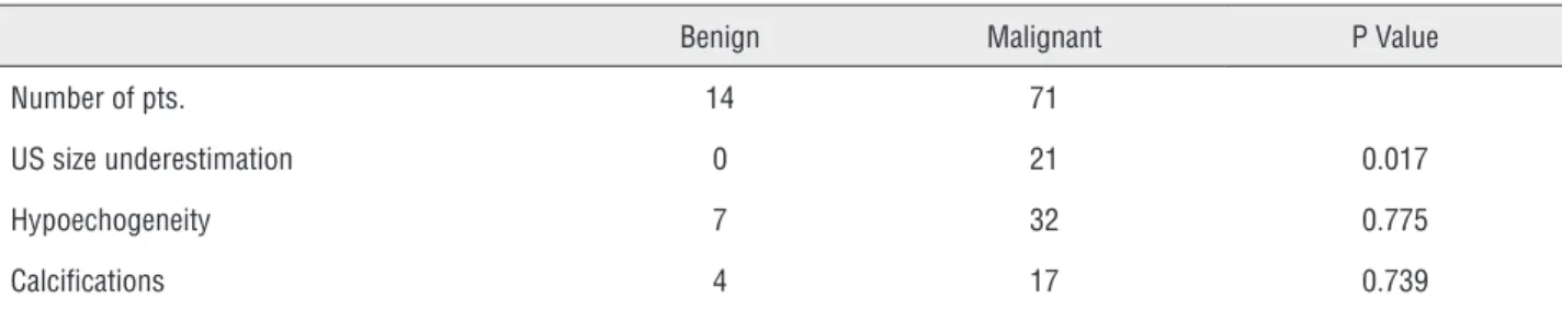
Table 1 - Collected ultrasonic results divided according to the type of tumor (benign vs. malignant).
Benign Malignant P Value
Number of pts. 14 71
US size underestimation 0 21 0.017
Hypoechogeneity 7 32 0.775
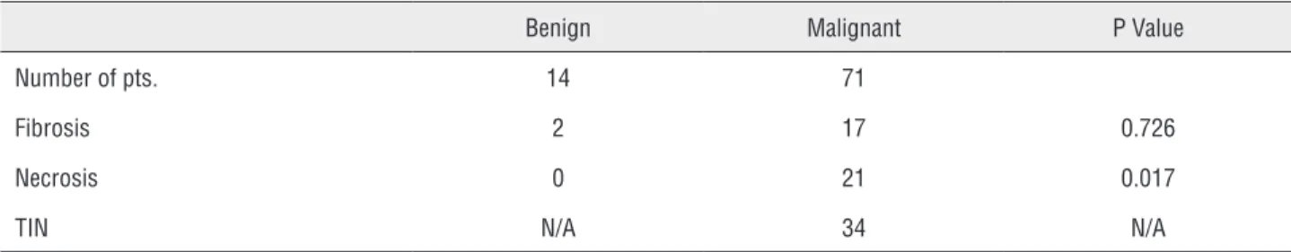
Table 2 - Collected histologic parameters divided according to the type of tumor (benign vs. malignant).
Benign Malignant P Value
Number of pts. 14 71
Fibrosis 2 17 0.726
Necrosis 0 21 0.017
TIN N/A 34 N/A
Table 3 - Collected ultrasonic results divided according to the histologic malignant subtype of tumor.
SGCT NSGCT P Value
Number of pts. 43 28
US size underestimation 11 10 0.429
Hypoechogeneity 28 7 0.001
Calcifications 13 6 0.584
Table 4 - Collected histologic parameters divided according to the histologic malignant subtype of tumor.
SGCT NSGCT P Value
Number of pts. 43 28
Fibrosis 14 3 0.047
Necrosis 13 9 1.00
TIN 23 12 1.00
benign tumors (p=0.017) and that hypoechogenei-ty and fibrosis were more frequent in SGCT than in NSGCT (p=0.001 and 0.047 respectively) (Figu-res 1 and 2).
COMMENTS
Testicular ultrasonography is usually per-formed with a high-frequency linear transducer; the echo texture of the two testicles is compared and areas of heterogeneity are searched for. Upon discovery of a lesion accurate dimensioning is crucial as clinicians must carefully consider the size of the lesion in their decision as to whether or not to perform testis preserving surgery,
espe-cially when facing a single testis (anatomical or functional) (13). General consensus is that a sono-graphic finding of any solid or mixed cystic lesion mass is an indication for surgical exploration (6, 8). However, there are only scant publications on the correlation between sonographic findings and type, local stage, size, and the histology of testicu-lar tumors (TT) (8).
and the presence of germ cell tumors, showing that lesions of 16-32 mm have a high relative risk for malignancy. Unfortunately this study cannot represent the general population of testicular tu-mors due to its small size of only 48 subjects and inclusion of only patients with impalpable lesions. Shilo et al. presented a larger group of 131 pa-tients concluding that benign lesions tend to be smaller than malignant lesions (15 mm vs. 41 mm respectively) and therefore a proper sonographic estimation can lead to consideration of partial
or-Figure 1 - Leydig cell tumor - Heterogeneous mass that was suspected for malignancy eventually found to be Leydig cell tumor with similar size estimated preoperatively.
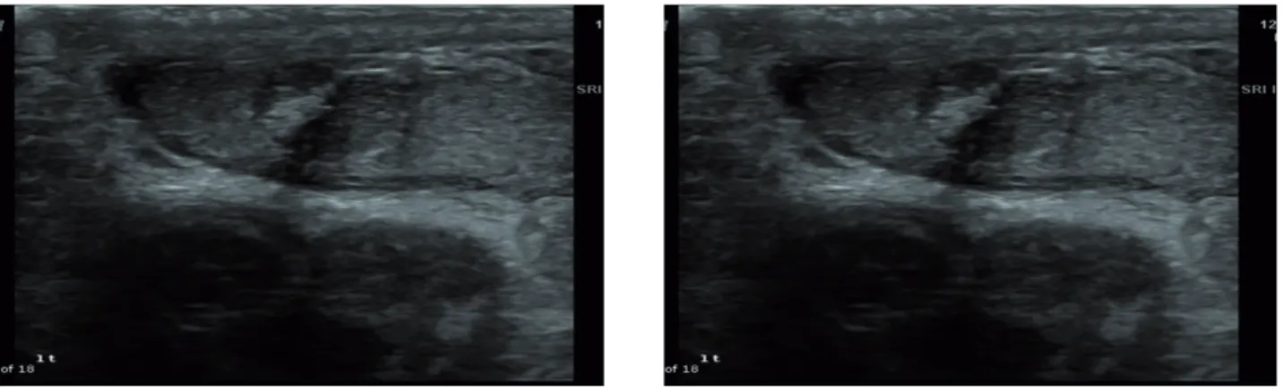
Figure 2 - Seminoma - Three hypo and anechoic lesions with variable diameters eventually found to be pure classical seminoma. The rest of the testis was fully indurate by seminoma nests, although homogeneous, non suspected preoperatively. Obviously, this case demonstrates sonographic underestimation of tumor size.
only 5 malignant masses (2 seminomas, 1 nonse-minoma and 2 cases of lymphoma). This attempt pointed to our goal but the low power of the study precluded any conclusive deductions. Schwerk et al. (6) has reported a prospective study on 57 le-sions, demonstrating a broad spectrum of texture patterns for malignancies of which 92% exhibi-ted hypoechogeneity, but could not differentiate between the histological subtypes. There is no doubt that this publication investigates a part of our discussion. However, the number of patients is inferior to our study and their study does not deal with the ability of the sonogram to evalu-ate the size of the tumor and therefore does not contribute to the planning of partial orchiectomy. Moreover, our data confirms the predominance of hypo-echogeneity in seminoma and therefore contributes to the preoperative evaluation.
Our study provides the percentage of preo-perative sonographic tumor size underestimations, an issue not yet addressed. Moreover, we attempt to support prior assumptions presented in uro-logical literature without sufficiently solid proof regarding the ability of sonographic findings to predict testicular tumor type. More than that, we have demonstrated another preoperative tool or attempt to distinguish benign from malignant tu-mors aside to other characteristics that have been described by Shilo and his colleagues (13).
This new data provides help in the surgi-cal consideration and planning of an orchiectomy, especially the consideration of a partial resection with or without a guided intraoperative biopsy. Herein we are adding another proof for the opi-nion that seminomas tend to be more hypoechoic than nonseminoma tumors. These facts combine to show that concentrating on the sonographic characteristics of the testicular lesion can vastly improve clinical judgment. A reason for underes-timation might be that sonography only shows the centralized body of the malignancy and cannot reliably pick up tendrils that are of clinical sig-nificance. To improve preoperative management and characterization of nonvascularized tissue, contrast-enhanced ultrasound, a new sonogra-phic technique, can be performed as an adjuvant to color Doppler ultrasound. Its role in evaluation of malignant suspected lesions is not well defined
and therefore not included in the official urologi-cal guidelines. The role for elastography, a mediurologi-cal imaging modality that maps the elastic properties of soft tissue, is limited to small testicular lesions, especially in surveillance. However, the combina-tion of these two techniques along with the clini-cal conclusions of our study might improve the future management of testicular lesions (19).
Before concluding we should note a few points about our study. We made the tradeoff of in-cluding more patients thereby increasing the power of our study and accepting the need for the require-ment of a non-centralized pathological and radio-logical review. Additionally, although our patients were heterogeneous, prior publications that contri-buted to our knowledge of the prevalence of diffe-rent types of tumors within groups of patients lead to the conclusion that this is acceptable (20,21).
CONCLUSIONS
ABBREVIATIONS
TT = Testicular tumor
TIN = Testicular Intraepithelial Neoplasia SGCT = Seminoma
NSGCT = Nonseminoma US = Ultrasound
CONFLICT OF INTEREST
None declared.
REFERENCES
1. Kennedy PT, Elliott JM, Rice PF, Kelly BE. Ultrasonography of intratesticular lesions: its role in clinical management. Ulster Med J. 1999;68:54-8.
2. Hamm B. Differential diagnosis of scrotal masses by ultrasound. Eur Radiol. 1997;7:668-79.
3. Rosenfield AT, Hammers LW. Imaging of the testicle: the painful scrotum and nonpalpable masses. Urol Radiol. 1992;14:229-33.
4. Horstman WG. Scrotal imaging. Urol Clin North Am. 1997;24:653-71.
5. Lesnik G, Nickl S, Kuschnig P, Sinzig M, Hausegger K, Jeschke K. Sonography of the scrotum. Rofo. 2006;178:165-79.
6. Schwerk WB, Schwerk WN, Rodeck G. Testicular tumors: prospective analysis of real-time US patterns and abdominal staging. Radiology. 1987;164:369-74.
7. Fowler RC, Chennells PM, Ewing R. Scrotal ultrasonography: a clinical evaluation. Br J Radiol. 1987;60:649-54.
8. Carmignani L, Morabito A, Gadda F, Bozzini G, Rocco F, Colpi GM. Prognostic parameters in adult impalpable ultrasonographic lesions of the testicle. J Urol. 2005;174:1035-8.
9. Steiner H, Höltl L, Maneschg C, Berger AP, Rogatsch H, Bartsch G, et al. Frozen section analysis-guided organ-sparing approach in testicular tumors: technique, feasibility, and long-term results. Urology. 2003;62:508-13.
10. Heidenreich A, Weissbach L, Höltl W, Albers P, Kliesch S, Köhrmann KU, et al. Group. Organ sparing surgery for malignant germ cell tumor of the testis. J Urol. 2001;166:2161-5.
11. Heidenreich A, Stark L, Derschum W, von Vietsch HV. Organ saving therapy of bilateral testicular tumor. Urologe A. 1993;32:43-8.
12. Weissbach L. Organ preserving surgery of malignant germ cell tumors. J Urol. 1995;153:90-3.
13. Lawrentschuk N, Zuniga A, Grabowksi AC, Rendon RA, Jewett MA. Partial orchiectomy for presumed malignancy in patients with a solitary testis due to a prior germ cell tumor: a large North American experience. J Urol. 2011;185:508-13.
14. Canda AE, Atmaca AF, Ozdemir AT, Akbulut Z, Balbay MD. Testis sparing surgery for sequential bilateral testicular tumors. Can J Urol. 2009;16:4677-81.
15. Shilo Y, Zisman A, Lindner A, Raz O, Strauss S, Siegel YI, Segal M, Sandbank J, Leibovici D. The predominance of benign histology in small testicular masses. Urol Oncol. 2012;30:719-22.
16. Carmignani L, Gadda F, Gazzano G, Nerva F, Mancini M, Ferruti M, et al. High incidence of benign testicular neoplasms diagnosed by ultrasound. J Urol. 2003;170:1783-6.
17. Wang W, Gong ZH, Dai YT. Value of ultrasonography in the diagnosis and differential diagnosis of testicular tumor. Zhonghua Nan Ke Xue. 2007;13:424-7.
18. Ye L, Wang X, Zhang Y, Ding Q, Wang Y. Nonpalpable testicular masses incidentally discovered by ultrasound. Zhonghua Wai Ke Za Zhi. 1999;37:168-70.
19. Grasso M, Blanco S, Raber M, Nespoli L. Elasto-sonography of the testis: preliminary experience. Arch Ital Urol Androl. 2010;82:160-3.
20. Zheng LW, Li FB, Liu RZ, Ji RG, Zhao ZW. Clinical analysis of 87 cases of testicular tumor. Zhonghua Nan Ke Xue. 2005;11:445-7.
21. Xu K, Fang ZJ, Zheng J, Lu Y, Li BK, Ding Q. Diagnosis of testicular cancer: review of 47 cases. Zhonghua Yi Xue Za Zhi. 2009;89:2140-1.