Emotional Stress Evaluation of Patients with
Moderate and Severe Sleep Apnea Syndrome
Micheli Aparecida Gomes dos Santos
1Tatiana de Cássia Nakano
1Felipe Almeida Mendes
2Bruno Bernardo Duarte
3Silvio Antonio Monteiro Marone
31Department of Psychology, Pontifícia Universidade Católica,
Campinas, São Paulo, Brazil
2Department of Otolaryngology, Pontifícia Universidade Católica,
Campinas, São Paulo, Brazil
3Department of Otorhinolaryngology, Pontifícia Universidade
Católica, Campinas, São Paulo, Brazil
Int Arch Otorhinolaryngol 2017;21:28–32.
Address for correspondence Felipe Almeida Mendes, Department of Otolaryngology, Pontifícia Universidade Católica, Av. John Boyd Dunlop Jardim Ipaussurama, Campinas, São Paulo 13060-904, Brazil (e-mail: [email protected]).
Introduction
Obstructive sleep apnea (OSA) is characterized by recurrent collapse of the pharynx during sleep, resulting in substantial reduction of airflow (apnea or hypopnea) leading to inter-mittent disorders of blood gases (hypoxemia and hypercap-nia) and activation of the sympatic system1.
Diagnosis of OSA is made by clinical history and poly-somnography, defined by an apnea-hypopnea index (AHI) greater than 15 or AHI greater than 5 with daytime and nighttime symptoms. The severity of the apnea is classified as mild (AHI 5 to 14.9), moderate (AHI 15 to 29.9), and severe (AHI greater than 30).1
Keywords
►
sleep apnea
►
obstructive
►
stress
►
psychological
►
evaluation studies
Abstract
Introduction
The scienti
fi
c literature has shown that the damage caused by sleep
fragmentation in people affected by Obstructive Sleep Apnea (OSA) can re
fl
ect
emotionally, generating not only physical symptoms such as drowsiness and tiredness,
but also psychical symptoms, such as stress.
Objective
This study aimed at comparing symptoms of stress in patients with
moderate or severe OSA, before and after two months of treatment (clinical or surgical).
Method
This is an Individual, prospective, longitudinal, and interventional study. All
patients underwent polysomnography before treatment. We collected data through the
application of Stress Symptoms Inventory for Adults Lipp (ISSL) before and after two
months of medical or surgical treatment for moderate or severe OSA.
Results
The sample consisted of 18 patients (72.2% male) with a mean age of 51.83
years. We found that 77.8% (
n
¼
14) of patients had stress in the
fi
rst evaluation. In the
second evaluation (after treatment), this reduced to 16.7% (
n
¼
3). The average stress
symptoms decreased from the
fi
rst to the second evaluation (M
¼
13.78 and M
¼
6.17,
respectively), being statistically signi
fi
cant (z
¼
-3.53;
p
<
0.000).
Conclusions
We found that moderate and severe apnea patients have signi
fi
cant
stress index and that, after two months of medical or surgical treatment, there is a
signi
fi
cant reduction of the symptom. In addition, the patients with severe OSA had a
better outcome regarding the reduction of stress index than patients with moderate
OSA.
received
February 14, 2016
accepted
June 18, 2016
published online
August 16, 2016
DOI http://dx.doi.org/ 10.1055/s-0036-1586251.
ISSN 1809-9777.
Copyright © 2017 by Thieme-Revinter Publicações Ltda, Rio de Janeiro, Brazil Original Research
Tuffik et al2showed in their study that, among 1042
volun-teers, 32.8% presented the criteria for OSA according to the American Academy of Sleep Medicine (discovered by overnight polysomnography). Prevalence estimates are higher among men and increases in both genders with the aging.3OSA was more
prevalent in overweight and obese men and women.2–4
Symptoms of stress are frequent in patients with OSAS. According to Lipp,5stress is a general wear and tear of the
body caused by psycho-physiological changes that occur when the individual is forced to face a situation that arouses a strong, good or bad emotion, which requires changes.
The most common symptoms of emotional stress are: tachycardia, excessive sweating, muscle tension, dry mouth, and feeling of being on alert.4In the course of its
develop-ment, the differences manifest according to individual genetic predispositions, potentiated by possible impairments devel-oped later in life, such as accidents and diseases.6
The scientific literature has shown that the damage caused
by sleep fragmentation in people affected by OSA can reflect emotionally, generating not only physical symptoms, such as drowsiness and tiredness, but also psychical symptoms such as stress.7Considering this fact, the aim of this study was to
compare stress symptoms in patients with OSA, moderate and severe before and two months after the start of clinical or surgical treatment.
Methods
The project received approval for execution from the Ethics Committee of the University that houses it. Patients signed an Informed Consent in which they agreed to participate. Sub-sequently, the data was disclosed for scientific purposes anonymously. Offering medical care or treatment was neces-sarily conditional to participation in our research.
We conducted the study at an outpatient clinic specialized in sleep medicine, a division of the Otorhinolaryngology Service of a university hospital in the state of São Paulo.
This study is characterized as individualized, prospective, longitudinal, and interventional. There were no control group or blinding of assessors.
All patients received treatment at the Otorhinolaryngol-ogy ward and were sent to sleep medicine clinic equipped with polysomnography type I, which always took place in the same laboratory.
We performed the data collection through the use of an identification questionnaire prepared to obtain demographic data of the participants and the Lipp’s Inventory of Stress
Symptoms for Adults (ISSL).8
The ISSL is an instrument that assesses symptoms of stress, the patient́s stress level (alarm, resistance, near-exhaustion, and exhaustion), and the kind of predominant symptoms present (physical or psychological). The instrument consists of 53 items, 34 concerning the physical conditions of psycho-logical and 19, divided into three quadrants. Thefirst frame
shows symptoms experienced in the last 24 hours, 12 being physical and three psychological. The second shows symp-toms in the last week, 10 physical and psychological 5. The third frame includes symptoms experienced in the last month, 12 physical and 11 psychological. Evaluation of responses is done through the use of the ASSI guide tables, which transforms the raw data into percentages.
In thefirst evaluation with the participants before the start of treatment for OSA, we applied the socio-demographic questionnaire and the inventory of stress. At the second meeting, after two months of treatment (medical or surgical), we only applied the inventory.
The initial sample consisted of 23 participants; however,five patients did not adhere to treatment and were excluded from the
final sample. Thus, thefinal sample consisted of 18 patients with
a mean age of 51.83 years, minimum 26 years and maximum of 74 years (SD¼13.46). In this group, 72.2% (n¼13) were men. Regarding education, there was a higher frequency of partic-ipants with incomplete primary education with 55.6% (n¼10) and, in relation to marital status, 83.3% (n¼15) were married. We determined type of OSA by overnight polysomnography and found that 72.2% (n¼13) of the sample suffered from severe OSA and 27.8% (n¼5) had moderate OSA.
We did not include patients who suffered from mild OSA because they did notfit the inclusion criteria.
Results
The evaluation of the results obtained in the ISSL allowed the identification of stress levels presented by individuals and we observed that 77.8% (n¼14) of patients had stress in thefirst evaluation. In the second evaluation after treatment, the amount fell to 16.7% (n¼3).
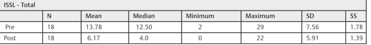
To better visualize the data, descriptive statistics from before and after treatment are shown in►Table 1. The average
stress symptoms drop significantly from thefirst to the second evaluation (M¼13.78 and F¼6.17, respectively).
The non-parametric Wilcoxon test was used to assess the most common symptoms of stress pre and post treatment. The results showed that patients experienced a statistically significant decrease in mean symptom of post-stress treat-ment (z¼-3.53,p<0.000).
Table 1 Descriptive statistics for stress pre and post treatment
ISSL - Total
N Mean Median Minimum Maximum SD SS
Pre 18 13.78 12.50 2 29 7.56 1.78
Post 18 6.17 4.0 0 22 5.91 1.39
To further investigate this situation, a second analysis sought to determine which stages of stress prevailed in both evaluations among patients who were stressed, as shown in►Fig. 1.
We noticed important changes in the second evaluation, given the fact that only the resistance phase was present in 100% of individuals who remained stressed after treatment (n¼3).
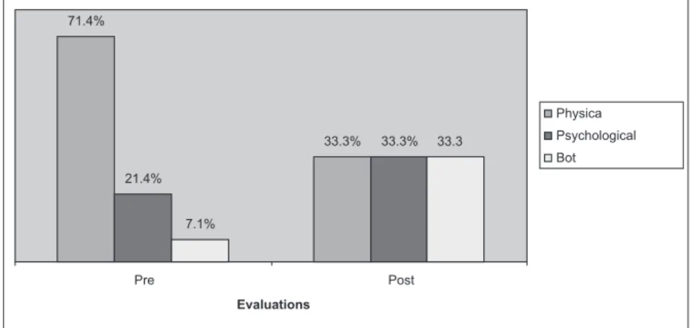
We performed a more detailed investigation to identify the predominant symptom (physical or psychological) in individ-uals with stress, whose data are presented in►Fig. 2.
The results from the ISSL showed that the feeling of constant physical wear is the most commonly reported by patients, persisting in the second assessment (in 66.7% of cases in thefirst evaluation and 33.3% in the second
evalua-tion). Memory problems also showed high prevalence, pres-ent in 55.6% of cases before treatmpres-ent and in 38.9% of patipres-ents after treatment. Approximately 44.4% of patients had muscle tension and xerostomia in the first evaluation. The most prevalent psychological symptoms claimed by responders in thefirst evaluation were feelings of anguish and daily anxiety (61.1%). Moreover, 55.6% of the responders reported
exces-sive emotional sensitivity and 44.4% reported excesexces-sive irritability. In the evaluation performed after two months of treatment, the psychological symptoms that prevailed were anxiety / daily anxiety (38.9%), constantly thinking or talking about one subject (22.2%), and loss of sense of humor (22.2%).
A second analysis compared the stress between the groups with moderate apnea (n¼13) and severe apnea group (n¼5). The results pointed to a lower average gift symptoms within the group with moderate OSA (M¼11.8, SD¼7.59) when compared with the severe OSAS group (M¼14.54; SD¼7.72) in initial assessment, pre-treatment. In the second evaluation after the beginning of treatment, the mean for decreased symptoms in both groups (mean¼4.6; SD¼4.33 for moderate and M¼6.77; SD¼6.47 for severe), although the decrease was higher in the severe OSAS group.
We used the non-parametric Mann-Whitney test to enable the differences between two test conditions and different participants.8 The results showed no significant inter-group
difference (moderately x severe) in the first evaluation (U¼24.5; z¼-0.790;p¼0.43), or in the evaluation performed after starting treatment (U¼26.5; z¼-0.594;p¼0.55). 7.1%
0% 71.4%
100%
14.2%
0% 7.1%
0%
Pre Post
Evaluations
Alert
Resistance
Near- Exhaustion
Exhaustion
Fig. 1 Percent stage of pre-treatment and post stress.
71.4%
33.3%
21.4%
33.3%
7.1%
33.3
t s o P e
r P
Evaluations
Physica
Psychological
Bot
We used a second analysis for intragroup comparison of scores from the same participants (moderate OSA before and after treatment, severe OSA before and after treatment). Results showed that patients with moderate OSA presented a significant decrease in mean symptom of post-stress treat-ment (z¼-2.971;p<0.003); the same happened to patients with severe OSA (z¼-2.023;p<0.043), demonstrating that the treatment was effective in improving symptoms of stress. Comparing the symptoms of stress among the women (n¼5) with those among the men (n¼13), there was a slight decrease in the frequency of stress symptoms in women (M¼15.4, SD¼8.26 in the first evaluation and M¼9.8, SD¼8.7 in the second evaluation) and a relatively greater reduction among the men (M¼13.1, SD¼7.53 in the pre-treatment and M¼4.77, SD¼4.08 in the post-treatment). By analyzing the differences between the genders in each of the assessments, the results of the Mann-Whitney test at the
first assessment (U¼28.5; z¼-, 395;p¼0.693) as well as the second (U¼22.0; z¼-1.039; p¼0.299) suggest that there is no significant intergroup differences in the two periods. On the other hand, the intra-group analysis showed that women showed no significant improvement in symp-toms of stress after treatment (z¼-1.633;p¼0.102). The men, on the other hand, showed a steep decrease in symp-toms (z¼-3.184;p¼0.001).
Finally, another exploratory analysis was to compare patients due to the treatment (CPAP or Lateral Pharyngo-plasty). The results showed a significant reduction in anxiety symptoms in the group that underwent treatment with CPAP (M¼14, SD¼7.57 before treatment and M¼6.8, SD¼6.23 after starting treatment). In the surgical group, the decrease was not as important (M¼12.67, SD¼9.07 before treat-ment and M¼3.0, SD¼2.64 after start of treatment).
Given the apparent mean differences, we applied the Wilcoxon non-parametric test. We compared results from patients who received treatment with CPAP in terms of the existence of significant differences in anxiety symptoms from the first to the second evaluation (z¼-3.21; p¼0.001), which did not occur in the group that underwent surgery (z¼-1.60;p¼0.109).
Discussion
In terms of psychological evaluation, this study found a significant decrease in stress symptoms in patients after treatment with CPAP, a result similar to the study conducted by Grunstein et al9based on the hypothesis that the
with-drawal of CPAP in patients suffering from severe OSA, would produce a measurable stress response.
Important to note that the stress measurements between the study results presented by Grunstein et al9 and the
present study are different mainly in resources used for the diagnosis of stress. Therefore, caution is recommended before any comparison may be performed to affirm the effect of CPAP treatment in patients with OSAS.
The correlation between cortisol and sleep is verified by Spiegel, Leproult, and Van Cauter.10Their study showed sleep
restriction (4 hours / day for 6 nights) increased cortisol levels in
normal individuals. Thisfinding confirms what has already been pointed out by Spath-Schwalbe, Gofferje, Jern, Born, and Fehm,11
who suggest that nocturnal awakenings in OSA are associated with alterations in the hypothalamic-pituitary-adrenal axis (HPA) and claim that intermittent hypoxia, fragmentation, and sleep deprivation causes cortisol release. Therefore, it expected a high level of stress in patients with OSA.11–13
We found that most of the sample was in the resistance phase in both evaluations. This phase, an intermediate one in the stress process, is characterized by physical and mental tiredness, memory difficulties, and increased vulnerability to diseases that occur due to poor functioning of the immune system. Productivity can also suffer as a result of the appear-ance of symptoms. If the body cannot reverse the process, the person enters the stage of exhaustion.14
Among the most prevalent physical symptoms during the
first evaluation is memory loss. This symptom persisted as the most common physical symptom in post-treatment evalua-tion. People with daytime sleepiness can present problems in the transfer of short-term memory to long-term, compromis-ing thus their daily activities.2,3,15
Regarding the psychological symptoms in addition to daily distress or anxiety, irritability and excessive emotional sen-sitivity were the most frequent in pretreatment. In the second evaluation, distress or daily anxiety remained the most prevalent complaints, followed by thinking or talking con-stantly in one subject and loss of sense of humor. However, it is important to consider the presence of daytime sleepiness and adaptation requirements to treatment.
When comparing the groups of moderate and severe apnea, the results point to the existence of statistically significant differences related to the stress levels after treat-ment. In this way, it was possible to verify that the treatment was effective in improving symptoms for both levels of the disease (moderate and severe). Franco,16 whose general
objective was to evaluate the correlation between the severity of OSA, oxidative stress markers, and the presence of depres-sive and anxiety symptoms, developed research with similar results. The conclusion was that the greater the severity of OSA, the greater the fragmentation of sleep, increasing the incidence of daytime symptoms, particularly somnolence, depressive and anxiety symptoms. Unlike this study, both in the first assessment and in the second there were no significant differences in the level of stress among the mod-erate and severe groups.
In this study, when comparing the prevalence of stress among genders, there was no statistically significant differences between groups in the incidence of stress symptoms, either in the assessment performed pre-treatment or post-treatment. However, it is noteworthy that, in the second evaluation, all patients who were stressed were female. In their survey on gender, education, and stress, Calais, Andrade, and Lipp17found
that women were more vulnerable to stress than men in all age groups. A similar result was found by Calais.18
Regarding the treatment used, we found that patients showed a significant reduction in symptoms after treatment with CPAP.19,20 However, patients in this study who have
not show a significant improvement in symptoms of stress, which does not correspond to some data in the literature. In Li et al,21for example, the authors concluded that surgery can
significantly improve psychological symptoms in patients with OSAS, although the effect of surgery was mild but clinically relevant. It is worth noting, however, that, due to the short time interval between evaluations, the results may have been influenced by other factors. It is worth mentioning, for instance, the need for further adjustment on the part of patients resulting from the use of CPAP as compared to disturbances from surgery, such as wound healing and side effects of medication.
Cahali, Formigoni, Gebrim, and Miziara22 stated that the
healing process occurs in 6 to 9 months. Moreover, in thefirst days after surgery, there is edema of the pharyngeal region, which can narrow the upper airways and worsen OSA frame-work in this period. The authors say that the results of Lateral Pharyngoplasty reach their peak after a period of three to six months of the procedure, including better clinical and poly-somnographic results than uvulopalatopharyngoplasty.22,23We
would recommend carrying out a new assessment 6 months after surgery to verify that these data remain the same.
Conclusion
The quest for understanding what happens emotionally in patients suffering from OSA is necessary. It is important to point out that this study was conducted with a small sample and, therefore, the considerations presented here should be considered cautiously, avoiding early spreads. We suggest pursuing further studies with a larger sample to obtain data from. This also raises the hypothesis that receiving a diagno-sis, as well as the possibility of receiving treatment, may have influenced stress reduction in patients, by which factors other than the treatment itself may have contributed to the reduc-tion in symptoms. It is important that future studies shed light onto other variables that deserve to be better investigat-ed in future studies, such as adaptation to treatment, use of an instrument to evaluate the quality of sleep, or offering an intervention program aimed at stress management and a better quality of life for such patients.
Furthermore, this study presented limitations regarding the absence of control variables, such as obesity and use of medications that could interfere with the results, which we consider intervening variables worth investigation in further studies.
References
1 Sleep-related breathing disorders in adults: recommendations for syndrome definition and measurement techniques in clinical research. The Report of an American Academy of Sleep Medicine Task Force. Sleep 1999;22(5):667–689
2 Tufik S, Santos-Silva R, Taddei JA, Bittencourt LRA. Obstructive sleep apnea syndrome in the Sao Paulo Epidemiologic Sleep Study. Sleep Med 2010;11(5):441–446
3 Young T, Palta M, Dempsey J, Skatrud J, Weber S, Badr S. The occurrence of sleep-disordered breathing among middle-aged adults. N Engl J Med 1993;328(17):1230–1235
4 Noal RB, Menezes AMB, Canani SF, Siqueira FV. Habitual snoring and obstructive sleep apnea in adults: population-based study in Southern Brazil. Rev Saude Publica 2008;42(2):224–233 5 Lipp MEN. Como Enfrentar o Stress. 5th Ed. São Paulo, Brazil: Ícone
Editora.; 1998
6 Lipp MEN. O Stress: Conhecer e Enfrentar. 5th Ed. São Paulo, Brazil: Ed. Contexto.; 2003
7 Głebocka A, Kossowska A, Bednarek M. Obstructive sleep apnea and the quality of life. J Physiol Pharmacol 2006;57(Suppl 4):111–117 8 Lipp MEN. Pesquisas Sobre Stress no Brasil: Saúde, Ocupações e
Grupos de Risco. Campinas, Brazil: Ed. Papirus.; 2001
9 Grunstein RR, Stewart DA, Lloyd H, Akinci M, Cheng N, Sullivan CE. Acute withdrawal of nasal CPAP in obstructive sleep apnea does not cause a rise in stress hormones. Sleep 1996;19(10):774–782 10 Spiegel K, Leproult R, Van Cauter E. Impact of sleep debt on metabolic
and endocrine function. Lancet 1999;354(9188):1435–1439 11 Späth-Schwalbe E, Gofferje M, Kern W, Born J, Fehm HL. Sleep
disruption alters nocturnal ACTH and cortisol secretory patterns. Biol Psychiatry 1991;29(6):575–584
12 Buckley TM, Schatzberg AF. On the interactions of the hypotha-lamic-pituitary-adrenal (HPA) axis and sleep: normal HPA axis activity and circadian rhythm, exemplary sleep disorders. J Clin Endocrinol Metab 2005;90(5):3106–3114
13 Tomfohr LM, Edwards KM, Dimsdale JE. Is obstructive sleep apnea associated with cortisol levels? A systematic review of the research evidence. Sleep Med Rev 2012;16(3):243–249
14 Lipp MEN, Tanganelli MS.Stresse Qualidade de Vida em Magis-trados da Justiça do Trabalho: Diferenças entre Homens e Mul-heres. Psicol Reflex Crit 2002;15(3):537–548
15 Bittencourt LRA, Silva RS, Santos RF, Pires MLN, Mello MT. Excessive daytime sleepiness. Rev Bras Psiquiatr 2005;27(Suppl 1):16–21 16 de Lima AM, Franco CM, de Castro CM, Bezerra Ade A, Ataíde L Jr,
Halpern A. Effects of nasal continuous positive airways pressure treatment on oxidative stress and adiponectin levels in obese patients with obstructive sleep apnea. Respiration 2010;79(5): 370–376
17 Calais SL, Andrade LMB, Lipp MEN. Diferenças de Sexo e Escolar-idade na Manifestação deStressem Adultos Jovens. Psicol Reflex Crit 2003;16(2):257–263
18 Calais SL. Diferenças entre homens e mulheres na vulnerabilidades ao stress. In: M.E.N. Lipp, (Org.). Mecanismos Neuropsicofi sioló-gicos do Stress: Teoria e Aplicações Clínicas. São Paulo, Brazil: Casa do Psicólogo.; 2003:87–89
19 Bardwell WA, Norman D, Ancoli-Israel S, et al. Effects of 2-week nocturnal oxygen supplementation and continuous positive air-way pressure treatment on psychological symptoms in patients with obstructive sleep apnea: a randomized placebo-controlled study. Behav Sleep Med 2007;5(1):21–38
20 Derderian SS, Bridenbaugh RH, Rajagopal KR. Neuropsychologic symptoms in obstructive sleep apnea improve after treatment with nasal continuous positive airway pressure. Chest 1988;94(5): 1023–1027
21 Li HY, Huang YS, Chen NH, Fang TJ, Liu CY, Wang PC. Mood improvement after surgery for obstructive sleep apnea. Laryngo-scope 2004;114(6):1098–1102
22 Cahali MB, Formigoni GGS, Gebrim EMMS, Miziara ID. Lateral pharyngoplasty versus uvulopalatopharyngoplasty: a clinical, polysomnographic and computed tomography measurement comparison. Sleep 2004;27(5):942–950