Predictive Value of Ventilatory and Metabolic
Variables for Risk of Death in Patients with Cardiac
Failure
Ana Maria F. Wanderley Braga, Maria Urbana P. B. Rondon,
Carlos Eduardo Negrão, Maurício Wajngarten
Instituto do Coração do Hospital das Clínicas – FMUSP e Escola de Educação Física da USP - São Paulo, SP - BrazilMailing Address: Ana Maria F. Wanderley Braga • InCor - Av. Dr. Enéas de Carvalho Aguiar, 44 – 05403-000 – São Paulo, SP - Brazil E-mail: [email protected] Received on 19/04/04 • Accepted on 17/03/06
O
BJECTIVETo analyze the predictive value of respiratory, metabolic, and hemodynamic variables obtained during the cardiopulmonary stress test for the risk of death in patients with heart failure.
M
ETHODSEighty-seven NYHA Functional Class II and III patients were analyzed, ages 51 ± 0.5 years, 26 of them with Chagas’ disease, 30 with coronary ischemia, and 31 with idiopathic etiology. The cardiopulmonary stress test consisted of a ramp-protocol with 5 to 15 W/min workload increments performed on a bicycle-ergonometer until exhaustion.
R
ESULTSIn this study, the multiple Cox regression analysis of age, height, weight, body surface, and gender showed that these parameters were not statistically signifi cant control factors. Oxygen uptake, ventilatory equivalent of oxygen, ventilatory equivalent of carbon dioxide production, oxygen pulse, and end-tidal partial pressure of carbon dioxide at the anaerobic threshold, respiratory compensation point, and peak exercise proved to be important death predictors in heart failure patients. The relationship between the increase in carbon dioxide output as a function of the increase in minute ventilation, and the association between the oxygen uptake increase and the elevation of the workload from the beginning of exercise to the anaerobic threshold were statistically signifi cant predictors of death in heart failure patients (p<0.05).
C
ONCLUSIONThe cardiopulmonary stress test makes it possible to evaluate ventilatory, metabolic, and hemodynamic variables that may be utilized as important markers of life prognosis in these patients.
K
EY WORDSHeart failure is a syndrome characterized by exertional dyspnea and intolerance to exercise. The cardiopulmonary stress test is a method frequently used to evaluate the degree of exercise intolerance and functional capacity of heart failure patients. Based on this information it has been possible to identify the seriousness and even prognosis of patients with this syndrome that is responsible for innumerable deaths, including in Brazil1-2.
Among the variables obtained with the cardiopulmonary exercise test, oxygen uptake, measured at the peak of exercise (peak VO2) is the most employed variable since it allows the determination of the functional capacity of patients with several degrees of heart failure. This is vital in important decisions such as heart transplant indications3-5, or evaluations of the prognostic value of
mortality3, 6-17, stratifi cation of the degree of heart failure
severity3,18-21, differential diagnosis between dyspnea
of cardiac origin or dyspnea of pulmonary origin, 21-24, or an appraisal of an intervention with a cardiac
pacemaker22, 25. It is also a useful means for assessment
of drug therapy results relative to physical capacity 21,
26-28, or even the prescription of physical exercise29. The
maximum oxygen uptake (VO2 max.) estimated by linear regression equations is only precise in its application for submaximal exercise30.
Nevertheless, the evaluation of the VO2 peak in patients with cardiac failure may have limitations. Early interruption of the test on the part of the patient because of lack of motivation or fear of continuing the exercise, or an early interruption by the examiner may underestimate the peak VO2 value and hinder an adequate diagnostic evaluation11,31.Moreover, a study by Chomsky et al32
showed that cardiac output seems to be a better predictor of mortality in patients with heart failure than the peak VO2. These authors observed that patients with a low cardiac output during exertion had a shorter survival, regardless of the peak VO2 value, compared to those patients with a normal cardiac output. More recently, Corrà et al6, demonstrated that patients with intermediate
physical capacity, i.e., peak VO2 between 10 and 18 ml.kg-1.min.-1 but with a ratio of increased carbon dioxide
as a function of an increased minute ventilation (VE/VCO2 slope) greater than or equal to 35, showed a similar mortality rate as those patients with a peak VO2 smaller than or equal to 10 ml.kg-1.min.–1, but a VE/VCO
2 slope
of less than 35. Therefore, these results suggest the need for further information obtained during cardiopulmonary stress tests that may identify functional and metabolic limitations in these patients.
For this reason, some authors have sought to evaluate the prognostic value of other metabolic and respiratory variables, such as the ventilatory equivalent for oxygen (VE/VO2) 10,33, the ventilatory equivalent for carbon dioxide
(VE/VCO2)10,33,34, oxygen pulse (VO
2/HR)
7,10, heart rate
(HR)34-37, VE/VCO
2 slope
6,11,38-39, and the relationship
between the increase in oxygen uptake and the increase
of work load (∆VO2/∆W slope) 6, 8-11.
Despite the many studies already conducted in this area, especially relating the prognostic value of VO2 and the VE/VCO2 slope, most of these authors assessed the prognostic value of the metabolic and respiratory variables at the peak of exercise3, 38.
However, since many times the patient with cardiac failure has diffi culties in performing at maximal exercise, the analysis of the prognostic value of mortality as to the risk of death during the submaximal phase of exercise in the cardiopulmonary stress test is extremely appealing. In fact, many patients with heart failure cannot perform a maximal test, and must interrupt their exercise because of muscular-skeletal limitations40,42 or because they
experience potentially lethal cardiac arrhythmias43-45.
Additionally, the comparison of metabolic variables at distinct time points, such as at the anaerobic threshold and the point of respiratory compensation, assure us that the metabolic stress during exercise is proving to be the same in all of the patients. When the different metabolic variables are compared by absolute loads, the percentage of exertion put forth is not necessarily the same and therefore the metabolic stress and the hemodynamic impact will be different for each patient. Actually, in a study carried out by Gitt et al11 a greater prognostic
value was noted for risk of death in patients with heart failure with the association of VO2 at the anaerobic threshold and the VE/VCO2 slope. Nevertheless, in spite of this important fi nding, the study of the prognostic value for the risk of death of the metabolic variables at the transition from predominantly aerobic metabolism to predominantly anaerobic metabolism exercise timepoint and the establishment of metabolic acidosis (respiratory compensation point)46 in patients with heart failure has
not been amply divulged.
Another interesting aspect to be considered is that the prognostic value for risk of death of the metabolic variables analyzed at ventilatory thresholds has not been adequately studied in populations that include patients with cardiac failure of Chagasic etiology. This peculiar and unfortunate characteristic of our population has been considered a signifi cant public health problem not only in Brazil, but in other countries of South America as well47.
Thus, the objective of this study was to test the hypothesis that the level of respiratory, metabolic, and hemodynamic responses during progressive physical exercise, at the anaerobic threshold, the point of respiratory compensation, and peak exercise can predict mortality in patients with heart failure.
M
ETHODSInstitute of the Faculdade de Medicina da Universidade de São Paulo between 1990 and 1997. These were individuals with heart failure according to Weber’s functional classifi cation18 and the group consisted of 74
men and 13 women, average age 51 ± 0.5 years and ejection fraction of <50%. Among the patients, 62% received digitalis drugs, 75% received furosemide, and 70% received angiotensin converting enzyme inhibitors (captopril or enalapril), and all were submitted to an echodopplercardiographic study.
Patients with physical exertion limitations because of muscular-skeletal disease, peripheral arteriopathy, cachexia, chronic obstructive pulmonary disease, or any reason other than dyspnea or fatigue of cardiac origin were excluded from the study. Also excluded were those patients with acute myocardial infarct with less than 2 months of disease progression, those with atrial fi brillation or fl utter, congenital cardiopathy, valvulopathy as baseline illness such as mitral stenosis, aortic stenosis, mitral insuffi ciency, and aortic insuffi ciency.
All patients were submitted to the ramp-protocol cardiopulmonar y stress test, characterized by a continuous load increment on a bicycle-ergonometer with electromagnetic brakes (Medifi t 400 I, Medical Fitness Equipment Maarn, Holland). The respiratory fl ow and the fractions of expired oxygen and carbon dioxide were collected at each respiratory cycle using the sixty-second average in the posterior analysis. This evaluation was made on a computed system (CAD/NET 2001 model, Medical Graphics Corporation, United States).
Corrections of gas volumes of the variables analyzed under BTPS conditions (body temperature, ambient pressure, saturated with water vapor), were made at a 37º C body temperature and pressure that corresponds to the barometric pressure. These corrections are made when we need to know the volume of air ventilated by the lungs. For this reason, ventilation, the product of the breathing rate by tidal volume, is analyzed under BTPS conditions. On the other hand, the corrections of gas volumes analyzed under STPD conditions (standard temperature and pressure, dry), were made at a standardized temperature of 0º C, barometric pressure of 760 mmHg at sea level, with the volume occupied by water vapor molecules corrected, i.e., dry. These corrections are made when we need to know the quantity of oxygen uptake and carbon dioxide output48.
After the patients were adapted to the surroundings and equipment, resting assessments were made for 2 minutes, followed by 3 minutes of warm-up with no load at a speed of 60 rotations per minute (rpm). From the fi fth minute on, the load was progressively and constantly increased, and the increment was individualized for each patient. The choice of load increment was based on the predicted maximal load for normal adults according to established formulas for each gender that take into account age and anthropometric characteristics49-50.
In patients with heart failure, 80% of the predicted maximal potency was calculated for normal individuals; to determine the load variation, this predicted value was divided by 10. The load increment in the ramp-protocol utilized in patients with heart failure varied between 5 and 15 W/min. All patients were encouraged to exert progressive effort until symptoms such as dyspnea, fatigue, or that represented a risk made them incapable of continuing. Recovery period lasted 4 minutes; during the fi rst minute, the load maintained was equivalent to 50% and during the second minute, equivalent to 25% of the maximal load attained in the test. During the third minute, the load was removed, but the 60 rpm speed was maintained, and during the fourth minute, the individual remained seated and still on the bicycle-ergonometer. The anaerobic threshold was determined by the V-slope method that consists of the infl exion point at which the carbon dioxide gas production raises faster than the oxygen uptake51, or at the point where the lowest values
and posterior elevation of the ventilatory equivalent curves for oxygen and partial pressure of oxygen are observed52.
The respiratory compensation point was determined through the lowest point of the ventilatory equivalent for carbon dioxide before its continuous increase, and by the greatest value of the partial pressure of carbon dioxide preceding its abrupt fall46. Both the anaerobic threshold
and the respiratory compensation point were determined by two experienced observers. When there was divergence between the two, a third observer was consulted in order to reach a consensus.
The arterial blood pressure (BP) was checked by auscultation using a mercury column while at rest, at every two minutes of exercise, and at the fi rst, second, and fourth minutes of the recovery period. The heart rate (HR) was continually monitored by electrocardiographic signal (Tecnologia Eletrônica do Brasil, SM, or Apex 2000) and recorded at the end of each minute during the entire cardiopulmonary stress test. Patients refrained from ingesting caffeinated beverages and smoking on the day of the test.
Statistical Analysis - For comparison of the sample characteristics, age, stature, weight, body mass index, body surface, and ejection fraction the 1 path variance analysis was used. In case of signifi cance, the Scheffé post-hoc test was used. A p<0.05 signifi cance level was considered.
The inclusion of prognostic factors for the time between the cardiopulmonary stress test and the event of interest, considered death, during the period of 1,060 ± 90 days, was made through multivariate Cox proportional risk models53 considering the sample stratifi ed by etiology. We
analysis. The prognostic factors considered were: VO2, in ml.kg-1.min.-1, VE/VO
2, VE/VCO2, VO2/HR in ml.bt -1,
end-tidal partial pressure of carbon dioxide (PetCO2) in mmHg, HR in beats per minute (bpm), and the systolic arterial pressure (SAP) in millimeters of mercury (mmHg). All these variables were analyzed at the anaerobic threshold, respiratory compensation point, and peak exercise.
The percentage of maximal HR attained on the test was analyzed at the anaerobic threshold and respiratory compensation point. The anaerobic threshold percentage was calculated from the HR obtained at the anaerobic threshold and divided by the maximal HR attained on the test. The respiratory compensation point percentage was calculated from the HR obtained at the respiratory compensation point divided by the maximal HR obtained on the test.
The VE/VCO2 slope was calculated by means of a linear regression equation, from the start of the test to the anaerobic threshold, using the values of the minute ventilation elevation relative to the output of carbon dioxide. The ∆VO2/∆W ratio slope was calculated by means of the linear regression equation, from the beginning of the test to the anaerobic threshold, using the oxygen uptake (∆VO2) values increase relative to the workload (∆W) elevation.
A signifi cance level of p<0.05 was considered.
R
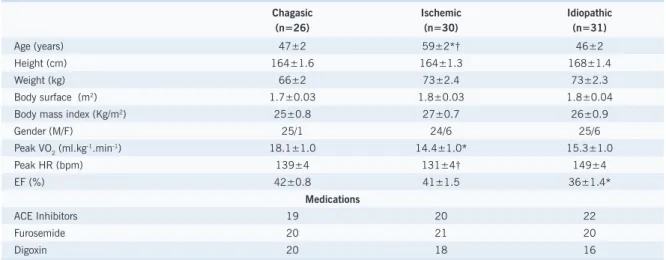
ESULTSThe characteristics of the sample analyzed are shown on Table 1. In the survival analysis by the Cox multivariate model, age (p=0.4), height (p=0.9), weight (p=0.1), body surface (p=0.3) and gender (p=0.6), were not variables that infl uenced the time to death. Nevertheless, the body mass index (p=0.05) showed a signifi cant prognostic value as to time to death. This variable was
considered the only control factor in the multivariate analysis performed for the prognostic factors.
The ejection fraction was signifi cantly smaller in patients with idiopathic etiology as compared to those of Chagasic etiology (p<0.05).
Tables 2, 3 and 4 show the metabolic, respiratory, and hemodynamic variables with their predictive risk values for death, at the anaerobic threshold, respiratory compensation point, and peak exercise, respectively.
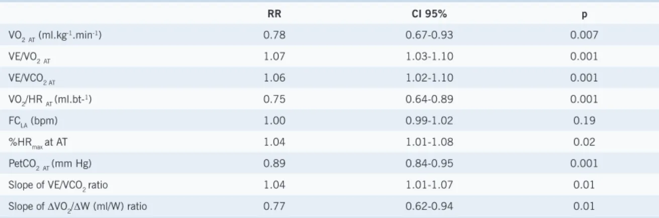
The VO2, VE/VO2, VE/VCO2, PetCO2, and VO2/HR held a statistically signifi cant correlation with the risk of death at the anaerobic threshold, respiratory compensation point, and peak exercise.
The HR did not show a statistically significant prognostic value at any phase of exercise. However, the maximal HR percentage showed a statistically signifi cant correlation with the risk of death at the anaerobic threshold and the respiratory compensation point. In addition, the SAP had a prognostic value for death at peak exercise.
The angular coeffi cients of the metabolic variables during the cardiopulmonary stress test were analyzed up to the anaerobic threshold, and both the VE/VCO2 slope and the ∆VO2/∆W slope showed a statistically signifi cant correlation to risk of death.
D
ISCUSSIONThe principal results of this study are: 1) metabolic and respiratory variables VO2, VE/VO2, VE/CO2, PetCO2 and VO2/HR at the anaerobic threshold, respiratory compensation point, and peak exercise obtained during the cardiopulmonary stress test may predict the relative mortality risk in patients with cardiac failure; 2) the degree of inclination of the VE/VCO2 and ∆VO2 /∆W slopes up to the anaerobic threshold during a
Table 1 – Sample Characteristics
Chagasic (n=26)
Ischemic (n=30)
Idiopathic (n=31)
Age (years) 47±2 59±2*† 46±2
Height (cm) 164±1.6 164±1.3 168±1.4
Weight (kg) 66±2 73±2.4 73±2.3
Body surface (m2) 1.7±0.03 1.8±0.03 1.8±0.04
Body mass index (Kg/m2) 25±0.8 27±0.7 26±0.9
Gender (M/F) 25/1 24/6 25/6
Peak VO2 (ml.kg-1.min-1) 18.1±1.0 14.4±1.0* 15.3±1.0
Peak HR (bpm) 139±4 131±4† 149±4
EF (%) 42±0.8 41±1.5 36±1.4*
Medications
ACE Inhibitors 19 20 22
Furosemide 20 21 20
Digoxin 20 18 16
Table 2 – Descriptive levels and relative risks of metabolic, respiratory, and hemodynamic variables associated to time to death at the anaerobic threshold in heart failure patients
RR CI 95% p
VO2AT (ml.kg-1.min-1) 0.78 0.67-0.93 0.007
VE/VO2AT 1.07 1.03-1.10 0.001
VE/VCO2 AT 1.06 1.02-1.10 0.001
VO2/HR AT (ml.bt-1) 0.75 0.64-0.89 0.001
FCLA (bpm) 1.00 0.99-1.02 0.19
%HRmax at AT 1.04 1.01-1.08 0.02
PetCO2AT (mm Hg) 0.89 0.84-0.95 0.001
Slope of VE/VCO2 ratio 1.04 1.01-1.07 0.01
Slope of ∆VO2/∆W (ml/W) ratio 0.77 0.62-0.94 0.01
RR – relative risk; CI 95% - 95% confi dence interval; p – value of p; VO2 AT – oxygen uptake at anaerobic threshold; VE/VO2AT – ventilatory equivalent
for oxygen uptake at anaerobic threshold; VE/VCO2 AT – respiratory equivalent for the production of carbon dioxide at anaerobic threshold; VO2 /HR
AT – oxygen pulse at anaerobic threshold; HR AT - heart rate at anaerobic threshold ; % HRmax at AT – percentage of maximal heart rate at which the
anaerobic threshold occurred; PetCO2 AT – end-tidal partial pressure of carbon dioxide at anaerobic threshold; Inclination of the VE/VCO2 ratio slope
– inclination of the ventilation and carbon dioxide production slope at anaerobic threshold; Inclination of the ∆VO2/∆W ratio – inclination of the
oxygen uptake increase to workload increase ratio at anaerobic threshold.
Table 3 – Descriptive levels and relative risks of metabolic, respiratory, and hemodynamic variables associated to time to death at the respiratory compensation point in patients with heart failure
RR CI 95% p
VO2RCP(ml.kg-1 .min-1) 0.86 0.72-0.95 0.002
VE/VO2 RCP 1.04 1.02-1.07 0.0004
VE/VCO2 RCP 1.05 1.01-1.09 0.003
VO2/HR RCP(ml.bt-1) 0.81 0.70-0.94 0.005
HR RCP (bpm) 1.00 0.99-1.02 0.37
%HRmax at RCP 1.08 1.01-1.15 0.02
PetCO2 RCP 0.91 0.86-0.97 0.002
RR – relative risk; CI 95% - 95% confi dence interval; p – value of p; VO2 RCP – oxygen uptake at respiratory compensation point; VE/VO2 RCP – ventilatory equivalent for the oxygen uptake at the respiratory compensation point; VE/VCO2 RCP – respiratory equivalent for the production of carbon dioxide at the
respiratory compensation point; VO2/HR RCP – oxygen pulse at the respiratory compensation point; HR RCP - heart rate at the respiratory compensation
point; %HRmax at RCP – percentage of maximal heart rate at which the respiratory compensation point occurred; PetCO2 RCP – end-tidal partial
pressure of carbon dioxide at the respiratory compensation point.
Table 4 – Descriptive levels and relative risks of metabolic, respiratory, and hemodynamic variables associated to time to death at peak exercise in patients with heart failure.
RR CI 95% p
VO2 peak (ml. kg-1 .min-1) 0.86 0.78-0.95 0.002
VE/VO2 peak 1.05 1.02-1.08 0.0007
VE/VCO2 peak 1.07 1.03-1.11 0.001
VO2/HR peak (ml.bt-1) 0.84 0.71-0.98 0.02
HR peak (bpm) 0.99 0.98-1.00 0.42
PetCO2 peak (mm Hg) 0.90 0.83-0.97 0.005
SAP peak (mm Hg) 0.97 0.96-0.98 0.001
RR – relative risk; CI 95% - 95% confi dence interval; p – value of p; VO2 peak – oxygen uptake at peak exercise; VE/VO2 peak – ventilatory equivalent for the oxygen uptake at peak exercise; VE/VCO2 peak- respiratory equivalent for the production of carbon dioxide at peak exercise; VO2 /HR peak-
oxygen pulse at peak exercise; HR peak – heart rate at peak exercise; PetCO2 - end-tidal partial pressure of carbon dioxide at peak exercise; SAP
progressive cardiopulmonary stress test may predict the relative mortality risk in patients with cardiac failure; 3) the percentages of maximal heart rate obtained at the anaerobic threshold and the respiratory compensation point were predictors of mortality during a progressive cardiopulmonary stress test in patients with cardiac failure; and 4) the systolic arterial blood pressure at peak exercise may predict the relative mortality risk in patients with cardiac failure.
The results of this study broaden our knowledge as they show that other respiratory and metabolic variables obtained during the cardiopulmonary stress test evaluation may estimate the mortality risk in patients with cardiac failure, not only at peak exercise, but also during submaximal physical exercise at the anaerobic threshold and at the respiratory compensation point, which are important moments of exercise. Besides clinical implications, these results have a practical importance as they point out the possibility of obtaining information on mortality risks, both at the peak exercise and during submaximal exercise, without necessarily exposing the patient to a very intense exertion. Currently, many heart failure patients may discontinue the procedure because of ischemia and/or arrhythmias10
or even because of an important functional limitation, hindering a maximal evaluation.
Additionally, the evaluation of PetCO2 has been the focus of interest during the cardiopulmonary stress test evaluation in patients with cardiac failure54-55. However,
as far as we know, this is the fi rst time the prognostic value of this variable at the anaerobic threshold, respiratory compensation point, and peak exercise is demonstrated. This fact is extremely important since PetCO2 has been recently correlated with cardiac output during exercise in patients with heart failure54-55. That is,
in patients with cardiac failure in whom a low cardiac output response is seen during exercise, the PetCO2 has lower values as well.
The level of metabolic and respiratory responses and even the altered ratio between them that leads to a predictive prognostic value of mortality in these patients raises the question as to which mechanisms direct the exaggerated ventilatory response during exercise in the presence of heart failure. Diminished perfusion in skeletal muscles because of a low cardiac output associated with a decrease in muscular oxidative capacity and even cachexia, leads to early metabolic acidosis. In patients with cardiac failure in whom muscular chemoreceptors are already hypersensitive56-58, a premature worsening
of metabolic acidosis may enormously potentiate the ventilatory drive during exercise. Our results clearly show this ventilatory behavior in heart failure. While in normal individuals the ventilatory equivalent for carbon dioxide at peak exercise is approximately 37 ± 0.2, in patients with cardiac failure it is 47 ± 0.2.
Another interesting fi nding in our study is that we did not detect that heart rate has a predictive value for
mortality in these patients. However, when a correlation is made of the percentage of maximal HR attained at peak exercise, we observe a signifi cant prognostic value both at the anaerobic threshold and at the respiratory compensation point. The fact that the heart rate at the peak exercise did not show a predictive value for mortality might be explained by the chronotropic incompetence observed in heart failure37,59. It is known that patients
with heart failure have a greater sympathetic activity as a result of elevated levels of circulating catecholamines60
that seems to be associated with desensitization, reduction of β-adrenergic receptors and down regulation of cardiac receptors61, and depressed inotropic and
chronotropic responses35, 37, 62.
In normal individuals, systolic arterial blood pressure increases progressively with progressive physical exercise, indirectly furnishing the evaluation of the inotropic response of the heart. In our results, it is clear that an inadequate increase of SAP is considered a risk factor for death in patients with cardiac failure.
Finally, we highlight a signifi cant aspect related to the sample analyzed. Contrary to other studies that also evaluated the prognostic value for mortality of the metabolic variables obtained during the ergospirometry test, racial miscegenation, a characteristic of our population, has been suggested as a factor that may infl uence the VO2 during exercise63.
Limitations - We recognize several limitations in this study. The cardiopulmonary stress test was carried out on a bicycle-ergonometer and we do not know if these results are reproducible on a treadmill. However, the test done on the bicycle-ergonometer with the ramp-protocol has proved to be very applicable in patients with cardiac failure, including for follow-up purposes of drug and non-drug treatments64-65. During the period in which this
study was conducted, the indication of beta-blockers in the treatment of patients with heart failure was not yet a decisive procedure. Today, treatment with beta-blockers is mandatory. Therefore, we do not know the importance of these metabolic and respiratory variables as predictors of a relative mortality risk while using beta-blockers.
Perspectives - A natural continuation of this study would be the analysis of the predictive value of respiratory, metabolic, and hemodynamic variables of exercise for the relative risk of mortality with the use of beta-blockers. Added to this, it would be interesting to verify if the type of ergonometer can infl uence the results attained in this study.
In closing, besides being safe and enabling an evaluation of functional capacity, the cardiopulmonary stress test, based on respiratory, metabolic, and hemodynamic responses, can provide important information as to the relative mortality risk in patients with heart failure.
Potencial Confl ict of Interest
R
EFERENCES1. Pereira-Barretto AC, Ramires JAF. Insuficiência cardíaca – um problema de Saúde Pública. Arq Bras Cardiol. 1998; 71:635-42.
2. Albanesi Fº FM. Insufi ciência cardíaca no Brasil. [Editorial]. Arq Bras Cardiol. 1998; 71:561-2.
3. Mancini DM, Eisen H, Kussmaul W, Mull R, Edmunds LH, Wilson JR. Value of peak exercise oxygen consumption for optimal timing of cardiac transplantation in ambulatory patients with heart failure. Circulation. 1991; 83:778-86.
4. Osada N, Chaitman BR, Miller LW, et al.Cardiopulmonary exercise testing identifies low risk patients with heart failure and severely impaired exercise capacity considered for heart transplantation. J Am Coll Cardiol. 1998; 31:577-82.
5. Koike A, Koyama Y, Itoh H, Adachi H, Marumo F, Hiroe M. Prognostic signifi cance of cardiopulmonary exercise testing for 10 year survival in patients with mild to moderate heart failure. Jpn Circ J. 2000; 64: 915-20.
6. Corrà U, Mezzani A, Bosimini E, Scapellato F, Imparato A, Giannuzzi P. Ventilatory response to exercise improves risk stratifi cation in patients with chronic heart failure and intermediate functional capacity. Am Heart J. 2002; 143:418-26.
7. Cohen-Solal A, Barnier P, Pessione F, et al. Comparison of the long-term prognostic value of peak exercise oxygen pulse and peak oxygen uptake in patients with chronic heart failure. Heart. 1997; 78:572-6.
8. Schalcher C, Rickli H, Brehm M, et al. Prolonged oxygen uptake kinetics during low-intensity exercise are related to poor prognosis in patientes with mild-to-moderate congestive heart failure. Chest. 2003; 124:580-6.
9. Rickli H, Kiowski W, Brehm M, et al.Combining low-intensity and maximal exercise test results improves prognostic prediction in chronic heart failure. J Am Coll Cardiol. 2003; 42:116-22.
10. De Vries RJM, Van Veldhuisen DJ, Dunselman PHJM, Van der Veer N, Crijns JGM. Physiological parameters during the initial stages of cardiopulmonary exercise testing in patients with chronic heart failure. Their value in the assessment of clinical severity and prognosis. Eur Heart J. 1997; 18:1921-30.
11. Gitt AK, Wasserman K, Kilkowski, C, et al.Exercise anaerobic threshold and ventilatory effi ciency identify heart failure patients for high risk of early death. Circulation. 2002; 106:3079-84.
12. Pilote L, Silberberg J, Lisbona R, Sniderman A. Prognosis in patients with low left ventricular ejection fraction: importance of exercise capacity. Circulation. 1989; 80:1636-41.
13. Corrà U, Mezzani A, Bosimini E, Giannuzzi P. Cardiopulmonary exercise testing and prognosis in chronic heart failure. Chest. 2004; 126:942-50.
14. Cohn JN, Rector TS. Prognosis of congestive heart failure and predictors of mortality. Am J Cardiol. 1988; 62:25A-30A.
15. Cleland JGH, Dargie HJ. Mortality in heart failure: clinical variables of prognostic value. Br Heart J. 1987; 58:572-82.
16. Cohn JN, Johnson GR, Shabetai R, et al.Ejection fraction, peak exercise oxygen consumption, cardiothoracic ratio, ventricular arrhythmias and plasma norepinephrine as determinants of prognosis in heart failure. Circulation. 1993; 87:VI- 5-VI-16.
17. Itoh H, Taniguchi K, Koike A, Doi M. Evaluation of severity of heart failure using ventilatory gas analysis. Circulation. 1990; 81(Suppl II): II31-II37.
18. Weber KT, Kinasewitz GT, Janicki JS, Fishman AP. Oxygen utilization and ventilation during exercise in patients with chronic cardiac failure. Circulation. 1982; 65:1213-23.
19. Janicki JS, Weber KT, McElroy PA. Use of the cardiopulmonary exercise
test to evaluate the patient with chronic heart failure. Eur Heart J. 1988; 9:H55-H8.
20. Weber KT, Janicki JS. Cardiopulmonary exercise testing for evaluation of chronic cardiac failure. Am J Cardiol. 1985; 55:22A-31A.
21. McElroy PA, Janicki JS, Weber KT. Cardiopulmonary exercise testing in congestive heart failure. Am J Cardiol. 1988; 62:35A-40A.
22. Higginbotham MB. The role of gas analysis in stress testing. Primary Care. 1994; 21:557-67.
23. Messner-Pellene P, Ximenes C, Brasileiro CF, Mercier J, Grolleau R, Préfaut CG. Cardiopulmonary exercise testing. Determinants of dyspnea due to cardiac or pulmonary limitation. Chest. 1994; 106:354-60.
24. Wasserman K. Diagnosing cardiovascular and lung pathophysiology from exercise gas exchange. Chest. 1997; 112:1091-101.
25. Braun MU, Rauwolf T, Zerm T, Schulze M, Schnabel A, Strasser RH. Long term biventricular resynchronization therapy in advanced heart failure: effect on neurohormones. Heart. 2005; 91:601-5.
26. Stevenson LW, Sietsema K, Tillisch JH, et al. Exercise capacity for survivors of cardiac transplantation or sustained medical therapy for stable heart failure. Circulation. 1990; 81:78-5.
27. Stevenson LW. Tailored therapy before transplantation for treatment of advanced heart failure: effective use of vasodilators and diuretics. J Heart Lung Transplant. 1991; 10:468-76.
28. Lowes BD, Higginbotham M, Petrovich L, et al.Low-dose enoximone improves execise capacity in chronic heart failure. J Am Coll Cardiol. 2000; 36:501-8.
29. Wasserman W. Short-term exercise training after cardiac surgery. In: Exercise gas exchange in heart disease. New York: Ed.Futura Publishing Company, Inc., 1996: 229-44.
30. American College of Sports Medicine – Appendix D: Metabolic calculations exercise testing and prescription. In: Guidelines for Exercise Testing and Prescription. 6th Ed. Philadelphia: Lippincott Williams & Wilkins, 2000: 300-12.
31. Clark AL, Poole-Wilson PA, Coats AJS. Effects on motivation of the patient on indices of exercise capacity in chronic heart failure. Br Heart J. 1994; 71:162-5.
32. Chomsky DB, Lang CC, Rayos GH. Hemodynamic exercise testing: a valuable tool in the selection of cardiac transplantation candidates. Circulation. 1996; 94:3176-83.
33. Mejhert M, Linder-Klingsell E, Edner M, Kahan T, Persson H. Ventilatory variables are strong prognostic markers in eldery patients with heart failure. Heart. 2002; 88:239-43.
34. Robbins M, Francis G, Pashkow FJ, et al. Ventilatory and heart responses to exercise. Better predictors of heart failure mortality than peak oxygen consumption. Circulation. 1999; 2411-7.
35. Samejima H, Omiya K, Uno M, et al. Relation ship between impaired chronotropic response, cardiac output during exercise, and exercise intolerance in patients with chronic heart failure. Jpn Heart J. 2003; 44:515-25.
36. Isnard R, Pousset F, Trochu JN, et al. Prognostic value of neurohormonal activation and cardiopulmonary exercise testing in patients with chronic heart failure. Am J Cardiol. 2000; 86:417-21.
37. Clark AL, Coats AJS. Chronotropic incompetence in chronic heart failure. Int J Cardiol. 1995; 49:225-31.
38. Chua TP, Ponikowski P, Harrington D, et al. Clinical correlates and prognosis signifi cance of the ventilatory response to exercise in chronic heart failure. J Am Coll Cardiol. 1997; 29:1585-90.
efficiency in heart failure prognostic impact. Circulation. 2000; 101:2803-9.
40. Drexler H, Riede U, Munzel T, Konig H, Funke E, Just H. Alterations of skeletal muscle in chronic heart failure. Circulation. 1992; 85:1751-9.
41. Mancini DM, Walter G, Reichek N, et al. Contribution of skeletal muscle atrophy to exercise intolerance and altered muscle metabolism in heart failure. Circulation. 1992; 85:1364-73.
42. Minotti JR, Pillay P, Oka R, Wells L, Christophy I, Massie BM. Skeletal muscle size: relationship to muscle function in heart failure. J Appl Physiol. 1993; 75:373-81.
43. Rostagno C, Lazzeri C, Gensini GF. Arrhythmia risk stratifi cation base on clinical and functional data. Ital Heart J. 2001; 2:1270-7.
44. Wilson JR, Schwartz JS, Sutton M, et al. Prognosis in severe heart failure: relation to hemodynamic measurements and ventricular ectopic activity. J Am Coll Cardiol. 1983; 2:403-10.
45. Gradman AH, Deedwania PC. Predictors of mortality in patients with heart failure.Cardiol Clin. 1994; 12:25-35.
46. Wasserman K, Hansen JE, Sue DY, Whipp BJ. Measurement of the physiological response to exercise. In: Principles of Exercise Testing and Interpretation. Philadelphia: Ed. Lea & Febiger, 1987: 27-46.
47. Mendes RP, Shimizu SH, Silva AMM, et al. Serological diagnosis of Chagas’ disease: a potencial confi rmatory assay using preserved protein antigens of Trypanosoma cruzi. J Clin Microbiol. 1997; 35:1829-34.
48. Fox EL, Mathews PF - Apêndice C: Lei dos gases. In: Bases fi siológicas da educação física e dos desportos. 3ª. Ed. Rio de Janeiro: Ed.
Interamericana Ltda, 1983: 435-9.
49. Wasserman H, Hansen JE, Sue DY, Whipp BJ. Normal values. In: Principles of exercise testing and interpretation . Philadelphia: Ed. Lea & Febiger, 1987: 72-86.
50. Medical Graphics Corporation System 2001. User’s Manual St. Paul, Normal Values, 1988; 1-8.
51. Beaver WL, Wasserman K, Whipp BJ. A new method for detecting the anaerobic threshold by gas exchange. J Appl Physiol. 1986; 60:2020-7.
52. Wasserman K, Whipp BJ, Koyal SN, Beaver WL. Anaerobic threshold and respiratory gas exchange during exercise. J Appl Physiol. 1973; 33:236-43.
53. Kleinbaum DF. Survival analysis: a self-learning text. New York: Ed:
Springer-Verlag 1996.
54. Matsumoto A, Itoh H, Eto Y, et al. End-tidal CO2 pressure decreases
during exercise in cardiac patients.Association with severity of heart failure and cardiac output reserve. Am Coll Cardiol. 2000; 36:342-9.
55. Tanabe Y, Hosaka Y, Ito Masahiro, Ito E, Suzuki K. Sinifi cance of end-tidal PCO2 response to exercise and its relation to functional capacity in patients with chronic heart failure. Chest. 2001; 119:811-7.
56. Clark AL, Piepoli M, Coats AJS. Skeletal muscle and the control of ventilation on exercise: evidence for metabolic receptors. Eur J Clin Invest. 1995; 25:299-305.
57. Piepoli M, Pinikowski P, Clark AL, et al. A neural link to explain the “muscle hypothesis” of exercise intolerance in chronic heart failure. Am J Cardiol. 1999; 137:1050-6.
58. Piepoli M, Clark AL, Volterrani M, Adamopoulos S, Sleight P, Coats AJS. Contribution of muscle afferents to the hemodynamic, autonomic and ventilatory responses to exercise in patients with chronic heart failure: effects of training physical. Circulation. 1996; 93:940-52.
59. Sullivan M, Higginbotham MB, Cobb Fr. Exercise training in patients with severe left ventricular dysfunction. Hemodynamic and metabolic effects. Circulation. 1988; 78:506-15.
60. Levine TB, Francis GS, Goldsmith SR, Simon AB, Cohn JN. Activity of the sympathetic nervous system and renin-angiotensin system assessed by plasma hormone levels and their relation to hemodynamic abnormalities in congestive heart failure. Am J Cardiol. 1982; 49:1659-65.
61. Bristow MR, Ginsburg R, Minobe W, et al. Decreased catecholamine sensitivity and beta-adrenergic receptor density in failing human hearts. N Engl J Med. 1982; 307:205-11.
62. Scheier RW, Abrahan WT. Hormones and hemodynamics in heart failure. N Engl J Med. 1999; 341:577-87.
63. Bitner V. Exercise testing in heart failure. Maximal, submaximal, or both? [Editorial]. J Am Coll Cardiol. 2003; 42: 123-125.
64. Roveda F, Middlekauff HF, Rondon MUPB, et al.The effects of exercise training on symathetic neural activation in advanced heart failure. J Am Coll Cardiol. 2003; 42:854-60.