Inflammatory mediators of coronary artery ectasia
Os mediadores inflamatórios de ectasia coronária
Shi-Min Yuan1
Abstract
he exact mechanisms underlying coronary artery ectasia (CAE) remain uncertain. his study aims to investigate whether and how inlammatory mediators play a role in the pathogenesis of CAE. he data sources of this study were located by literature searches on MEDLINE, Highwire Press and Google search engine for the year range 2000-2013. he most sensitive of the four types of plasma inlammatory mediators were cell adhesion molecules and systemic inlammatory markers followed by cytokines, while proteolytic substances were the least sensitive indicators of CAE. Hypersensitive C-reaction protein, homocysteine, intercellular adhesion molecule 1, vascular cell adhesion molecule 1, matrix metalloproteinase-9, tissue inhibitor of metalloproteinase-2, vascular endothelial growth factor and neopterin levels were signiicantly higher in CAE and coronary artery disease (CAD) patients than in controls without CAE. he percentage of granulocytes was higher in CAE, in comparison with individuals with normal coronary arteries. Polymerase chain reaction determination of angiotensin converting enzyme genotypes showed that the DD genotype was more prevalent in CAE patients than in CAD patients, while prevalence of the I allele was higher in CAD than in CAE patients. CAE is more a result of inlammatory processes than of extracellular matrix degradation, as demonstrated by investigations of plasma inlammatory mediators, activation markers and angiotensin converting enzyme genotypes. Contemporary theories are unable to explain CAE’s predilection for the right coronary artery or the occurrence of multi-vessel and multi-segment involvement.
Keywords: coronary aneurysm; extracellular matrix; inlammation mediators.
Resumo
Os mecanismos exatos da ectasia de artérias coronárias (EAC) não são completamente compreendidos. Este estudo busca veriicar, em detalhes, se e como os mediadores inlamatórios funcionam na pathogenesis de EAC. A fonte de dados do presente estudo veio da recuperação de literatura das investigações relevantes em MEDLINE, na Prensa de Highwire e na ativação de pesquisa do Google, do ano 2000 para 2013. Dos quatro tipos de mediadores inlamatórios do plasma, as moléculas de adesão de célula e os marcadores inlamatórios sistêmicos foram os mais sensíveis, sendo que cytokines foram mais sensíveis e substâncias de protease foram menos sensíveis na indicação da presença de EAC. A proteína C reativa hipersensível, o homocysteine, a molécula de adesão intercelular 1, a molécula de adesão de célula vascular 1, a matriz metalloproteinase-9, o nervo inibidor de tecido de metalloproteinase-2, o fator de crescimento endothelial vascular e os níveis de neopterin foram mais altos nos pacientes com EAC do que nos controles sem EAC. A porcentagem de granulocytes foi mais alta no grupo EAC, comparando-se com os indivíduos com a artéria coronária normal. A determinação de genótipo de enzima do angiotensin-conversão utilizando-se a técnica de reação em cadeia da polimerase revelou que o genótipo DD foi prevalecente na EAC, mas não nos pacientes de DAC, enquanto a presença do alelo I foi maior na DAC do que no EAC. O EAC é mais um resultado do processo inlamatório do que da degradação da matriz extracelular, como evidenciado por investigações dos mediadores inlamatórios de plasma, marcadores de ativação e genótipos de enzima do angiotensin a conversão. A predileção de EAC na artéria coronária direita e nos envolvimentos de multinavio e de multissegmento não é apurada por teorias contemporâneas.
Palavras-chave: aneurisma coronário; matriz extracelular; mediadores de inlamação.
1Department of Cardiothoracic Surgery, he First Hospital of Putian, Teaching Hospital, Fujian Medical University, Putian, Fujian Province, People’s Republic of China.
Financial support: None.
Conlicts of interest: No conlicts of interest declared concerning the publication of this article. Submitted: 01.23.14. Accepted: 04.30.14.
INTRODUCTION
Coronary artery ectasia (CAE) has been deined as
localized or diffuse dilation of the coronary arteries seen on coronary angiography and exceeding by 1.5 times the diameter of an adjacent and normal segment.1,2 Prevalence of CAE among patients who
underwent angiographic studies was 0.3-5.3%.3
Coronary artery ectasia may be the result of etiologies that are atherosclerotic (50%), congenital (20-30%), related to inflammatory or connective tissue diseases (10-20%), or iatrogenic following coronary interventions (3-4%).4 Inflammatory processes
play important roles in innate host defenses against infections, and therefore elevated inflammatory cytokine and C-reactive protein (CRP) levels are
associated with systemic inlammatory response.5
Over the years the number of investigations into CAE has been increasing, with similar metrological but different morphological interpretations presented by many authors. The topographical extent of CAE
has been classiied into four types: type I, diffuse
ectasia of two or three vessels; type II, diffuse disease in one vessel and localized disease in another vessel; type III, diffuse ectasia in one vessel; and type IV, localized or segmental ectasia.6 The average
diameter of ectatic segments has been reported as 5.87±0.78 mm;7 and the maximum diameters
of ectatic segments were 5.51±1.83 mm for the left anterior descending coronary artery (LAD),
4.82±1.13 mm for the circumlex artery (Cx) and
5.65±0.95 mm for the right coronary artery (RCA).6
A sample of CAE patients had much larger coronary artery diameter indices for LAD, Cx and RCA than were observed in a normal coronary artery (NCA) group.6 Zografos et al.6 have reported that the most
often involved arteries are the RCA (67.6%) and LAD (64.7%), followed by the Cx (35.3%), while the left main coronary artery (8.8%) was the least often involved. Ozbay et al.8 reported an ectasia
distribution of 60% in RCA, 57% in Cx and 50% in LAD, with 1-, 2- and 3-vessel ectasia accounting for 42.5%, 45% and 12.5%, respectively. Turhan et al.7
reported CAE distribution of 81% in RCA, 78% in LAD and 75% in Cx, with 1-, 2- and 3-vessel ectasia proportions of 19%, 28% and 53%, while rates of 1- to 6-segmental ectasia were 3%, 9%, 41%, 16%, 22% and 9%, respectively, with a mean of 3.4±1.2 ectatic segments per case. In general, ectasia often presents in 3-vessel and 3-segment forms and is most commonly found in the RCA.
It is well-known that atherosclerosis is an inflammatory process, as confirmed by recent
studies of atherosclerosis focusing in particular on the role of chemokines in atherosclerotic leukocyte accumulation.9 The coronary slow low phenomenon
has been observed angiographically in patients with CAE, potentially indicating that endothelial dysfunction is involved and that there is a link to subclinical atherosclerosis or inflammation.10
However, the exact links between inlammatory mediators and CAE remain to be clariied.
MATERIALS AND METHODS
A comprehens ive literature s earch w as conducted on MEDLINE, Highwire Press and Google search engine for the year range 2000-2013. Search terms included “coronary artery ectasia”, “inflammatory mediators”, “tumor necrosis factor (TNF)-a”,“interleukins (ILs)”, “selectin”, “homocysteine”, “intercellular adhesion molecule 1 (ICAM-1)”, “vascular cell adhesion molecule 1 (VCAM-1)”,“hypersensitive C-reactive protein (hsCRP)”, “matrix metalloproteinases (MMPs)”, “tissue inhibitors of metalloproteinases (TIMPs)”, “vascular endothelial growth factor (VEGF)”, “neopterin”, “cathepsins” and “cystatin C.” Additionally, articles describing “activation markers”, “percentages of leukocyte members” and “angiotensin converting enzyme (ACE) genotype” in connection to ACE were also identified and retrieved. The search yielded 22 potentially relevant nonrandomized and retrospective studies published from 2000 to 2013.6-8,11-29 Exclusion criteria described
in the articles selected included the following:
recent or current myocardial infarction, acute coronary syndromes, left ventricular dysfunction, left ventricular hypertrophy, cardiomyopathies, congenital heart disease, valvular heart disease,
inlammatory arrhythmias or immunologic diseases,
active infection, hepatic, renal or thyroid functional abnormalities, immunosuppressive therapy and statin use.
Data were extracted from the text, igures and/
or tables, with details of the study population, demographics, causative coronary artery disorders, types of mediators investigated, investigation methods and relationships between nature of the coronary artery disorders and types of mediators.
Patients with isolated CAE were the main study
subjects and were deined as the CAE group. Patients
with coronary artery disease (CAD) and individuals with normal coronary arteries (NCA) according to
angiography were taken as controls, and were deined
Quantitative data were collected, calculated and compared across CAE, CAD and NCA patients. Results were illustrated in bar graphs. Linear correlations between plasma levels of biomarkers and CAE morphology were summarized. Results for expression of activation markers and ACE genotypes were also compiled.
Measurement data were expressed as mean ± standard deviation and compared using the t test; while enumerative data were expressed as frequencies and compared using Fisher’s exact test. Two-tailed p<0.05 values were considered
statistically signiicant.
RESULTS
Patient data
A total of 22 relevant research articles6-8,11-29 on
inlammatory mediators of CAE were identiied and
analyzed, with an overall population of 1743 patients, breaking down as 759 (43.5%), 504 (28.9%) and 480 (27.5%) patients recruited into CAE, CAD and NCA groups respectively. All patients were adults aged over 50. No gender difference between groups
was noted. Plasma inlammatory mediators related
to CAEs were discussed in 17 articles, and could
be categorized into 4 types: cytokines (TNF-a,13,14
IL-611,12,14,16,19 and IL-1813), proteolytic substances
(cathepsins,6 cystatin,6 MMP-2,17 MMP-3,16,17
MMP-9,16 TIMP-116,17 and TIMP-225), cell adhesion
molecules (selectins,21,23 ICAM-1,15,21,24 VCAM-121,24
and VEGF25) and systemic inlammatory markers
(homocysteine,7,18 hs-CRP8,12,13,17,22 and soluble
neopterin20). Additionally, there were 5 articles that
studied expression of activation markers in peripheral blood13,26,27 and expression of ACE genotypes.28,29
Where methods used to test for plasma biomarkers were reported, enzyme-linked immunosorbent assay was used in 20 groups of patients (71.4%), immunonephelometry in 4 (14.3%), florescence polarization immunoassay (FPIA) in 2 (7.1%) and particle enhanced turbidimetric assay (for hsCRP) and sequential immunometric assay (for IL-6) were each used in 1 (3.6%) patient group (c2=59.196,
p<0.0001).
Plasma Inflammatory Mediators
Cytokines
Plasma TNF-α and IL-6 levels were signiicantly
higher in CAE than in NCA groups; whereas IL-18
levels did not differ signiicantly between the two
groups (Figure 1).
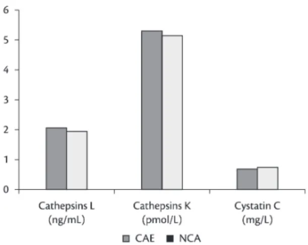
Proteolytic substances
There were no intergroup differences in cathepsins L and K or in cystatin C (Figure 2), MMP-2 or -3 (Figure 3), or TIMP-1 (Figure 4). MMP-9 levels were higher in CAE than in CAD and NCA groups, and were much higher in CAD than in the NCA group (Figure 3). TIMP-2 was signiicantly reduced in CAE patients compared with NCA subjects (Figure 4).
Cell adhesion molecules
E-selectin was significantly elevated in CAE patients compared with CAD and NCA groups (p<0.001 for CAE vs. CAD; and p<0.001 for CAE vs. NCA) (Figure 5). Patients with CAE were associated
Figure 1. Comparison of plasma tumor necrosis factor-a,13,14
interleukin-611,12,14,16,19 and interleukin-18 levels13 between
groups with coronary artery ectasia or normal coronary arteries. *p <0.05 vs. normal coronary artery group; CAE: coronary artery ectasia; IL: interleukin; NCA: normal coronary artery; TNF: tumor necrosis factor.
Figure 2. A comparison of plasma cathepsins L and K6 and
highest in CAE, higher in CAD and lowest in NCA groups, with all differences between groups
signiicant (Figure 7).
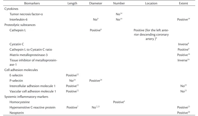
Correlations between plasma inlammatory mediators and CAEs
Different ectatic morphologies, in terms of length, diameter, number, location and extent, exhibited
Figure 3. A comparison of plasma matrix metalloproteinases16,17 between groups with coronary artery
ectasia, coronary artery disease or normal coronary arteries. **p <0.01 vs. normal coronary artery group; *, †p <0.05 vs. coronary artery disease group, and p <0.001 vs. normal coronary artery group; CAE: coronary artery ectasia; CAD: coronary artery disease; NCA: normal coronary artery.
Figure 4. A comparison of plasma tissue inhibitors of matrix metalloproteinases16,17,25 between groups with coronary artery
ectasia, coronary artery disease or normal coronary arteries. **p <0.01 vs. normal coronary artery group; CAE: coronary artery ectasia; CAD: coronary artery disease; NCA: normal coronary artery; TIMP: tissue inhibitors of matrix metalloproteinases.
Figure 5. A comparison of plasma selectins21,23 between
groups with coronary artery ectasia, coronary artery disease or normal coronary arteries. †p <0.001 vs. normal coronary artery group; †, †p <0.001 vs. coronary artery disease group, and p <0.001 vs. normal coronary artery group; CAE: coronary artery ectasia; CAD: coronary artery disease; NCA: normal coronary artery.
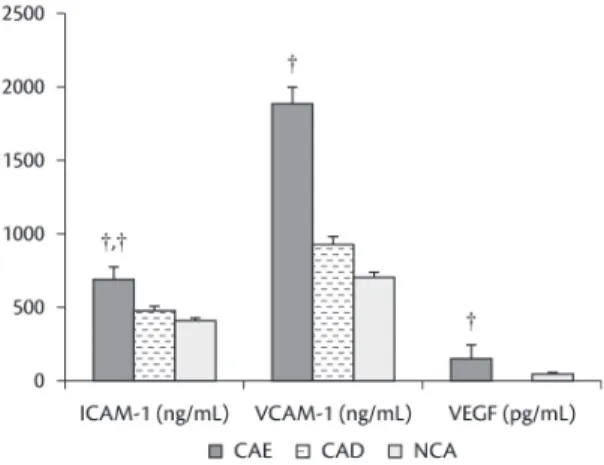
with significantly higher levels of P-selectin compared with NCA (Figure 5). Moreover, patients with CAE had much higher ICAM-1, VCAM-1 and VEGF levels than patients with NCA. Furthermore,
a signiicant difference in ICAM-1 was detected
between CAD and NCA groups (Figure 6).
Systemic inlammatory markers
Homocysteine levels were much higher in CAE than in NCA groups (Figure 7). Both hs-CRP and neopterin tapered off in all three groups, and were
Figure 6. A comparison of plasma intercellular adhesion molecule 1,15,21,24 vascular cell adhesion molecule 121,24 and
vascular endothelial growth factor25 between groups with
(localized or diffuse) and IL-6 levels, the cathepsin L to cystatin C ratio, and hs-CRP, MMP-3 and neopterin levels. Cystatin C and TIMP-1 exhibited an inverse relationship with extent (localized or diffuse) of CAE (Table 1).
Activation markers in peripheral blood
The percentage of granulocytes was higher in CAE than in the NCA group, whereas the percentage of monocytes was higher in NCA than in the CAE group.13 Mean low cytometry luorescence
intensities for cluster of differentiation (CD)11a on granulocytes, monocytes and lymphocytes and for CD45 on granulocytes and monocytes were both
signiicantly higher in CAE than in NCA group.
In CAE group patients, TNF-a levels signiicantly correlated with mean fluorescence intensity levels of CD45+ on granulocytes, monocytes and lymphocytes. Most of the CAE patients had multivessel CAEs, and the CAD patients with
CAE had signiicantly elevated activation markers
including CD11b, CD11c, CD54, CD83 and CD86, and major histocompatibility complex (MHC) class II molecules on the surface of mature dendritic cells, in comparison with CAD patients without CAE and with NCA subjects.26 Mean luorescence intensities
of CD45 and CD11b on monocyte and lymphocyte
surfaces were signiicantly higher in CAE patients
than in NCA subjects.27
Figure 7. A comparison of plasma homocysteine, hypersensitive C-reactive protein8,12,13,17,22 and neopterin20
between groups with coronary artery ectasia, coronary artery disease or normal coronary arteries. † p<0.001 vs. normal coronary artery group; †, †p <0.001 vs. coronary artery disease, and p <0.001 vs. normal coronary artery group; CAE: coronary artery ectasia; CAD: coronary artery disease; hsCRP: hypersensitive C-reactive protein; NCA: normal coronary artery.
Table 1. Correlations between plasma inlammatory mediators and morphology of coronary artery ectasia.
Biomarkers Length Diameter Number Location Extent
Cytokines
Tumor necrosis factor-a No14
Interleukin-6 No9 No14 Positive19
Proteolytic substances
Cathepsin L Positive8 Positive (for the left
ante-rior descending coronary artery )8
Cystatin C Inverse8
Cathepsin L to Cystatin C ratio Positive8
Matrix metalloproteinase-3 Positive16
Tissue inhibitor of metalloprotein-ase-1
Inverse16
Cell adhesion molecules
E-selectin Positive22
P-selectin No24 Positive24
Intercellular adhesion molecule 1 Positive22 No25
Vascular cell adhesion molecule 1 Positive22 No25
Systemic inlammatory markers
Homocysteine Positive6
Hypersensitive C-reactive protein Positive7 No7,23 Positive23
Neopterin Positive20
TNF-a levels were significantly correlated with mean fluorescence intensities of CD45+ on granulocytes, monocytes and lymphocytes.13
Additionally, CAE patients exhibited increased platelet activation, with higher levels of plasma P-selectin, b-thromboglobulin and platelet factor 4, in comparison with NCA subjects.23C. pneumoniae
IgG levels were the only marker of infection, among those that were studied, that were significantly higher in CAE patients than in NCA subjects. C. pneumoniae IgG tests were positive in 98.9% of CAD and 98.5% of CAE patients, compared to 83.5% in NCA subjects.12
ACE genotype
Determination of ACE genotypes by polymerase chain reaction revealed that the DD genotype was more prevalent in CAE than in CAD patients, while prevalence of the I allele was higher in the CAD than in CAE group.28,29
DISCUSSION
Inlammatory mediators are substances, which
can be endogenous or exogenous, that are released by immune cells when harmful agents impact on
the human body, leading to inlammatory reactions
through specific receptors.30 There are various
inflammatory mediators covered by a range of
different classiication systems, but members of the class of mediators of acute inlammation mainly
include vasoactive amines, plasma protein systems, prostaglandins and leukotrienes (eicosanoids), acetyl glycerol ether phosphocholine (PAF), cytokines, phagocyte products and nitric oxide.31 Nonetheless,
chronic inlammation is often caused by persistent
infections, prolonged exposure to toxic agents, or autoimmunity, and it is often pathologically
present with iniltration of mononuclear cells due to
persistent reaction to injury. Therefore, in chronic
inlammation, inlammatory mediators prevail with
T-lymphocyte and macrophage products including cytokines, growth factors, proteases, oxygen free radicals, complements and lipid mediators, etc.32
CAE is more likely to be involved in a chronic inflammation. Nowadays, there is increasing evidence to support this hypothesis.33 It has become
obvious that chronic inlammatory mediators are
associated with development of CAE, including cytokines, proteolytic substances, cellular adhesion
molecules and systemic inlammatory mediators, in
addition to the activation markers in peripheral blood and ACE genotypes, as indicated in the present study.
The etiology of CAE can vary, from congenital to inflammatory, but since it is most frequently seen in relation to atherosclerosis, a predominantly
inlammatory process is implied.34 Atherosclerotic
changes were observed to be more common among patients with aneurysms of the thoracic and abdominal aorta, popliteal arteries and pulmonary artery.35 Increased lumen and also circumferential
intimal thickening of ectatic coronary artery segments suggests that CAE and CAD share a common pathogenesis.36 Carotid intimal-medial
thickness was signiicantly higher in both CAE and
CAD patients with histological changes compatible with atherosclerosis than in NCA subjects.15,37 CAE
can also be associated with various conditions, such as exposure to herbicides,38 inflammatory
disorders (such as Kawasaki disease,39 Behçet’s
disease,40 Takayasu aortitis,41 polyarteritis nodosa42
and Mediterranean fever43), connective tissue
disorders (such as Ehler-Danlos syndrome)44 and
genetic disorders like Noonan syndrome.45 Coronary
vasculitis can even be present in the acute phase of acute renal failure and rheumatic heart disease, and may also be associated with CAE.46
Aydin et al.14 reported elevated plasma TNF-α
levels, whereas Adiloglu et al.13 recorded lower
TNF-α levels in CAE patients in comparison with
NCA subjects. The lower TNF-a levels were explained as predominance of TH2 and lack of TH1 type immunity in CAE patients, similar to aortic aneurysm patients. The absence of any
signiicant correlation between the dimensions of
ectatic segments and IL-6 levels might be due to the narrower range of the diameters of the coronary arteries, compared with the abdominal aorta.13
One-vessel, 2-vessel and diffuse CAE had different IL-6
levels but statistical signiicance was not attained.11
Proteolytic enzymes, such as cathepsins K and L, participate in the non-caspase pathway involved in apoptosis and atherosclerotic lesions.47 Apoptotic
pathways may be activated in the mitochondria by cathepsins, which cleave Bcl-2 interacting protein Bid and degrade the anti-apoptotic members of the Bcl-2 family, including Bcl-2, Bcl-xL and Mcl-1. Cathepsins also contribute to monocyte and macrophage differentiation and migration.48 CAE
is characterized by irregular, diffuse, saccular, or fusiform dilation of the coronary arteries, and the major pathophysiologic process involved in ectasia is most likely vascular remodeling in response to atherosclerosis.49 Experimental data show
increased inlammatory response and activation of
of the renin-angiotensin system. Additionally,
an insertion/deletion polymorphism of ACE is
closely correlated with coronary vascular tone and development of aneurysms.28 It was also found that
CAE patients had an increased prevalence of the
5A/5A polymorphism of MMP-3, compared with
CAD patients, implying overexpression of MMP-3 with increased extracellular matrix degradation.35
Overexpression of MMPs and imbalanced MMP/
TIMP in CAE patients,16 as well as significant
correlations between pro-brain natriuretic peptide
and MMP-2, TIMP-1 and MMP-2/TIMP-1 in CAE
but not in CAD and NCA groups, indicates that matrix remodeling is involved in pathogenesis of CAE.17
The concurrence of decreased MMP inhibition and increased angiogenetic activity suggests accelerated and persistent extracellular matrix remodeling predisposing to aneurysm formation and increased risks of thrombosis formation.25
Patients with isolated CAE have elevated levels of plasma soluble ICAM-1, VCAM-1 and E-selectin in comparison with patients with obstructive CAD but without CAE and in comparison with NCA subjects, with ICAM-1 being the only independent variable associated with isolated CAE, suggesting that ectasia
develops in an intensively inlammatory vascular
wall that predisposes to plaque instability.15 VEGF is
a key regulator of physiological angiogenesis during embryogenesis, skeletal growth and reproductive functions. VEGF, which increases in response to
inlammation, may play a role in the pathogenesis of
coronary artery lesions.50 Furthermore, transforming
growth factor-β1 overexpression in patients with
CAE and CAD in addition to signiicant correlation
between plasma cystatin C levels and transforming
growth factor-β1 strongly support this hypothesis. 51
Thus, CAE may be a destructive inlammatory lesion
of the vascular wall.52 However, it is not clear why
some patients with coronary atherosclerosis develop CAE while most do not.
Homocysteine enhances production of several
pro-inlammatory cytokines. Hyperhomocysteinemia
is an important risk factor for atherosclerosis and thrombotic disease.53 Patients with isolated CAE had
signiicantly higher levels of plasma homocysteine
than controls and 59% of patients with isolated CAE had elevated plasma homocysteine, compared to 7% of NCA subjects.7 This phenomenon is evidence to
support an inlammatory etiology of CAE. Plasma hs-CRP levels were signiicantly higher in CAE group than in CAD group at baseline, but had signiicantly
decreased from baseline 3 months later in both CAE and CAD patients. There was a positive correlation
between hs-CRP and low density lipoprotein cholesterol in both CAE and CAD groups.8 Neopterin
is produced by activated macrophages performing immune and macrophage activities.20 Patients
with isolated CAE had increased neopterin level compared with NCA subjects, indicating a possible
role for neopterin in inlammatory processes in CAD
patients.2
The source and mechanism of immune activation in CAEs remain unknown. T-cells from patients with congestive heart failure had enhanced surface expression of the activation markers CD69 and CD25, while there was no upregulation of the monocyte activation marker CD32.54 Patients with
elevated plasma thiols homocysteine and cysteine levels had increased risk of atherosclerosis. Total cysteine concentration, but not total homocysteine, CRP, or neopterin, was higher in CAD patients with stepwise increases relative to the extent of CAD.55
Mean serum neopterin levels were significantly higher in patients with adverse cardiac events than in those without. Multiple regression analysis revealed that neopterin levels, severity of CAD and a history of previous myocardial infarction were independent predictors of adverse cardiac events.56
The first phase of inflammation is adhesion of leukocytes to the endothelium, mediated by several adhesive molecules.57 Elevated cellular
adhesion molecule levels in CAE patients may be an
indicator of endothelial activation and inlammatory
processes.27 The neutrophil-lymphocyte ratio was
signiicantly higher in the CAE group compared
with control, and this ratio was also positively correlated with the number of ectatic segments.58,59
Furthermore, CAE patients had higher mean platelet and eosinophil volumes. Increased concentration of eosinophils might be explained by vascular destruction, endothelial dysfunction60 and thrombosis
in CAE patients.61 Activated cells express “activation
markers,” which is a class with many members, including immunoglobulins, T cells, natural killer cells, monocytes and other antigen-presenting cells that trigger immune reactions by entering cell cycles. Activated cells may also enter the cell cycle by means of T cell receptors encountering antigens or by “bystander” mechanisms via exposure to certain cytokines.62
The ACE DD genotype was more prevalent in patients with CAE.29 Most patients with CAE
gene polymorphism and CAD.63 The D allele of an
ID polymorphism was associated with higher plasma ACE concentrations. Therefore, the deleterious effect of the DD genotype might be attributable to overexpression of ACE. Angiotensin II may
promote CAE formation by enhancing inlammatory
reactions, promoting smooth muscle cell migration, inducing extracellular matrix remodeling and MMP generation, or by stimulating production of reactive oxygen species.64
The etiology of CAE’s predilection for the RCA has not been well-described.65 An insertion/deletion
polymorphism of the ACE was found to be associated with coronary vascular tone and the development of aneurysms.28 In contrast with discrete saccular CAE,
diffuse fusiform CAE is often bilateral and is often associated with abdominal aortic aneurysms rather than with concurrent CAD. The absence of CAD in CAE patients did not preclude patients from having left ventricular function impairment.66 The ACE DD
genotype might be a potential risk factor for CAE,28
and the role that the renin-angiotensin system might play in the genesis of CAE suggests use of ACE-modulating agents could reduce the risk of CAE.29
To date there is no data on the anatomical changes that may occur over time in CAE, nor
is there suficient comparative research into the
different anatomical forms of CAE involvement. In order to further clarify the underlying etiologies, experimental investigations designed to reveal the precise molecular mechanisms involved are needed.
In conclusion, the pathogenesis of CAE is more reliant on a strong inflammatory reaction than on extracellular matrix remodeling as has been demonstrated by investigation of inflammatory mediators. Activation markers and ACE genotypes may also play an important role in the development of CAE. However, contemporary theories are unable to explain CAE’s predilection for the RCA or the occurrence of multi-vessel and multi-segment involvements. Further investigations of different CAE morphologies designed to reveal the precise underlying pathogenesis are essential if effective antagonists of the causative mediators responsible
for CAE formation are to be identiied.
REFERENCES
1. Swaye PS, Fisher LD, Litwin P, et al. Aneurysmal coronary artery disease. Circulation. 1983;67(1):134-8. http://dx.doi. org/10.1161/01.CIR.67.1.134. PMid:6847792
2. Falsetti HL, Carrol RJ. Coronary artery aneurysm. A review of the literature with a report of 11 new cases. Chest. 1976;69(5):630-6. http://dx.doi.org/10.1378/chest.69.5.630. PMid:1083790
3. Pinar-Bermúdez E, López Palop R, Lozano Martínez-Luengas I, et al. [Coronary ectasia: prevalence, and clinical and angiographic characteristics]. Rev Esp Cardiol. 2003;56(5):473-9. http://dx.doi. org/10.1016/S0300-8932(03)76902-4. PMid:12737785
4. Mavrogeni S. Coronary artery ectasia: diagnosis and treatment. E-Journal of Cardiology Practice. 2009;8(15). http://www. escardio.org/communities/councils/ccp/e-journal/volume8/ Pages/Coronary-artery-ectasia-Mavrogeni.aspx.
5. Li JJ, He JG, Nan JL, He ZX, Zhu CG, Li J. Is systemic inlammation responsible for coronary artery ectasia? Int J Cardiol. 2008;130(2):69-70. http://dx.doi.org/10.1016/j.ijcard.2007.11.078. PMid:18207258
6. Zografos TA, Haliassos A, Korovesis S, Giazitzoglou E, Serelis J, Katritsis DG. Serum cathepsin levels in coronary artery ectasia. Int J Cardiol. 2010;145(3):606-7. http://dx.doi.org/10.1016/j. ijcard.2010.08.061. PMid:20837372
7. Turhan H, Erbay AR, Yasar AS, et al. Plasma homocysteine levels in patients with isolated coronary artery ectasia. Int J Cardiol. 2005;104(2):158-62. http://dx.doi.org/10.1016/j. ijcard.2004.10.025. PMid:16168808
8. Ozbay Y, Akbulut M, Balin M, Kayancicek H, Baydas A, Korkmaz H. The level of hs-CRP in coronary artery ectasia and its response to statin and angiotensin-converting enzyme inhibitor treatment. Mediators Inlamm. 2007;2007(1):89649.
9. Tuttolomondo A, Di Raimondo D, Pecoraro R, Arnao V, Pinto A , Licata G. Atherosclerosis as an inf lammatory disease. Curr Pharm Des. 2012;18(28):4266-88. http://dx.doi. org/10.2174/138161212802481237. PMid:22390643
10. Wang X, Nie SP. The coronary slow flow phenomenon: characteristics, mechanisms and implications. Cardiovasc Diagn her. 2011;1(1):37-43. PMid:24282683.
11. Tokgozoglu L, Ergene O, Kinay O, Nazli C, Hascelik G, Hoscan Y. Plasma interleukin-6 levels are increased in coronary artery ectasia. Acta Cardiol. 2004;59(5):515-9. http://dx.doi.org/10.2143/ AC.59.5.2005226. PMid:15529557
12. Adiloglu AK, Can R , Nazli C, et al. Ectasia and severe atherosclerosis: relationships with chlamydia pneumoniae, helicobacterpylori, and inlammatory markers. Tex Heart Inst J. 2005;32(1):21-7. PMid:15902817.
13. Adiloglu AK, Ocal A, Tas T, Onal S, Kapan S, Aridogan B. Increased expression of CD11a and CD45 on leukocytes and decreased serum TNF-alpha levels in patients with isolated coronary artery ectasia. Clin Lab. 2011;57(9-10):703-9. PMid:22029185.
14. Aydin M, Tekin IO, Dogan SM, Yildirim N, Arasli M, Sayin MR, et al. The levels of tumor necrosis factor-alpha and interleukin-6 in patients with isolated coronary artery ectasia. Mediators Inlamm. 2009;2009:106145. 10.1155/2009/106145. Epub 2009 Jun 17.
15. Daoud EM, Abdelaziz AA, Hassan NA. Isolated coronary artery ectasia debate: Inlammation versus atherosclerosis. Egypt Heart J. 2012;64(4):185-90. http://dx.doi.org/10.1016/j.ehj.2012.06.001.
16. Dogan A, Tuzun N, Turker Y, Akcay S, Kaya S, Ozaydin M. Matrix metalloproteinases and inlammatory markers in coronary artery ectasia: their relationship to severity of coronary artery ectasia. Coron Artery Dis. 2008;19(8):559-63. http://dx.doi.org/10.1097/ MCA.0b013e3283109079. PMid:19005290
18. Kosar F, Sincer I, Aksoy Y, Ozerol I. Elevated plasma homocysteine levels in patients with isolated coronary artery ectasia. Coron Artery Dis. 2006;17(1):23-7. http://dx.doi.org/10.1097/00019501-200602000-00004. PMid:16374137
19. Ateia MY, Azmy AM, El-Shafy SA, El-Naggar WM, Abdel-Latif I. Evaluation of serum levels of C-reactive protein (CRP) and interleukin-6 (Il-6) in coronary artery ectasia. Heart Mirror J. 2007;1(2):75-81.
20. S ahin M , Varol E , Oz aydin M , et al . Comparison of neopterin levels in patients with coronary artery ectasia versus patients with obstructive coronary artery disease. South Med J. 2008;101(5):476-9. http://dx.doi.org/10.1097/ SMJ.0b013e31815d22f4. PMid:18414153
21. Turhan H, Erbay AR, Yasar AS, et al. Plasma soluble adhesion molecules; intercellular adhesion molecule-1, vascular cell adhesion molecule-1 and E-selectin levels in patients with isolated coronary artery ectasia. Coron Artery Dis. 2005;16(1):45-50. http://dx.doi.org/10.1097/00019501-200502000-00009. PMid:15654200
22. Turhan H, Erbay AR, Yasar AS, Balci M, Bicer A, Yetkin E. Comparison of C-reactive protein levels in patients with coronary artery ectasia versus patients with obstructive coronary artery disease. Am J Cardiol. 2004;94(10):1303-6. http://dx.doi. org/10.1016/j.amjcard.2004.07.120. PMid:15541253
23. Yasar AS, Erbay AR, Ayaz S, et al. Increased platelet activity in patients with isolated coronary artery ectasia. Coron Artery Dis. 2007;18(6):451-4. http://dx.doi.org/10.1097/ MCA.0b013e3282a30665. PMid:17700216
24. Yilmaz H, Tayyareci G, Sayar N, et al. Plasma soluble adhesion molecule levels in coronary artery ectasia. Cardiology. 2006;105(3):176-81. http://dx.doi.org/10.1159/000091414. PMid:16490963
25. Savino M, Parisi Q, Biondi-Zoccai GG, Pristipino C, Cianlone D, Crea F. New insights into molecular mechanisms of diffuse coronary ectasiae: a possible role for VEGF. Int J Cardiol. 2006;106(3):307-12. http://dx.doi.org/10.1016/j. ijcard.2005.01.025. PMid:16337037
26. Yildirim N, Tekin IO, Arasli M, Aydin M. Further increase in the expression of activation markers on monocyte-derived dendritic cells in coronary artery disease patients with ectasia compared to patients with coronary artery disease alone. Mediators Inlamm. 2010;2010:748919. 10.1155/2010/748919.
27. Yildirim N, Tekin IO, Dogan SM, et al. Expression of monocyte and lymphocyte adhesion molecules is increased in isolated coronary artery ectasia. Coron Artery Dis. 2007;18(1):49-53. http://dx.doi. org/10.1097/MCA.0b013e32801104d4. PMid:17172930 28. Uyarel H, Okmen E, Tartan Z, et al. he role of angiotensin
converting enzyme genotype in coronary artery ectasia. Int Heart J. 2005;46(1):89-95. http://dx.doi.org/10.1536/ihj.46.89. PMid:15858940
29. Gülec S, Aras O, Atmaca Y, et al. Deletion polymorphism of the angiotensin I converting enzyme gene is a potent risk factor for coronary artery ectasia. Heart. 2003;89(2):213-4. http://dx.doi. org/10.1136/heart.89.2.213. PMid:12527685
30. Chauhan LS. Inlammation mediators: a review. http://www. pharmainfo.net/reviews/inlammation-mediators-review.
31. Halfman CJ. Mediators of Inlammation. Laboratory Medicine and PathoPhysiology. http://pro2services.com/lectures/fall/infmeds/ infmed.htm.
32. Chronic inlammation and mediators. Illinois: University of Illinois. http://www.life.illinois.edu/mcb/493.bhp/private/lectures/ ppt_pdf/Chronic%20Inlammation.pdf.
33. Balin M, Celik A, Kobat MA. he association between soluble lectin-like oxidized low-density lipoprotein receptor-1 levels and patients with isolated coronary artery ectasia. J hromb Thrombolysis. 2012;33(3):239-45. http://dx.doi.org/10.1007/ s11239-011-0668-4. PMid:22271373
34. Díaz-Zamudio M, Bacilio-Pérez U, Herrera-Zarza MC, et al. Coronary artery aneurysms and ectasia: role of coronary CT angiography. Radiographics. 2009;29(7):1939-54. http://dx.doi. org/10.1148/rg.297095048. PMid:19926755
35. Manginas A, Cokkinos DV. Coronary artery ectasias: imaging, functional assessment and clinical implications. Eur Heart J. 2006;27(9):1026-31. http://dx.doi.org/10.1093/eurheartj/ehi725. PMid:16415301
36. Sudhir K, Ports TA, Amidon TM, et al. Increased prevalence of coronary ectasia in heterozygous familial hypercholesterolemia. Circulation. 1995;91(5):1375-80. http://dx.doi.org/10.1161/01. CIR.91.5.1375. PMid:7867176
37. Markis JE, Joffe CD, Cohn PF, Feen DJ, Herman MV, Gorlin R. Clinical significance of coronary arterial ectasia. Am J Cardiol. 1976;37(2):217-22. http://dx.doi.org/10.1016/0002-9149(76)90315-5. PMid:1108631
38. Sorrell VL, Davis MJ, Bove AA. Current knowledge and signiicance of coronary artery ectasia: a chronologic review of the literature, recommendations for treatment, possible etiologies, and future considerations. Clin Cardiol. 1998;21(3):157-60. http://dx.doi. org/10.1002/clc.4960210304. PMid:9541758
39. Baer AZ, Rubin LG, Shapiro CA, et al. Prevalence of coronary artery lesions on the initial echocardiogram in Kawasaki syndrome. Arch Pediatr Adolesc Med. 2006;160(7):686-90. http://dx.doi.org/10.1001/archpedi.160.7.686. PMid:16818833
40. Tatli E, Surucu H, Aktoz M, Buyuklu M. Coronary artery ectasia in a patient with Behcet’s disease. Saudi Med J. 2007;28(8):1281-2. PMid:17676219.
41. Suzuki H, Daida H, Tanaka M, et al. Giant aneurysm of the left main coronary artery in Takayasu aortitis. Heart. 1999;81(2):214-7. PMid:9922363.
42. Pick RA, Glover MU, Vieweg WV. Myocardial infarction in a young woman with isolated coronary arteritis. Chest. 1982;82(3):378-80. http://dx.doi.org/10.1378/chest.82.3.378. PMid:6125346
43. Cascio A, Maggio MC, Cardella F, et al. Coronary involvement in Mediterranean spotted fever. New Microbiol. 2011;34(4):421-4. PMid:22143818.
44. Di Mario C, Zanchetta M, Maiolino P. Coronary aneurysms in a case of Ehlers-Danlos syndrome. Jpn Heart J. 1988;29(4):491-6. http://dx.doi.org/10.1536/ihj.29.491. PMid:3184455
45. Hakim FA, Gruden JF, Panse PM, Alegria JR. Coronary artery ectasia in an adult Noonan syndrome detected on coronary CT angiography. Heart Lung Circ. 2013;22(12):1051-3. http://dx.doi. org/10.1016/j.hlc.2013.03.079. PMid:23608065
46. Eleftheriadis D, Eleftheriadis N. (2012). Coronary artery disease and systemic vasculitis: case report and review. In: Chaikovsky I, editor. Coronary artery diseases. Rijeka; 2012. p. 281-300. http://www.intechopen.com/books/coronary-artery-diseases/ coronary-artery-diseaseand-systemic-vasculitis.
47. Garg NJ. Inflammasomes in cardiovascular diseases. Am J Cardiovasc Dis. 2011;1(3):244-54. PMid:22254202.
49. Antoniadis AP, Chatzizisis YS, Giannoglou GD. Pathogenetic mechanisms of coronary ectasia. Int J Cardiol. 2008;130(3):335-43. http://dx.doi.org/10.1016/j.ijcard.2008.05.071. PMid:18694609 50. Chakrabarti S, homas E, Wright JG, Vettukattil JJ. Congenital
coronary artery dilatation. Heart. 2003;89(6):595-6. http://dx.doi. org/10.1136/heart.89.6.595. PMid:12748209
51. Yetkin E, Acikgoz N, Sivri N, et al. Increased plasma levels of cystatin C and transforming growth factor-b1 in patients with coronary artery ectasia: can there be a potential interaction
between cystatin C and transforming growth factor-b1.
Coron Artery Dis. 2007;18(3):211-4. http://dx.doi.org/10.1097/ MCA.0b013e328087bd98. PMid:17429295
52. Turhan H, Yetkin E. Coronary artery ectasia: is it a destructive inflammatory lesion of the vascular wall? Int J Cardiol. 2007;118(2):241. http://dx.doi.org/10.1016/j.ijcard.2006.07.009. PMid:16959337
53. Gokkusu C, Tulubas F, Unlucerci Y, Ozkok E, Umman B, Aydin M. Homocysteine and pro-inlammatory cytokine concentrations in acute heart disease. Cytokine. 2010;50(1):15-8. http://dx.doi. org/10.1016/j.cyto.2009.12.015. PMid:20129796
54. Yndestad A, Holm AM, Müller F, et al. Enhanced expression of inf lammator y cytokines and activation markers in T-cells from patients with chronic heart failure. Cardiovasc Res. 2003;60(1):141-6. http://dx.doi.org/10.1016/S0008-6363(03)00362-6. PMid:14522416
55. Schroecksnadel K, Walter RB, Weiss G, Mark M, Reinhart WH, Fuchs D. Association between plasma thiols and immune activation marker neopterin in stable coronary heart disease. Clin Chem Lab Med. 2008;46(5):648-54. http://dx.doi.org/10.1515/ CCLM.2008.121. PMid:18839466
56. Avanzas P, Arroyo-Espliguero R, Quiles J, Roy D, Kaski JC. Elevated serum neopterin predicts future adverse cardiac events in patients with chronic stable angina pectoris. Eur Heart J. 2005;26(5):457-63. http://dx.doi.org/10.1093/eurheartj/ehi111. PMid:15684278
57. Dejana E, Breviario F, Caveda L. Leukocyte-endothelial cell adhesive receptors. Clin Exp Rheumatol. 1994;12(Suppl 10):S25-8. PMid:7955623.
58. Ayhan SS, Oztürk S, Erdem A, et al. Nötroil/lenfosit oranının koroner ektazisi varlığı ve yaygınlığı ile ilişkisi. Turk Kardiyol Dern Ars. 2013;41(3):185-90. http://dx.doi.org/10.5543/ tkda.2013.83030. PMid:23703551
59. Sarli B, Baktir AO, Saglam H, et al. Neutrophil-to-lymphocyte ratio is associated with severity of coronary artery ectasia. Angiology. 2013;65(2):147-51. PMid:23657176
60. Syal SK, Kapoor A, Bhatia E, et al. Vitamin D deiciency, coronary artery disease, and endothelial dysfunction: observations from a coronary angiographic study in Indian patients. J Invasive Cardiol. 2012;24(8):385-9. PMid:22865308.
61. Demir M, Keceoglu S, Melek M. The relationship between plasma eosinophil count and coronary artery ectasia. Cardiol Rev. 2013;4(4-5):159-64.
62. Landay A, Desai S. Immune activation and inlammation: role in co-morbidities in HIV disease. http://link.springer.com/content/ pdf/10.1007%2F978-1-4615-3736-6_51.pdf.
63. Marković BB, Bergovec M, Reiner Z, Sertić J, Vincelj J, Marković M. Deletion polymorphism of the angiotensin I-converting enzyme gene in elderly patients with coronary heart disease. Coll Antropol. 2007;31(1):179-83. PMid:17598398.
64. Chen Q, Jin M, Yang F, Zhu J, Xiao Q, Zhang L. Matrix metalloproteinases: inlammatory regulators of cell behaviors in vascular formation and remodeling. Mediators Inlamm. 2013;2013(2013):1-14. 928315. 10.1155/2013/928315.
65. Wuyts B, Delanghe J, De Buyzere M. Angiotensin I-converting enzyme insertion/deletion polymorphism: clinical implications. Acta Clin Belg. 1997;52(6):338-49. PMid:9489129.
66. Ceyhan K, Koc F, Ozdemir K, et al. Coronary ectasia is associated with impaired left ventricular myocardial performance in patients without signiicant coronary artery stenosis. Med Princ Pract. 2012;21(2):139-44. http://dx.doi.org/10.1159/000333390. PMid:22123194
Correspondence
Shi-Min Yuan Department of Cardiothoracic Surgery, he First Hospital of Putian, Teaching Hospital, Fujian Medical University 389 Longdejing Street, Chengxiang District Putian 351100, Fujian Province, People’s Republic of China Tel: 86 594 6923117 E-mail: [email protected]
Author information Shi-Min Yuan is a cardiac surgeon at he First Hospital of Putian, Teaching Hospital, Fujian Medical University.
Author contributions
Conception and design: S-MY Analysis and interpretation: S-MY Data collection: S-MY Writing the article: S-MY Critical revision of the article: S-MY Final approval of the article*: S-MY Statistical analysis: S-MY Overall responsibility: S-MY Obtained funding: None.