Expression, production, and renaturation
of a functional single-chain variable
antibody fragment (scFv)
against human ICAM-1
H. Sun
1,2, G.M. Wu
1, Y.Y. Chen
3,4, Y. Tian
1, Y.H. Yue
1and G.L. Zhang
1 1Institute of Military Veterinary, Academy of Military Medical Sciences, Changchun, China 2Department of Biological Pharmacy, Heilongjiang Vocational College of Biology Science and Technology, Harbin, China 3Institute of Medicinal Biotechnology, Peking Union Medical College and Chinese Academy of Medical Sciences, Beijing, China 4College of Animal Husbandry and Veterinary Medicine, Jilin University, Changchun, ChinaAbstract
Intercellular adhesion molecule-1 (ICAM-1) is an important factor in the progression of inflammatory responsesin vivo. To develop a new anti-inflammatory drug to block the biological activity of ICAM-1, we produced a monoclonal antibody (Ka=4.19610–8M) against human ICAM-1. The anti-ICAM-1 single-chain variable antibody fragment (scFv) was expressed
at a high level as inclusion bodies in Escherichia coli. We refolded the scFv (Ka=2.35610–7M) by ion-exchange
chromatography, dialysis, and dilution. The results showed that column chromatography refolding by high-performance Q Sepharose had remarkable advantages over conventional dilution and dialysis methods. Furthermore, the anti-ICAM-1 scFv yield of about 60 mg/L was higher with this method. The purity of the final product was greater than 90%, as shown by denaturing gel electrophoresis. Enzyme-linked immunosorbent assay, cell culture, and animal experiments were used to assess the immunological properties and biological activities of the renatured scFv.
Key words: Intercellular adhesion molecule-1; Single-chain variable antibody fragment; Expression; Purification; Renaturation; Biological activity
Introduction
Intercellular adhesion molecule-1 (ICAM-1) is a member of the immunoglobulin supergene family and is a cell surface ligand for lymphocyte function-associated antigen-1 (LFA-1). ICAM-1 is mainly expressed on the surface of endothelial cells. It is also expressed on activated lymphocytes in inflamed regions; however, the expression in peripheral blood lymphocytes is normally very low. The level of ICAM-1 is upregulated in the presence of various stimuli (e.g., inflammatory mediators, oxidative stress, and viral infection) (1). The interaction of ICAM-1 with LFA-1 plays an important role in leukocyte adhesion and in the performance of immunological and inflammatory functions mediated by leukocyte adhesion (2). Increased ICAM-1 levels result in the transmigration of neutrophils during the initial phase of inflammation. Inhibition of this process could decrease the inflammatory response and tissue damage (3). Many diseases are
associated with overexpressed ICAM-1, such as acute pancreatitis (4), inflammatory bowel disease and colonic neoplasms (5), inflammation associated with organ transplantation (6-8), angiocardiopathy (9,10), ischemia-reperfusion injury (11), and cancer (12-14). These reports suggested that the anti-ICAM-1 strategy has a potential application in the treatment of ICAM-1-mediated immu-nological and inflammatory diseases.
A monoclonal antibody (mAb) with biological activity against human ICAM-1 was prepared in our laboratory by immunizing BALB/c mice with recombinant human ICAM-1. The secreted mAb was purified by caprylic acid-ammonium sulfate precipitation and affinity chromatogra-phy. Its subclass and titer were determined by indirect enzyme-linked immunosorbent assay (ELISA), specificity by Western blot, and neutralizing activity by cell adhesion tests.
Correspondence: Y.H. Yue and/or G.L. Zhang, Institute of Military Veterinary, Academy of Military Medical Sciences, Changchun, 130122, China. Fax: ++86-0431-8698-5961. E-mail: [email protected] and/or [email protected]
The single-chain variable antibody fragment (scFv) strategy has become one of the most popular methods in antibody engineering because of its lower level of immunogenicity, and its small molecular size endows scFv with better tissue penetration (15). Thus, scFv has a wide range of applications in diagnosis and therapy. It is possible for anti-ICAM-1 scFv to block the biological activity of ICAM-1, and it may be effective in preventing the progression of the above-mentioned diseases. Large-scale production of anti-ICAM-1 scFv protein is required for carrying out further research and application.
In this study, we expressed anti-ICAM-1 scFv at a high level in the form of inclusion bodies inEscherichia coli. We successfully refolded the denatured scFv using ion exchange chromatography (IEC). This study paves the way for preparing large quantities of anti-ICAM-1 scFv for application against diseases associated with inflammation.
Material and Methods
Material
The following reagents were used in this study: isopropyl-b-D-thiogalactoside (IPTG; Merck, Germany); horseradish peroxidase (HRP)-conjugated goat anti-rabbit IgG antibody (KeyGen Biotech, China); reduced glu-tathione (GSH), oxidized gluglu-tathione (GSSG), lipopolysac-charide (LPS), and bovine serum albumin (BSA; Sigma, USA); Ficoll-Paque (Amersham Biosciences, China); recombinant human ICAM-1 and rat ICAM-1 (R&D, USA); XK16/20 Q Sepharose high performance (Q HP; 17-1064-01; Pharmacia Biotech, USA); A¨KTA prime protein purification system (18-1137-18; Amersham Biosciences, Sweden); fermenter (10L, East China University Science and Technology); and anti-ICAM-1 mAb (prepared in our laboratory) (16). A human umbilical vein endothelial cell line (ECV-304) and Kunming mice (weight ,18-22 g) were purchased from Maisha Biotechnology Limited Company (China) and Changchun Institute of Biological Products (China), respectively.
All the other chemicals used were of analytical grade. Anti-scFv rabbit polyclonal antibodies (antisera) were obtained following hypodermic injection of rabbits with the purified protein from inclusion bodies.
The animal experiments were conducted under applicable laws and guidelines and after approval by the Animal Care and Use Committee of Jilin University.
Plasmids and strains
The pET22b plasmid and host E. coli BL-21(DE3) were obtained from Novagen (Germany). The expression plasmid pET22b-(ICAM-1)scFv was constructed in our laboratory (17).
Expression of anti-ICAM-1 scFv
Bacterial cells with the expression plasmid pET22b-(ICAM-1)scFv were grown overnight in 5 mL Luria-Bertani
broth (LB) with 50 mg/L ampicillin at 376C. The overnight culture was transferred into 1 L LB with 50 mg/L ampicillin and grown at 376C. Expression was induced by adding IPTG to a final concentration of 0.8 mM when the culture was grown to absorbance600 of ,0.6-0.8. The mixture was further incubated for another 4 h. Cells were harvested by centrifugation at 3000gfor 20 min at 46C.
The fermentation procedure for the large-scale scFv production in a bioreactor was as follows. The prepared strain was added to the fermentation medium, pH , 7.0-7.3, with 400 rpm rotation and aeration rate 20 L/min; meanwhile, the dissolved oxygen was controlled at levels of ,20-30%. Production of scFv was then induced by addition of 0.8 mM IPTG, and the culture was allowed to incubate at 376C for 4 h.
Preparation of samples
ScFv was expressed as described earlier. After extraction from E. coli cells using a combination of lysozyme and sonication, the inclusion bodies were washed three times with 100 mL 0.5% Triton X-100 (v/v) and 2 M urea for 30 min each time. Two grams of the pellet were suspended in 10 mL of denaturing buffer (50 mM Tris-HCl, 50 mM NaCl, 1 mM EDTA, 8 M urea, and 10 mM dithiothreitol, pH 8.0) and kept at room temperature for 2-4 h to dissolve the inclusion bodies. Residual insoluble matter was removed by centrifuging at 4000gfor 30 min. The supernatant was filtered through a 0.22mm filter (Millipore, USA) before chromatography.
Refolding of scFv
Refolding by dilution. Seven milliliters of solubilized inclusion bodies with a concentration of 7 mg/mL were slowly dropped into the refolding buffer (30 mM Tris-HCl, 1 mM EDTA, 1 mM GSH, 0.2 mM GSSG, pH 8.0) and adjusted to a protein concentration of 100mg/mL. The solution was stirred for 2 h at room temperature, followed by incubation at 46C for more than 48 h (18).
Purification by IEC
A volume of 500 mL diluted supernatant was applied to a 10 mL IEC column (Q HP), which was pre-equilibrated with Buffer B (30 mM Tris-HCl, 1 mM EDTA, pH 8.0). The A¨KTA Prime Protein Purification System was used, with the column eluted with a 50 mL linear gradient of Buffer B to Buffer B containing 0.5 M NaCl. The final protein concentration was determined with the Bradford assay.
concentration was reduced as follows: 6R4R2R1R 0.5R0 M (19). After centrifugation, the supernatant was applied to a Q HP column for further purification, as described above.
Refolding by IEC. An IEC system was used with a XK16/20 column containing 10 mL of Q HP of the A¨KTA Prime Protein Purification System. The column was equilibrated with denaturing buffer (30 mM Tris-HCl, pH 8.0, with 6 M urea, 1 mM EDTA, 1 mM GSH, and 0.2 mM GSSG). Following equilibration, the urea concentration of the solubilized inclusion bodies (7 mL; 7 mg/mL) was adjusted to 6 M urea and the samples were loaded at 0.5 mL/min. After sample loading, the refolding procedure was performed with a linear gradient of 25 column volumes by decreasing urea concentration from 6 M urea to without urea, maintaining a flow rate at 0.5 mL/min. Protein was gradually refolded within the column. Following the refolding process, another buffer (30 mM Tris-HCl, pH 8.0, 1 mM EDTA) was used to elute the column. A linear gradient from 0 to 0.5 M NaCl was utilized with a gradient length of six column volumes. Eluate fractions were collected and analyzed by polyacrylamide gel electrophoresis (SDS-PAGE) at 46C. IEC refolding with buffers at different pH values (7.0, 7.5, 8.0, 8.5) was performed to determine the effect of pH. All experiments were repeated three times.
Protein determination
Relative protein concentration of denatured and purified scFv was determined using the Bradford assay with BSA as standard protein. Refolding yield was calculated as the percentage of soluble protein after refolding vs total protein of inclusion bodies before refolding.
Indirect cellular ELISA for antigen-binding activity of anti-ICAM-1 scFv
Antigen-binding activity of refolded scFv was detected and identified routinely by noncompetitive ELISA. Cultured ECV-304 cells were seeded overnight on 96-well culture plates at 105 cells/well. Cells were fixed in 10% formalin-PBS, pH 7.4, for 15 min at room tempera-ture, washed three times with 1% BSA-PBS, and blocked by 3% BSA-PBS for 2 h at 376C. After it was washed, refolded scFv was serially diluted and added to the plate. The control well was prepared without scFv, and the plate was incubated for 1 h at 376C. The secondary antibody (rabbit anti-scFv IgG, made in our laboratory) and HRP-conjugated goat anti-rabbit IgG were added in turn and incubated for 1 h at 376C. Finally, tetramethylbenzidine was used as color developer, and absorbance was measured at 450 nm (20).
The binding of purified scFv to rat ICAM-1 was also determined by indirect ELISA. For this, 96-well plates were coated with 1mg/mL rat ICAM-1, and the following steps were performed as previously mentioned.
Cell adhesion assay for peripheral mononuclear cells (MCs) to ECV-304
ECV-304 cells grown in monolayers on 96-well culture plates were treated with LPS (100 ng/mL) for 24 h at 376C (21), then either anti-ICAM-1 mAb (5mg/well) or purified scFv (5mg/well) was added or they were left untreated (PBS control), and the 96-well plate was cultured for 0.5 h. Human MCs were isolated from healthy human peripheral blood using Ficoll-Paque according to the manufacturer’s instructions. MCs were adjusted to a concentration of 106/mL with RPMI 1640 and added to the monolayers of MCs in a final volume of 100mL. After a 0.5 h co-culture in a CO2 incubator, the population of
nonadherent cells was removed from the plate and counted manually under the microscope at 206 magni-fication (22). The percentage inhibition was calculated as follows: 1006[1––(the cell population of nonadhesion)/ (total cellular score)].
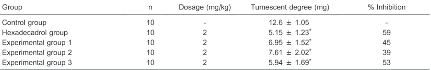
Inhibitory effect on the swelling of mouse auricle that was induced by dimethyl benzene
Kunming mice weighing,18-22 g (female and male) were randomly divided into five experimental groups: group 1 (2 mg/kg scFv, intraperitoneally (ip)), group 2 (2 mg/kg scFv,iv), group 3 (2 mg/kg mAb,iv), hexadeca-drol group (2 mg/kg hexadecahexadeca-drol, ip), and a control group (0.9% saline). Dimethyl benzene was dropped onto the left ear concha in all animals 1 h after injection. After another 2 h, the mice were killed, and both ears were removed with scissors. The ear pieces were obtained with a punch at the same location and weighed. The percentage inhibition of engorgement was calculated as follows: 1006[(average tumescence of control group–– average tumescence of experimental group)/average tumescence of control group].
Results
Expression of scFv
The constructed expression vector was transferred into BL21(DE3) and induced with IPTG at 376C. A 30 kDa protein was strongly expressed after 4 h incubation with 0.8 mM IPTG. It was found that scFv was expressed as insoluble inclusion bodies. The wet weight of the cells was about 5 g in 1 L E. coli flask culture. After large-scale production of anti-ICAM-1 scFv in the fermenter, the overall yield of the harvested cells (wet weight) was about 10 g/L.
Preparation of the samples
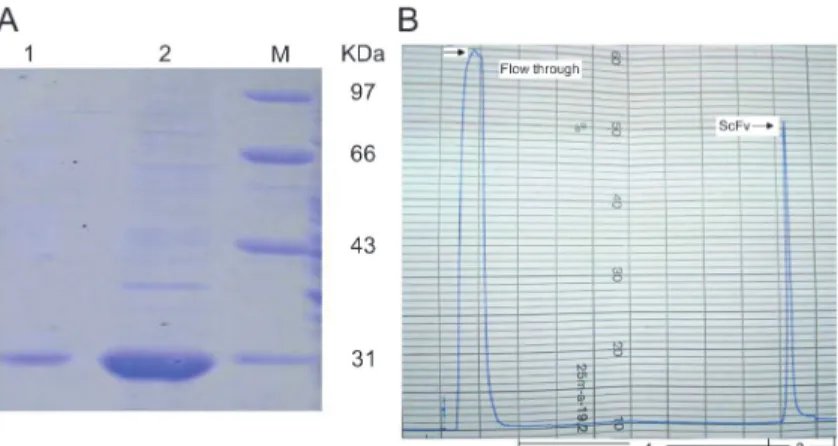
Washing can remove nontarget proteins and may also remove proteases that could degrade the expressed product. In this study, we washed the inclusion bodies three times with buffer containing 0.5% Triton X-100 buffer and 2 M urea. Finally, 346 mg of inclusion body protein per liter of culture were obtained using a fermenter, and the purity of inclusion bodies was up to 70%, as determined by SDS-PAGE (Figure 1A).
Refolding of scFv
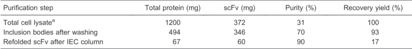
For comparison of the methods, both dilution and dialysis were carried out. During the two processes, a slight protein aggregation was found. Perhaps due to the prolonged experimental time, the activities of scFv proteins refolded by dilution and dialysis were lower than that of proteins refolded by IEC. Comparing the renatured scFv using three refolding methods (Table 1), and considering purity, activity, time required, as well as consumption of reagents, the column chromatography method for refolding was found to be the most suitable for large-scale production. After refolding by IEC, we obtained a single protein peak during the elution procedure, and collected approximately 20 mL of the protein (Figure 1B). The overall purification procedure is summarized in Table 2. The purity of product was about
90%, as shown by 12% SDS-PAGE (Figure 1A).
Effect of pH on refolding by IEC
Protein yield was significantly affected by pH. To study the influence of pH on refolding recovery, denatured scFv was loaded and eluted with buffers having various pH values (7.0, 7.5, 8.0, and 8.5). As shown in (Figure 2), the pH 8.0 elution buffer led to a significantly increased yield of functional anti-ICAM-1 scFv (17%). It is suggested that this condition is suitable for native disulfide bond forma-tion, so pH 8.0 was used throughout the experiment.
Activity of refolded scFv
ELISA results demonstrated that refolded anti-ICAM-1 scFv was able to bind specifically to human ICAM-1-expressing cells in a dose-dependent manner. Specifically, when gradually increasing the concentrations of refolded scFv, the extent of ICAM-1 and scFv binding increased (Figure 3). Meanwhile, indirect ELISA showed the specific antigen-binding activity of refolded scFv to rat ICAM-1.
Cell adhesion assay for peripheral MCs to ECV-304
Statistical analysis was carried out using SPSS 13.0 (USA) and statistical significance was set at P,0.05. It Figure 1. A, Analysis of purified and refolded scFv on SDS-PAGE.Lane 1, scFv refolding by Q Sepharose HP; Lane 2, solubilized inclusion bodies. M: protein molecular weight markers.B, Chromatographic elution profile of refolding scFv by Q Sepharose HP column. Protein peaks were observed at 280 nm. Process 1: refolding proce-dure; Process 2: gradient elution. The arrow indicates flow through protein and interest protein.
Table 1. Comparison of three refolding methods.
Refolding method Dilution Dialysis IEC
Protein yield (%) 22 28 17
Protein concentration (mg/mL) 0.03 0.15 0.4-0.5
Purity (%) .70 .70 .90
Activity (absorbance at 490 nm) 1.86 2.05 2.53
Time required (h) .48 .72 ,10
Reagent consumption (relative amount) 2 100 1
can be seen from the data that the adhesion of MCs to LPS-stimulated ECV-304 monolayers was largely inhib-ited at 30 min in the presence of a neutralizing ICAM-1 mAb and scFv compared with the untreated condition (with PBS). As a result, the ratios for adhesion in the presence of mAb and refolded scFv were 31 and 36% (data not shown), respectively, which was significantly different compared to the control (42%; P,0.05). Although both mAb and scFv could inhibit the adhesion of MCs to ECV-304, scFv was less effective than the mAb (P.0.05).
Analysis of the inflammation depressant effect
Statistical analysis was carried out as described earlier. We found that injection of anti-ICAM-1 scFv or mAb reduced the severity of auricle swelling to different extents, but the depressant effect of scFv and mAb was a little weaker than that of hexadecadrol. As shown in Table 3, there was a statistically significant difference in the percent of inhibition between the experimental groups and the control group.
Discussion
In previous studies, it has been shown that increasing ICAM-1 in vivo contributed to the pathogenesis of inflammation-related diseases. Many reports had sug-gested the protective effects of anti-ICAM-1 mAb, which could block the inflammation responsein vitroorin vivo.
Results in our laboratory also indicated that ICAM-1 and its receptors exhibit a high expression level in highly pathogenic avian influenza (H5N1) and viral pneumonia (HPAIV), and may play an important role in the pathogenesis (23). Moreover, we achieved excellent results in curing mice of avian influenza with the anti-ICAM-1 antibody. We applied for a patent in 2007 in which we named the anti-ICAM-1 antibody as a treatment for avian influenza. However, mAb molecules are large and have greater immunogenicity, so it may be advisable to use anti-ICAM-1 scFv in diagnostic and therapeutic applications. In our present study, active scFv against human ICAM-1, successfully prepared from the inclusion bodies by chromatography renaturation, had a significant effect on aseptic inflammation.
To obtain the active protein, refolding the expressed products from the inclusion bodies is the most important and fundamental procedure. If we can develop a refolding strategy in vitro at lower cost and with higher yield and activity, then less expensive, and easier to achieve, prokaryotic expression systems, namely, bacterial fer-mentation systems, may become feasible for the produc-tion of inclusion body proteins.
In the past, dilution and dialysis were convenient and traditional refolding strategies. At this time, there are many reports about these two methods being used for refolding Table 2. Purification summary of single-chain variable antibody fragment (scFv).
Purification step Total protein (mg) scFv (mg) Purity (%) Recovery yield (%)
Total cell lysatea 1200 372 31 100
Inclusion bodies after washing 494 346 70 93
Refolded scFv after IEC column 67 60 90 17
aApproximately 10 g of wet-weight cells and a total cell lysate containing 1200 mg protein were obtained from 1000 mL cell culture.
IEC: ion exchange chromatography.
Figure 2. Effect of pH on refolding yield. Refolding was performed at different pHs, 7.0, 7.5, 8.0, 8.5, to measure pH dependence of refolding. Data indicated that the optimal pH for refolding of scFv may be 8.0.
many proteins. Most of them were laboratory preparations, not industrial scale production. These refolding techniques have some disadvantages, such as that the dialysis procedure requires large amounts of reagents, long treatment times (24), and can also cause the adhesion of protein to the membranes used (25). Occasionally protein aggregates formed (26). The disadvantages of the dilution method are the large processing volumes involved, higher costs, and the ‘‘step-change’’ in denaturant concentration to native conditions may result in aggregation (27). Moreover, the concentration of denatured protein during the two refolding processes has to be controlled at a low level to prevent aggregate formation, which restricted their application on a large-scale production.
In recent years, chromatographic methods have been developed for refolding of inclusion body proteins fromE. coli. In many cases, these methods appeared to be more effective than traditional refolding strategies (28-30). IEC has the characteristics of simple operation, good biologi-cal compatibility, and high capacity. Furthermore, the medium can be reused, which decreases the cost of materials. In addition, the IEC method provides the concomitant purification of the target protein during the refolding process. Therefore, considered from all per-spectives, the IEC process is the most suitable method for refolding of the engineered proteins.
In the case of proteins containing cysteine, the isolated inclusion bodies usually contain a certain amount of interchain disulfide bonds (31). There are two disulfide bonds in anti-ICAM-1 scFv. For disulfide-containing proteins, the refolding yields are strongly dependent upon the redox environment, which helps to form the proper disulfide bonds and to associate different domains (32). In our study, 1 mM GSH and 0.2 mM GSSG were included in the first gradient buffers during the refolding procedure. In the second gradient, we investigated the two conditions and found that introducing redox conditions to the buffer refolding system did not lead to a significantly increased yield of functional anti-ICAM-1 scFv (data not shown). Therefore, GSH-GSSG was not added in the second gradient. It may be that the first gradient helps to form the disulfide bonds. That stage is a critical refolding period for
native disulfide bond formation, unlike the post-refolding stage.
Because of the lack of an Fc domain in the scFv structure, the refolded scFv did not bind with the HRP-conjugated secondary antibody. In the noncompetitive ELISA experiment, we prepared a secondary antibody (rabbit anti-mouse IgG antibody) that could bind to scFv. In conclusion, a high production level of scFv in theE. coliexpression system was successfully established, and an inexpensive, convenient refolding strategy for scFv recovery was also developed. We could obtain 60 mg of active scFv from 1 liter of cultivatedE. colicells by fermentation. Guo et al. (33) reported that the overall yield of IP10-scFv with bioactivity inE. coliflask culture was more than 40 mg/L. Yuasa et al. (34) reported that 3 liters of culture produced 217 mg of crude protein, yielding 44 mg of purified protein. However, for anti-intimin scFv, the protein yield was 1 mg protein per 100 mL of bacterial culture (33-35).
The established on-column refolding procedure for the efficient recovery of anti-ICAM-1 scFv from inclusion bodies has a practical significance for further research on other scFv or recombinant proteins. The advantages of this method include the biophysical and biochemical characteristics and the fact that purified scFv can markedly suppress the adhesion of MCs to LPS-stimu-lated ECV-304 monolayers. Moreover, in this mouse model of aseptic inflammation, scFv significantly inhibited the inflammatory swelling of auricles induced by dimethyl benzene.
The renaturation process was significant, and it is possible that IEC could be very useful in refolding inclusion body proteins on a large scale. However, refolding efficiency still needs to improve so as to extend its commercial application to other recombinant proteins.
Acknowledgments
We express our appreciation to Cairong Jiang, who revised the manuscript. Research supported by a grant from the Academy of Military Medical Sciences of the People’s Liberation Army (#20060929).
Table 3. Effect of anti-ICAM-1 scFv and mAb on dimethyl benzene-induced swelling of the auricle.
Group n Dosage (mg/kg) Tumescent degree (mg) % Inhibition
Control group 10 - 12.6 ± 1.05
-Hexadecadrol group 10 2 5.15 ± 1.23* 59
Experimental group 1 10 2 6.95 ± 1.52* 45
Experimental group 2 10 2 7.61 ± 2.02* 39
Experimental group 3 10 2 5.94 ± 1.69* 53
References
1. Pipkin ME, Sacks JA, Cruz-Guilloty F, Lichtenheld MG, Bevan MJ, Rao A. Interleukin-2 and inflammation induce distinct transcriptional programs that promote the differen-tiation of effector cytolytic T cells.Immunity2010; 32: 79-90, doi: 10.1016/j.immuni.2009.11.012.
2. Chen CL, Liang CM, Chen YH, Tai MC, Lu DW, Chen JT. Glucosamine modulates TNF-alpha-induced ICAM-1 expression and function through O-linked and N-linked glycosylation in human retinal pigment epithelial cells.Invest Ophthalmol Vis Sci 2012; 53: 2281-2291, doi: 10.1167/ iovs.11-9291.
3. Basit A, Reutershan J, Morris MA, Solga M, Rose CE Jr, Ley K. ICAM-1 and LFA-1 play critical roles in LPS-induced neutrophil recruitment into the alveolar space.Am J Physiol Lung Cell Mol Physiol2006; 291: L200-L207, doi: 10.1152/ ajplung.00346.2005.
4. Kaufmann P, Tilz GP, Smolle KH, Demel U, Krejs GJ. Increased plasma concentrations of circulating intercellular adhesion molecule-1 (cICAM-1) in patients with necrotizing pancreatitis. Immunobiology 1996; 195: 209-219, doi: 10.1016/S0171-2985(96)80040-4.
5. Vainer B, Horn T, Nielsen OH. Colonic epithelial cell expression of ICAM-1 relates to loss of surface continuity: a comparative study of inflammatory bowel disease and colonic neoplasms.Scand J Gastroenterol2006; 41: 318-325, doi: 10.1080/00365520510024241.
6. Domanski L, Gryczman M, Pawlik A, Sulikowski M, Romanowski M, Ostrowski M, et al. Circulating adhesion molecules during kidney allograft reperfusion. Transpl Immunol2006; 16: 172-175, doi: 10.1016/j.trim.2006.08.002. 7. Zhao X, Song GM, Xu JL, Wang XJ, Song HM, Qu X. [Expression of intercellular adhesion molecule-1 and vas-cular cell adhesion molecule in cardiac allografts and significance thereof: experiment with rats]. Zhonghua Yi Xue Za Zhi2007; 87: 2657-2659.
8. Aronni S, Cortes M, Sacchetti M, Lambiase A, Micera A, Sgrulletta R, et al. Upregulation of ICAM-1 expression in the conjunctiva of patients with chronic graft-versus-host disease.Eur J Ophthalmol2006; 16: 17-23.
9. Hajilooi M, Sanati A, Ahmadieh A, Ghofraniha A, Massoud A. Circulating ICAM-1, VCAM-1, E-selectin, P-selectin, and TNFRII in patients with coronary artery disease. Immunol Invest2004; 33: 263-275, doi: 10.1081/IMM-120037275. 10. Bowes MP, Zivin JA, Rothlein R. Monoclonal antibody to the
ICAM-1 adhesion site reduces neurological damage in a rabbit cerebral embolism stroke model. Exp Neurol1993; 119: 215-219, doi: 10.1006/exnr.1993.1023.
11. Souza-Moraes MR, David-Filho R, Baptista-Silva JC, Ullian M, Franco MF, Gabriel A Jr, et al. Effect of antibodies to intercellular adhesion molecule type 1 on the protection of distant organs during reperfusion syndrome in rats.Braz J Med Biol Res2003; 36: 605-612.
12. Alexiou D, Karayiannakis AJ, Syrigos KN, Zbar A, Kremmyda A, Bramis I, et al. Serum levels of E-selectin, ICAM-1 and VCAM-1 in colorectal cancer patients: correla-tions with clinicopathological features, patient survival and tumour surgery.Eur J Cancer 2001; 37: 2392-2397, doi: 10.1016/S0959-8049(01)00318-5.
13. Coskun U, Sancak B, Sen I, Bukan N, Tufan MA, Gulbahar
O, et al. Serum P-selectin, soluble vascular cell adhesion molecule-I (s-VCAM-I) and soluble intercellular adhesion molecule-I (s-ICAM-I) levels in bladder carcinoma patients with different stages.Int Immunopharmacol2006; 6: 672-677, doi: 10.1016/j.intimp.2005.10.009.
14. Thomas GJ, Speight PM. Cell adhesion molecules and oral cancer. Crit Rev Oral Biol Med 2001; 12: 479-498, doi: 10.1177/10454411010120060301.
15. Yokota T, Milenic DE, Whitlow M, Schlom J. Rapid tumor penetration of a single-chain Fv and comparison with other immunoglobulin forms.Cancer Res1992; 52: 3402-3408. 16. Sun H, Wan ZH, Zhang GL, Wu GM, Zhu P, Yue YH.
Preparation of monoclonal antibody against human ICAM-1 and identification of its activity.Chinese J Biol 2008; 21: 308-311.
17. Sun H, Wan ZH, Zhang GL, Wu GM, Zhu P, Yue YH. Construction and expression of anti-human ICAM-1 scFv in E. coli.Chinese J Biol2008; 21: 82-86.
18. Takai T, Takaoka M, Yasueda H, Okumura K, Ogawa H. Dilution method to refold bacterially expressed recombinant Der f 2 and Der p 2 to exhibit the secondary structure and histamine-releasing activity of natural allergens. Int Arch Allergy Immunol2005; 137: 1-8, doi: 10.1159/000084607. 19. Tan PH, Sandmaier BM, Stayton PS. Contributions of a
highly conserved VH/VL hydrogen bonding interaction to scFv folding stability and refolding efficiency.Biophys J1998; 75: 1473-1482, doi: 10.1016/S0006-3495(98)74066-4. 20. Chen LH, Huang Q, Wan L, Zeng LY, Li SF, Li YP, et al.
Expression, purification, and in vitro refolding of a huma-nized single-chain Fv antibody against human CTLA4 (CD152). Protein Expr Purif 2006; 46: 495-502, doi: 10.1016/j.pep.2005.09.002.
21. Beck-Schimmer B, Madjdpour C, Kneller S, Ziegler U, Pasch T, Wuthrich RP, et al. Role of alveolar epithelial ICAM-1 in lipopolysaccharide-induced lung inflammation.Eur Respir J 2002; 19: 1142-1150, doi: 10.1183/09031936.02.00236602. 22. Li H, Nord EP. CD40/CD154 ligation induces mononuclear cell adhesion to human renal proximal tubule cells via increased ICAM-1 expression.Am J Physiol Renal Physiol 2005; 289: F145-F153, doi: 10.1152/ajprenal.00317.2004. 23. Zhao HX, Xia XZ, Lu JR, Yue YH, Yang ST, Zhu P, et al.
Roles of ICAM-1 and it’s receptors in HPAIV pneumonia. Virol Sin2006; 21: 38-42.
24. Yamaguchi H, Miyazaki M, Briones-Nagata MP, Maeda H. Refolding of difficult-to-fold proteins by a gradual decrease of denaturant using microfluidic chips.J Biochem2010; 147: 895-903, doi: 10.1093/jb/mvq024.
25. Feng Y, Liu L, Wang J, Liu J, Hu W, Wang X, et al. Integrated refolding techniques forSchistosoma japonicum MTH1 overexpressed as inclusion bodies in Escherichia coli. Protein Expr Purif 2012; 84: 181-187, doi: 10.1016/ j.pep.2012.05.005.
26. Clark EDB. Refolding of recombinant proteins.Curr Opin Biotechnol1998; 9: 157-163, doi: 10.1016/S0958-1669(98) 80109-2.
protein refolding.J Chromatogr A2004; 1022: 103-113, doi: 10.1016/j.chroma.2003.09.013.
29. Nian R, Tan L, Yoo IK, Choe WS. Chaperone-assisted column refolding of gloshedobin with the use of refolding cocktail.J Chromatogr A2008; 1214: 47-58, doi: 10.1016/ j.chroma.2008.10.076.
30. Freydell EJ, van der Wielen LA, Eppink MH, Ottens M. Size-exclusion chromatographic protein refolding: fundamentals, modeling and operation.J Chromatogr A2010; 1217: 7723-7737, doi: 10.1016/j.chroma.2010.10.038.
31. Schoemaker JM, Brasnett AH, Marston FA. Examination of calf prochymosin accumulation in Escherichia coli: disul-phide linkages are a structural component of prochymosin-containing inclusion bodies.EMBO J1985; 4: 775-780. 32. Huang YJ, Chen IC, Yu CM, Lee YC, Hsu HJ, Ching AT,
et al. Engineering anti-vascular endothelial growth factor single chain disulfide-stabilized antibody variable fragments
(sc-dsFv) with phage-displayed sc-dsFv libraries. J Biol Chem 2010; 285: 7880-7891, doi: 10.1074/jbc.M109.061 457.
33. Guo JQ, Li QM, Zhou JY, Zhang GP, Yang YY, Xing GX, et al. Efficient recovery of the functional IP10-scFv fusion protein from inclusion bodies with an on-column refolding system.Protein Expr Purif2006; 45: 168-174, doi: 10.1016/ j.pep.2005.05.016.
34. Yuasa N, Ogawa H, Koizumi T, Tsukamoto K, Matsumoto-Takasaki A, Asanuma H, et al. Construction and expression of anti-Tn-antigen-specific single-chain antibody genes from hybridoma producing MLS128 monoclonal antibody. J Biochem2012; 151: 371-381, doi: 10.1093/jb/mvs007. 35. Menezes MA, Aires KA, Ozaki CY, Ruiz RM, Pereira MC,