Incidence of Cervical Human Papillomavirus and
Cervical Intraepithelial Neoplasia in Women
with Positive and Negative HIV Status
Incidência de infecção cervical pelo vírus papiloma
humano e neoplasia intraepitelial cervical em mulheres
com sorologia positiva e negativa para o HIV
Benito Pio Vitorio Ceccato Junior
1Mark Drew Crosland Guimarães
2Ana Paula Ceccato Lopes
3Lorena Fiorini Nascimento
4Luísa Magalhães Novaes
3Dora Méndez del Castillo
5Victor Hugo Melo
61Department of Gynecology and Obstetrics, Faculdade de Ciencias Médicas de Minas Gerais - FCCMG, Belo Horizonte, Minas Gerais, Brazil 2Department of Preventive and Social Medicine, Faculdade de
Medicina, UFMG, Belo Horizonte, Minas Gerais, Brazil
3Faculdade de Medicina de Barbacena, Barbacena, Minas Gerais, Brazil 4Faculdade de Ciencias Médicas de Minas Gerais - FCCMG, Belo
Horizonte, Minas Gerais, Brazil
5Center for Action and Research in Support Diagnostics, UFMG, Belo Horizonte, Minas Gerais, Brazil
6Department of Gynecology and Obstetrics, Faculdade de Medicina, Universidade Federal de Minas Gerais - UFMG, Belo Horizonte, Minas Gerais, Brazil
Rev Bras Ginecol Obstet 2016;38:231–238.
Address for correspondence Victor Hugo Melo, DSc, Department of Obstetrics and Gynecology, Faculdade de Medicina, Universidade Federal de Minas Gerais–UFMG, Avenida Alfredo Balena, 190, 30130-100–Belo Horizonte, MG, Brazil (e-mail: [email protected]).
Keywords
►
HIV infections
►
HPV DNA probes
►
papillomavirus
infections
►
cervical
intraepithelial
neoplasia
►
polymerase chain
reaction
Abstract
Objectives
To evaluate the incidence and factors associated with cervical
intra-epithelial neoplasia (CIN) and cervical infection by human papillomavirus (HPV) among
HIV-positive and HIV-negative women.
Methods
A cohort of 103 HIV positive and 113 HIV negative women were monitored
between October 2008 and February 2012, for at least one year. Procedures included
cervical cytology, DNA/HPV detection by polymerase chain reaction, colposcopy with
biopsy if necessary, followed by an interview for exposure characteristics data. CIN was
based on the histopathological results.
Results
The incidence of CIN was of 8.8 and 4.6 cases/100 women-years in
HIV-positive and HIV-negative women, respectively. HIV-HIV-positive women presented a
hazard ratio (HR) of 2.8 for CIN and developed lesions earlier (0.86 year) than
HIV-negative women (2 years) (p
¼
0.01). The risk of developing CIN decreased with age
(HR
¼
0.9) and marital status (HR
¼
0.4). HPV patients presented a higher incidence of
CIN when compared HIV-positive and HIV-negative women (p
¼
0.01). The incidence
of HPV cervical infection was 18.1 and 11.4 cases/100 women-years in HIV-positive and
HIV-negative women, respectively. Those HIV-positive presented earlier HPV infection
(p
¼
0.002). The risk of developing HPV infection decreased with age and was higher
received
November 25, 2015 accepted
February 2, 2016 published online April 28, 2016
DOIhttp://dx.doi.org/ 10.1055/s-0036-1583294. ISSN 0100-7203.
Copyright © 2016 by Thieme Publicações Ltda, Rio de Janeiro, Brazil
Introduction
Cervical cancer is the second most frequent tumor in the Brazilian female population (8.1%) after breast cancer (20.6%), and is the fourth leading cause of cancer death in women in Brazil. This type of cancer is associated with infection by HPV, and some of its subtypes entail a high risk for malignant tumors.1
HPV infection is considered the most prevalent sexually transmitted disease (STD) in the world.2 The relationship between cervical cancer and HPV infection is well established, and high-risk HPV has been detected in almost all cases of cervical cancer.3Neoplastic transformation is mediated by the action of HPV, and is a direct consequence of viral infection.4 The persistent infection by high-risk HPV is a strong predictor of risk for CIN.5Meta-analysis have demonstrated that HPV 16 is the most prevalent type, followed by type 18, and both types are responsible for 65.9% of all cases of cervical cancer.3
HPV infection is an essential factor for the development of cervical cancer but is not sufficient; the risk of acquiring
cervical infection by HPV during one’s lifetime is 84%, but the incidence of cervical cancer is only 0.7%.6Other risk factors, including immune factors, co-infection with HIV and STDs, as well as behavioral factors, such as the early initiation of sexual life, multiple sex partners and smoking, may explain the progress of these infections.6,7
HIV-positive women have a higher risk of acquiring cervical infection by HPV, and those co-infected with HPV have a greater chance of developing CIN, probably because of low immunity. Cohort studies have shown that the incidence of cervical lesions varied between 4.9 and 9% in HIV-negative women, and between 20 and 35% in HIV-positive women.8,9 A meta-analysis of 15 studies has shown that cervical infections by HPV and HIV were factors independently associated with cervical cancer. The interaction between HPV, HIV and the development of cervical cancer has also been demonstrated. Immunosuppression was not associated with cervical disease. The conclusion was that HIV seems to be a risk cofactor in the association between infection by HPV and CIN.10A recent study has demonstrated that the most
among HIV-positive women. HPV 16 was the most common type in HIV-positive
women, and also the type most closely associated with CIN in HIV-negative women.
Conclusions
HIV-positive women had a greater incidence of HPV and CIN, and in a
shorter time interval. More rigorous and timely clinical control is required for this
group.
Resumo
Objetivos
Avaliar a incidência e fatores associados com neoplasia intraepitelial
cervical (NIC) e infecção cervical pelo Papiloma Vírus Humano (HPV) entre mulheres
HIV positivas e negativas.
Métodos
Coorte de 103 mulheres positivas para o HIV e 113 negativas, que foram
acompanhadas entre outubro de 2008 a fevereiro de 2012, com seguimento mínimo
de um ano. Os procedimentos realizados foram coleta de material cervical para
citologia oncótica e detecção do DNA/HPV pela reação em cadeia da polimerase,
colposcopia seguida de biópsia, se necessário, e entrevista para obter dados e
carac-terísticas de exposição. O diagnóstico de NIC foi baseado no resultado histopatológico
das biópsias.
Resultados
A incidência pessoas-tempo de NIC foi de 8,8 e 4,6 casos/100
mulheres-ano para as mulheres HIV-positivas e HIV-negativas, respectivamente. As HIV-positivas
apresentaram uma razão de risco (HR) de 2,8 para NIC e desenvolveram lesões mais
precocemente (0,86 ano) do que as negativas (2 anos) (p
¼
0,01). O risco de
desenvolver NIC diminuiu com a idade (HR
¼
0,9) e o estado civil (HR
¼
0,4). Pacientes
com HPV apresentaram maior incidência de NIC, quando comparadas as mulheres
HIV-positivas e as negativas (47,6
10,5%) (p
¼
0,01). A incidência de infecção cervical
pelo HPV, por pessoa/tempo, foi de 18,1 e 11,4 casos/100 mulheres-ano,
respecti-vamente para mulheres HIV-positivas e negativas. As HIV-positivas apresentaram HPV
mais precocemente (p
¼
0,002). O risco de apresentar HPV diminuiu com a idade e foi
maior entre as positivas. O HPV 16 foi o tipo mais comum entre as mulheres
HIV-positivas.
Conclusões
As mulheres HIV-positivas tiveram maior incidência de HPV e NIC, e um
menor intervalo de tempo. Controle clínico mais rigoroso e oportuno é requerido para
este grupo.
Palavras-chave
►
infecções por HIV
►
sondas de DNA de
HPV
►
infecções por
papillomavirus
►
neoplasia
intraepitelial cervical
►
reação em cadeia da
important factor for contracting an HPV infection was co-infection with HIV.11
The present study aims to evaluate and compare the incidence of CIN and cervical infection by HPV among HIV-positive and HIV-negative women. The HPV types and their correlation with the development of CIN and the factors that influenced these diseases were also evaluated.
Methods
A prospective cohort of HIV-positive or HIV-negative women was monitored in two public medical care referral centers in Belo Horizonte, Brazil, between 2006 and 2011. The study was approved by the Ethical Review Boards of all the in-stitutions involved (Universidade Federal de Minas Gerais–
UFMG, under Opinion No. 289 ETIC/2008 and Faculdade de Ciências Médicas de Minas Gerais–FCMMG, under Research Protocol No. 87/2007).
Population
Two groups of women were monitored according to their HIV status (positive or negative). Non-pregnant women over 18 years old and without CIN at thefirst consultation were included in the two groups. The absence of CIN was defined as uterine cervix without cervical intraepithelial lesion upon smear cytology and with normal colposcopy, or as negative biopsy for CIN, when performed. Hysterectomized women were excluded. All patients signed a free informed consent form.
HIV-negative women were selected HIV-negative women were selected on routine first-come first-served basis at FCMMG Affonso Silvianao Brandão Outpatient Service with enrollment between October 2008 and February 2011. HIV-positive women were selected at the Gynecology Outpatient Service of the Clinical Hospital of UFMG between Octo-ber 2006 and February 2011. All participants had a minimum follow-up of one year, until February 2011 or until the occurrence of any events, and the intervals between con-sultations ranged between six months and one year.
Similarly, HIV positive women were recruited on a routine
first-come first-served basis, regardless of antirretroviral use, clinical stage of infection, or morbidities that might have appeared before or during the study period.
Events and Data Collection
An event was defined as the occurrence of CIN and/or cervical infection by HPV. In all consultations, a gynecological exami-nation was performed, as well as the collection of material for cervical cytology and confirmation of cervical infection by HPV. After the collection, colposcopy was performed. Directed biopsies were performed for all colposcopy changes visualized. Participants were asked to answer a standardized question-naire on demographic and behavioral data in thefirst and subsequent consultations, and to assess their correlation with the occurrence of events. There was no interruption of the follow-up with the occurrence of a single event, and two events did not occur simultaneously during the study period.
The occurrence of CIN was defined according to the result of the histopathological examination of the biopsies of the
colposcopy alterations identified during the gynecological examination and it was based on Richart’s Classification.
The occurrence of cervical infection by HPV was defined by the detection of cervical HPV in women with previous negative diagnosis. All patients underwent the same method for detection and typing of HPV. Initially, HPV was detected via PCR using a nested-PCR strategy. Subsequently, the amplification product of the nested-PCR was sequenced directly. The samples were purified and sequenced using the BigDye Terminator kit (version 3.1; Byosystems, Foster City, USA) and the GP6þprimer set. The
sequences were analyzed in an ABI Prism 3100-Avant genetic analyzer (Byosystems, Foster City, USA). The sequences obtained were edited by selecting 30 nucleotide segments and aligning them with HPV sequences retrieved from the ICTVdB database using the Bioedit software version 7.0, Carlsbad, USA.12In cases in which this technique identified the presence of two or more viral types, PCR was performed for the other 11 frequent types in our region (6, 11, 16, 18, 31, 33, 35, 39, 45, 56 and 58). HPV was detected by PCR with a nested-PCR approach using the MY09/11 and GP5þ/6þ primer sets.12 The combination of the two
methods had a sensitivity of 87% for the identification of type-specific HPV, which was higher than the sensitivity of PCR and sequencing alone (36 and 75% respectively), indicating that the two methods are complementary.12
The presence of cervical HPV was assessed in thefirst and in all subsequent consultations. The occurrence of cervical infection by HPV was defined by the detection of cervical HPV by PCR. Inhibition of the amplification of HPV DNA was assessed by inhibition of the amplification of the human globin gene by viral DNA. In these cases, it was not possible to evaluate the presence of HPV. Based on the available pub-lished data, the following viral types were considered as high-risk HPV: 16, 18, 31, 33, 35, 39, 45, 51, 52, 53, 55, 56, 58, 59, 66, 67, 68, 69, 70, 73, 82 and 83.13,14
HHIV-negative women were assessed for HIV status shift and only those who remained HIV negative throughout the study period were kept in this group until the end of the study or the appearance of the events of interest (such as HPV and CIN). There were no cases of serologic shift in this sample. HIV-positive women had previously confirmatory HIV diagnosis according to the standard Ministry of Health Protocol adopted in Brazil at the time of the study.15
The following sociodemographic and behavioral data possibly involved in the development of CIN and cervical infection by HPV were collected: age; marital status (stable and unstable relationships); age atfirst sexual intercourse; education (number of years of regular schooling); number of pregnancies, deliveries, and abortions; number of sexual partners (stratified into lower or higher than 3); number of sexual partners in the past year (stratified as without a partner or with 1 or more partners); smoking status (yes or no); type of employment (from home or working outside of home); and use of condom as a contraceptive (yes or no).
women would be required in each group, totaling 120 patients.
Statistical Analysis
An initial descriptive analysis was conducted to assess socio-demographic characteristics and data consistency. Statistical significance of categorial variables was assessed by Pearson Chi-square (2) test while for continuous variables Student’s
t-test was used. The cumulative incidence of CIN or cervical infection by HPV was calculated by dividing the number of new cases of CIN or HPV by the total number of women who had not yet develop CIN or cervical infection by HPV. Person-year incidence of CIN or cervical infection by HPV was calculated by dividing the number of new cases by the sum of the periods of risk for each of these two events. Kaplan–Meier analysis was used to estimate the median time of onset of CIN and cervical infection with HPV, and the log-rank test was used to compare the curves for the two groups. Cox proportional hazards model was used in the univariate and multivariate analysis to identify factors associated with the incidence of HPV and CIN. The magnitude of the associ-ations was estimated using the Hazard Ratio (HR) with 95% confidence interval (95% CI). Predictive variables with a
p-value<0.15 in the univariate analysis were selected for
inclusion in the multivariate model. Cox multivariate regres-sion was adjusted using the backward algorithm and only variables withp-value<0.05 remained infinal models. The
software used in the analysis was R version 2.13.0.
Results
The present study is part of a cohort study aimed at deter-mining the incidence of CIN and cervical infection by HPV. Among the HIV-positive women, 63.7% used antiretroviral drugs.
The incidence of CIN was evaluated in 103 HIV-positive women and 113 HIV-negative women, and none of the women evaluated presented CIN in thefirst consultation and were monitored for at least one year. The overall cumulative inci-dence of CIN was 8.0% in HIV-negative women (9 of 113) and 17.4% in HIV-positive women (18 of 103) (p¼0.08). The
incidence of CIN was 4.6 and 8.8 cases/100 person-years for the HIV-positive and HIV-negative women, respectively. Over-all, CIN was detected in 10% of the cases after 0.86 year and 2.03 years of follow-up for, respectively, positive and HIV-negative women (p¼0.01) (►Fig. 1).
The univariate analysis (p<0.15) included potential
pre-dictors. The univariate analysis (p<0.15) included potential
predictors of CIN in the multivariate regression model: infection by HIV (HIV-positive patients presented a higher risk of developing CIN than those HIV-negative); age (the incidence of CIN decreased for every year of life and every year after the initiation of sexual life); marital status (those who were married or in a stable relationship had a lower risk of developing CIN than single, separated and divorced women).
Cox multivariate analysis indicated that the risk of devel-oping CIN was higher among HIV-positive women (HR¼2.9;
95% CI: 1.27–6.55) and in younger women (decreased5.0%
for each year of life; HR¼0.95; 95% CI: 0.91–0.98), and was
lower in women in a stable relationship (HR¼0.4; 95% CI:
0.18–0.91) (►Table 1).
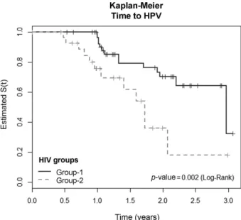
To evaluate the incidence of cervical HPV infection, 70 HIV-positive and 93 HIV-negative women were assessed and monitored for at least one year. Women with cervical infec-tion by HPV in the first consultation and those whose amplification reactions were inconclusive were excluded from this analysis. During the entire study period, 58 incon-clusive reactions were identified (35 in HIV-negative and 23 in HIV-positive women) and were thus excluded. Moreover, one sample was inappropriate and it was therefore dis-carded. The overall cumulative incidence of cervical infection by HPV was 19.4% in HIV-negative women (18 of 93) and 18.5% in HIV-positive women (13 of 70) (p¼0.899). The Fig. 1 Kaplan–Meier model for the time of progression to CIN (group 1, HIV-negative women; group 2, HIV-positive women).
Table 1 Cox multivariate analysis of the time of progression to CIN according to HIV status, sociodemographic factors, sexual behavior, and possible progression factors
Variables P-Value HR 95% CI
HIV-positive 0.01 2.88 1.27 - 6.55
Age 0.01 0.96 0.91 - 0.98
Marital status - stable relationship 0.03 0.41 0.18 - 0.91
incidence of cervical infection by HPV was of 11.4 and 18.1 cases/100 person-years among HIV-positive and HIV-nega-tive women, respecHIV-nega-tively. The median time of onset of cervical infection by HPV was1.65 years for the
seroposi-tive patients and 2.6 years for the seronegaseroposi-tive patients (p¼0.002) (►Fig. 2).
The univariate analysis (p<0.15) included potential
pre-dictors of the presence of HPV in the multivariate regression model: infection by HIV (HIV-positive patients had a higher risk of contracting HPV than HIV-negative patients) and age (the incidence of cervical infection by HPV decreased for every year of life and for every year after the initiation of sexual life).
Cox multivariate analysis indicated that the risk of cervical infection by HPV was higher among HIV-positive women (HR¼2.1; 95% CI: 1.02–4.93), and among younger women
the risk decreased 17.2% for each year after the initiation of sexual life (HR¼0.8; 95% CI: 0.73–0.93) (►Table 2).
A total of 34 different types of HPV were detected in this study in the two study groups, 16 of which were high-risk types. HIV-positive women had a higher incidence of CIN, and those who developed CIN had a higher incidence of cervical infection by HPV; accordingly, the incidence of the latter was 47.8 and 10.5% (p¼0.015) in HIV-positive and
HIV-negative women, respectively. Furthermore, HIV-posi-tive women had a higher incidence of cervical infection by high-risk HPV than HIV-negative women (14.9 and 7.9% respectively), although this difference was not statistically significant (p¼0.127). HPV type 16 was the type with the
highest incidence in both groups: 9.2 and 5.9% of the total cases of cervical infection in HIV-positive and HIV-negative women, respectively.
Discussion
The overall incidence of CIN in this study was high and it was higher among HIV-positive women. These results differ from those in the literature, possibly because of differences in methodology. One study that used a similar methodology, that is, biopsy directed by colposcopy for the diagnosis of CIN, found rates of 5 and 20% for positive and HIV-negative women, respectively,8which were similar to our results of 8.0 and 15.5% respectively. Other studies revealed results of 9 and 35%10and of 2.7 and 4.9%.17However, these studies only used smear cytology in the diagnosis of CIN. Of the two studies that included only HIV-positive women, one of them found a cumulative incidence of 13.5% with a mean follow-up of 40 months,18which is similar to our results, whereas the authors of the other study did notfind any new case of CIN after 12 months of follow-up.19 A more recent cohort study showed that for high-grade lesions (CIN 2 and 3) the incidence was also higher in positive than in HIV-negative women (12 and 4% for CIN 2 and 5 and 2% for CIN 3 respectively).20
The cumulative incidence of CIN in our study was 4.6 and 8.8/ 100 women-years for HIV-positive and HIV-negative women, respectively. Other studies with different methodologies found rates of 2.6 and 11.5 person-years (using only diagnostic cytology)20and 1.5 and 15.1 person-years (using cytology for screening, and colposcopy followed by biopsy of the cytological alterations).21Another study performed in HIV-positive women revealed an incidence of 4.1 person-years.18
In the present study, HIV-positive women developed CIN in a shorter interval than HIV-negative women (0.9 and 2 years respectively), and presented a relative risk (RR) of 2.9, whereas other authors found a relative risk (RR) of 4.5 for the development of CIN among HIV-positive women.10
The data on the cumulative incidence of cervical infection by HPV in HIV-positive and HIV-negative women are diverse: 73.4 and 90.2%,2255.0 and 80%,23and 32 and 54%24for HIV-negative and HIV-positive women respectively, and one
Fig. 2 Kaplan–Meier models for the time of progression to HPV infection (group 1, HIV-negative women; group 2, HIV-positive women).
Table 2 Cox multivariate analysis of the presence of cervical infection by HPV according to HIV status, sociodemographic factors, sexual behavior, and possible progression factors
Variables P-Value HR 95% CI
HIV-positive 0.04 2.12 1.02–4,93
Initiation of sexual life 0.01 0.82 0.73–0.93
study did notfind any new case of cervical infection by HPV in HIV-positive women after 12 months of follow-up.19Our results indicated an incidence of 19.5 and 18.5% for HIV-positive and HIV-negative women, respectively. The differ-ences found in the literature for positive and HIV-negative women may be related to the selection criteria of patients: the HIV-negative women selected in this study had a healthy sexual behavior, whereas other studies recruited HIV-negative women with a risky sexual behavior, who are known to be more prone to infection by HPV and other STDs.8,9,25,26Another factor that may have interfered with the results is the average period of enrollment in the study, because the longer the follow-up period, the higher the exposure and the probability of acquiring an HPV infection. The follow-up period ranged between 1.019and 5.5 years27in previous studies, and it was of 1.6 years in the present study. HIV-positive women presented a higher incidence of cervical infection by low and high-risk HPV,24,28and HPV type 16 had the strongest association with the develop-ment of CIN.5,6,8,23,26,29Cervical infection with HPV and infection persistence were associated with the develop-ment of CIN, and infection by types 16 and 18 was associated with higher risk.8 Other studies also showed that HPV types 16 and 18 were strongly associated with the persistence and progression of cervical lesions com-pared with other high-risk types.5,6In this study, 34 types of HPV were identified, 16 of which presented the highest risk. The incidence of cervical infection by high-risk HPV and by CIN was higher in HIV-positive women; in addi-tion, the incidence of HPV 16 was higher in the two study groups, with a rate of 9.2% and 5.9% in HIV-positive and HIV-negative women respectively.
Genital infections with HPV and CIN are associated with risky sexual behaviors, number of sexual partners and age at
first sexual intercourse,30 and younger age and a higher number of sexual partners were the main risk factors.8,31 Younger women tend to have a higher number of sexual partners.29,32 In the present study, women with positive serology for HIV had RR of 2.9 and 2.2 for cervical infection by HPV and CIN, respectively. Among the demographic and behavioral factors analyzed, those that influenced the devel-opment of CIN were age (younger women had a higher risk, and this risk declined by 4.8% for each year of life) and the marital status of women in stable relationships, such as younger women, and those who were not in stable relation-ships had a higher risk of developing CIN. Age significantly influenced cervical infection by HPV, such that younger women had a higher risk of infection, and this probability decreased18% for every year after the beginning of sexual
life.
The limitations of this study include the fact that the selected women did not represent the general population. HIV-negative women with low socioeconomic status from various regions of Belo Horizonte and satellite cities were recruited on afirst-come,first-served basis at a gynecology outpatient clinic; they all had a low risk for cervical infection by HPV and CIN. The women infected with HIV were part of a cohort of women monitored in a referral medical care
facility. Another limitation was the large number of losses. Several women did not attend the scheduled appointments, despite attempts to contact them. Because no remuneration was provided, many may have missed their appointments due tofinancial hardship or because they could not be absent from work. Strategies such as the donation of transportation coupons and the justification of absences at work could minimize these losses. Although the rate of losses was high (28.7 and 34.6% for HIV-positive and HIV-negative women respectively), they were similar to those reported in the literature (between 22 and 35%).31–33
On the other hand, one of the strengths of this study was the performance of colposcopy in all consultations, with directed biopsies of the colposcopy alterations when neces-sary; this allowed the histopathological study of the uterine cervix, which is the gold standard for the diagnosis of neoplastic cervical changes. Colposcopy had a good agree-ment (92%) with the results of the biopsy when a degree of difference between the histological interpretations of CIN 2 and 3 in situ was allowed.33Another strength of our study is that a large part of the cohort studies published used the smear cytology for diagnosis of squamous intraepithelial lesion (SIL) and/or used colposcopy with directed biopsy only when alterations in smear cytology were observed. Some authors have indicated that screening methods for cervix cancer based on cytology are currently recognized as ineffective because of their low sensitivity, and that other screening techniques are desirable.34A meta-analysis com-pared smear cytology and the DNA test for HPV and found that cytology presented a low sensitivity, with a high inci-dence of cancer in apparently well-screened women.35 In our study, the sensitivity of smear cytology for the diagnosis of CIN was of 46.7%. One should note that because of antiretroviral therapy, data of viral and CD4 cell count in the progression to CIN and cervical infection by HPV were only available among HIV-positive women, and no compar-ison was carried out for these variables.
incidence was higher and closely associated with the devel-opment of CIN in both groups.
Acknowledgments
We are grateful to the Research Support Foundation of Minas Gerais (FAPEMIG) and the National Council for Scientific and Technological Development (CNPq) for thefinancial support.
References
1 Brasil. Ministério da Saúde. Instituto Nacional de Câncer José Alencar Gomes da Silva [Internet]. Estimativa 2012: incidência de câncer no Brasil. Rio de Janeiro: INCA; 2011 [citado 2012 Aug 21]. Disponível em: http://portal.saude.sp.gov.br/resources/ses/per-fil/gestor/homepage/estimativas-de-incidencia-de-cancer-2012/ estimativas_incidencia_cancer_2012.pdf
2 Roccio M, Dal Bello B, Gardella B, et al. HPV infection and intra-epithelial lesions: comparison between HIV positive and negative women. Curr HIV Res 2012;10(7):614–619
3 Tricco AC, Ng CH, Gilca V, Anonychuk A, Pham B, Berliner S. Canadian oncogenic human papillomavirus cervical infection prevalence: systematic review and meta-analysis. BMC Infect Dis 2011;11:235
4 zur Hausen H. Papillomaviruses in the causation of human cancers - a brief historical account. Virology 2009;384(2): 260–265
5 Trottier H, Mahmud SM, Lindsay L, et al; GSK HPV-001 Vaccine Study Group. Persistence of an incident human papillomavirus infection and timing of cervical lesions in previously unexposed young women. Cancer Epidemiol Biomarkers Prev 2009;18(3): 854–862
6 Thomison J III, Thomas LK, Shroyer KR. Human papillomavirus: molecular and cytologic/histologic aspects related to cervical intraepithelial neoplasia and carcinoma. Hum Pathol 2008; 39(2):154–166
7 Kirwan JM, Herrington CS. Human papillomavirus and cervical cancer: where are we now? BJOG 2001;108(12):1204–1213 8 Ellerbrock TV, Chiasson MA, Bush TJ, et al. Incidence of cervical
squamous intraepithelial lesions in HIV-infected women. JAMA 2000;283(8):1031–1037
9 Six C, Heard I, Bergeron C, et al. Comparative prevalence, inci-dence and short-term prognosis of cervical squamous intraepi-thelial lesions amongst HIV-positive and HIV-negative women. AIDS 1998;12(9):1047–1056
10 Mandelblatt JS, Kanetsky P, Eggert L, Gold K. Is HIV infection a cofactor for cervical squamous cell neoplasia? Cancer Epidemiol Biomarkers Prev 1999;8(1):97–106
11 Hessol NA, Holly EA, Efird JT, et al. Concomitant anal and cervical human papillomavirusV infections and intraepithelial neoplasia in HIV-infected and uninfected women. AIDS 2013;27(11): 1743–1751
12 Carvalho NO, del Castillo DM, Perone C, Januário JN, Melo VH, Brasileiro Filho G. Comparison of HPV genotyping by type-specific PCR and sequencing. Mem Inst Oswaldo Cruz 2010; 105(1):73–78
13 Fuessel Haws AL, He Q, Rady PL, et al. Nested PCR with the PGMY09/11 and GP5(þ)/6(þ) primer sets improves detection of HPV DNA in cervical samples. J Virol Methods 2004;122(1): 87–93
14 Weaver B, Shew M, Qadadri B, et al. Natural history of multiple human papillomavirus infections in female adolescents with prolonged follow-up. J Adolesc Health 2011;48(5):473–480
15 Brasil. Ministério da Saúde. Secretaria de Vigilância em Saúde [Internet]. Portaria n. 151, de 14 de outubro de 2009. 2009 [citado 2013 Abr. 10]. Disponível em: http://bvsms.saude.gov.br/bvs/ saudelegis/svs/2009/prt0151_14_10_2009.html
16 Diggle PJ, Heagerty PJ, Liang K, Zeger SL. Analysis of longitudinal data. 2nd ed. Oxford: Oxford University Press; 2002
17 Araújo AC, Carvalho NO, Teixeira NC, et al. Incidence of cervical intraepithelial neoplasia in a cohort of HIV-infected women. Int J Gynaecol Obstet 2012;117(3):211–216
18 Isaakidis P, Pimple S, Varghese B, et al. HPV infection, cervical abnormalities, and cancer in HIV-infected women in Mumbai, India: 12-month follow-up. Int J Womens Health 2013; 5:487–494
19 Schuman P, Ohmit SE, Klein RS, et al; HIV Epidemiology Research Study (HERS) Group. Longitudinal study of cervical squamous intraepithelial lesions in human immunodeficiency virus (HIV)-seropositive and at-risk HIV-seronegative women. J Infect Dis 2003;188(1):128–136
20 Massad LS, Xie X, D’Souza G, et al. Incidence of cervical precancers among HIV-seropositive women. Am J Obstet Gynecol 2015; 212(5):606.e1–606.e8
21 Singh N, Bansal B, Singh U, Qureshi S, Tripathi AK. Comparative study of preinvasive and invasive lesions of the cervix in positive and HIV-negative women. Clin Cancer Invest J. 2015;4(1):39–42
22 Branca M, Rossi E, Alderisio M, et al. Performance of cytology and colposcopy in diagnosis of cervical intraepithelial neoplasia (CIN) in HIV-positive and HIV-negative women. Cytopathology 2001; 12(2):84–93
23 Strickler HD, Burk RD, Fazzari M, et al. Natural history and possible reactivation of human papillomavirus in human immu-nodeficiency virus-positive women. J Natl Cancer Inst 2005; 97(8):577–586
24 Ahdieh L, Klein RS, Burk R, et al. Prevalence, incidence, and type-specific persistence of human papillomavirus in human immu-nodeficiency virus (HIV)-positive and HIV-negative women. J Infect Dis 2001;184(6):682–690
25 Ng’andwe C, Lowe JJ, Richards PJ, Hause L, Wood C, Angeletti PC. The distribution of sexually-transmitted Human Papillomavi-ruses in HIV positive and negative patients in Zambia, Africa. BMC Infect Dis 2007;7:77
26 Almonte M, Silva IdosS, Asare A, et al. Sexual behavior and HPV infection in British women, by postal questionnaires and tele-phone interviews. J Med Virol 2011;83(7):1238–1246
27 Giuliano AR, Harris R, Sedjo RL, et al. Incidence, prevalence, and clearance of type-specific human papillomavirus infec-tions: The Young Women’s Health Study. J Infect Dis 2002; 186(4):462–469
28 Vaccarella S, Franceschi S, Herrero R, et al; IARC HPV Prevalence Surveys Study Group. Sexual behavior, condom use, and human papillomavirus: pooled analysis of the IARC human papillomavi-rus prevalence surveys. Cancer Epidemiol Biomarkers Prev 2006; 15(2):326–333
29 Minkoff H, Feldman J, DeHovitz J, Landesman S, Burk R. A longitudinal study of human papillomavirus carriage in human immunodeficiency virus-infected and human immunodeficiency virus-uninfected women. Am J Obstet Gynecol 1998;178(5): 982–986
30 Nielsen A, Iftner T, Munk C, Kjaer SK. Acquisition of high-risk human papillomavirus infection in a population-based cohort of Danish women. Sex Transm Dis 2009;36(10): 609–615
31 Franco EL, Villa LL, Sobrinho JP, et al. Epidemiology of acquisition and clearance of cervical human papillomavirus infection in women from a high-risk area for cervical cancer. J Infect Dis 1999;180(5):1415–1423
intervals in a high-risk population: experience from the LAMS study. J Med Screen 2008;15(2):97–104
33 Stoler MH, Vichnin MD, Ferenczy A, et al; FUTURE I, II and III Investigators. The accuracy of colposcopic biopsy: analyses from the placebo arm of the Gardasil clinical trials. Int J Cancer 2011; 128(6):1354–1362
34 Castle PE, Stoler MH, Wright TC Jr, Sharma A, Wright TL, Behrens CM. Performance of carcinogenic human
papilloma-virus (HPV) testing and HPV16 or HPV18 genotyping for cervical cancer screening of women aged 25 years and older: a subanalysis of the ATHENA study. Lancet Oncol 2011;12(9): 880–890