Abstract
Submitted: December 12, 2016 Modiication: April 10, 2017 Accepted: May 5, 2017
Evaluation of the sealing ability
of different root canal sealers: a
combined SEM and micro-CT study
Objective: The purpose of this study was to analyze the ability of multiple compounds to seal the dental tubules using scanning electron microscopy (SEM) and micro-computed tomogra-phy (micro-CT). Material and Methods: Twenty-four single-root human mandibular premolars were selected and instrumented with nickel-titanium rotary ile and the inal ile size was # 40/06. They were then randomly allocated into 2 groups, and all samples were illed with single cone gutta-percha (#40/06) and one of the tested sealers (AH Plus and EndoSequence BC sealers). All specimens were scanned using micro-CT and then three from each group were randomly selected for SEM analysis. Results: According to SEM, both root canal sealers showed suficient adaptation to dentin along the whole length of the root canal, though the coronal sections presented superior sealing than the apical sections. Micro porosity analyses revealed that the volume of closed pores and the surface of closed pores had the largest values in the coronal sections, followed by the middle and the apical sections for both sealants (p<0.05). However, no signiicant difference was observed for those two parameters between AH Plus and EndoSequence BC sealers in any of the three sections (p>0.05), whereas they were larger in the apical section when the AH Plus sealer was used. Conclusions: By using the single cone technique, neither EndoSequence or AH Plus pro-vides a porosity-free root canal illing. The EndoSequence BC sealer may have similar sealing abilities regarding the whole root canal as the AH Plus sealer. A better sealing effect could be obtained in the coronal and middle sections of a root canal than the apical part by using the tested sealers.
Keywords: Scanning electron microscopy. Microcomputed tomography.
Root canal. Sealer. Yan HUANG1,2
Kaan ORHAN3 Berkan CELIKTEN4 Ayşe Işıl ORHAN5 Pelin TUFENKCI6 Semra SEVIMAY4
1Sichuan University, West China Hospital of Stomatology, State Key Laboratory of Oral Diseases &
National Clinical Research Center for Oral Diseases, Chengdu, China.
2KU Leuven, University Hospitals Leuven, Faculty of Medicine, Department of Imaging & Pathology,
Leuven, Belgium.
3Ankara University, Faculty of Dentistry, Department of Dentomaxillofacial Radiology Ankara, Turkey. 4Ankara University, Faculty of Dentistry, Department of Endodontics, Ankara, Turkey.
5Ministry of Health, 75th Year Ankara Oral and Dental Health Centre, Division of Pedi-atric Dentistry,
Ankara, Turkey.
6Mustafa Kemal University, Faculty of Dentistry, Department of Endodontics, Hatay, Turkey. Corresponding address:
Introduction
The long-term success of endodontic therapies
relies on complete illing after root canal obturation12. Microleakage is one of the signiicant causes for
endodontic failure, which occurs due to poor contacts
between the gutta-percha and the sealer, the sealer
and the dentin, or through voids within the sealer16,26.
In general, the oral bacteria could contaminate
the entire length of root canal within 30 days of
obturation16, while endotoxins from Actinobacillus
actinomycetemcomitans could be observed in
obturated root canals within 20 days26.
Due to the complexity of root canal systems, pulp
tissue and inorganic debris remain in areas instruments
and irrigation solutions cannot easily access after root
canal treatments. Thus, microorganisms surviving
in the root canal will subsequently grow and spread
to the periradicular areas between the sealer and
dentin27. Permanent coronal restorations also provide
seals equally as important as the apical seal after
the root canals are illed3. When insuficient coronal sealing occurs or the root canal remains open (e.g.,
when sealing is delayed for permanent illings, broken illings, or secondary caries formation; etc), oral
bacteria will access the apical foramen28.
It is not easy to achieve a complete illing with the current root-illing materials used in the clinic, due to the dimensional changes and lack of adhesion
from gutta-percha, which is also the reason to use
endodontic sealers in combination of gutta-percha.
Thus, the adaptability of a sealer to the dentin is the
primary factor inluencing microleakage and reinfection
of the root canal19. Many endodontic sealers are used
in clinical practice, including the recently-introduced
calcium silicate-based sealers. The EndoSequence
BC sealer (Brasseler USA, Savannah, Georgia, USA; also named the iRoot SP; (Innovative. BioCeramix
Inc, Vancover, Brtish Columbia, Canada) has been
introduced as an ideal premixed and injectable
biomaterial in the clinical, exhibiting excellent
radiopaque, zero-shrinkage, insoluble, and hydrophilic
(using moisture from the dentinal tubules to initiate
and complete its setting reaction) characteristics9.
The adaptation of a sealant to the dentin has
generally been evaluated using stereo-microscopy,
confocal laser microscopy, scanning electron
microscopy (SEM), leakage tests, and digital
imaging13,20,25. Compared to other two-dimensional
(2D), time-consuming, and destructive evaluation
ways, micro-computed tomography (micro-CT) is
one kind of advanced imaging modality used to scan
illed roots and reconstruct them three-dimensionally
(3D) for the assessment of the sealant’s adaptation to
the root canal walls13. To the best of our knowledge,
however, no research has been performed on 3D
micro porosity by using micro-CT to assess the sealing
ability of BC sealer in the whole root canal system and
its circumferential dentin area. Moreover, there is no
study yet using SEM and micro-CT for this purpose.
Therefore, the aim of this study was to quantitatively
evaluate and compare the sealing ability of BC sealer
and AH Plus at the apical, middle, and coronal
dental tubules using SEM and micro-CT. The study
was performed under the null hypothesis that no
differences in the ability of sealing dental tubules
would be observed between the two tested root canal
sealants.
Material and methods
In this study, similarly sized, single-rooted, human
mandibular premolars, extracted by orthodontic
reasons, were collected from patients in the clinic,
after their verbal informal consent for the use of these
teeth in the lab with the ethical approval from the
Ethics Committee of Mustafa Kemal University (No.
20012017/4919). All teeth were decontaminated in
5.25% sodium hypochlorite for 2 hours. They were
then stored in distilled water until further testing.
Teeth were examined using an operating microscope
(Carl Zeiss Meditec AG, Oberkochen, Germany) at
20× magniication, and those with immature apices,
caries, restorations, fractures, or cracks were excluded
from the study, and only teeth with oval canals were
included in the standardization procedure for the
experiment. Preoperative radiographs were obtained
in the mesiodistal and buccolingual directions to
conirm the presence of a single unmanipulated root canal without root caries, resorption, or calciication.
In total 24 teeth were included and decoronated at the
cemento-enamel junction, and each root was adjusted
to about 12 mm in length. Subsequently, a #10 K-File
(Dentsply Maillefer, Ballaige, Switzerland) was inserted
into the root canal until the tip was at the apex. The
working length was determined by subtracting 0.5
All samples had a taper of 0.06. Accordingly, all
teeth were instrumented to a size of 40/06 using a
crown-down technique with an EndoSequence 0.06
taper NiTi rotary instrument (Brasseler). Irrigation
was performed with 2 mL 2.5% NaOCl between each
instrument use. Following tooth manipulation, a inal
1 min rinse with 2 mL 2.5% NaOCl, 2 mL 17% EDTA
(Ethylenediaminetetraacetic Acid, Patterson Dental
Supply, Fort Worth, Texas, USA), and 10 mL distilled
water was performed to eliminate the smear layer.
Canals were then dried with paper points (Dentsply
Tulsa Dental, Johnson City, Tennessee, USA), and
those samples were randomly divided into two groups
(n=12 for each group). After the micro-CT tests, three
teeth from each group were randomly selected for the
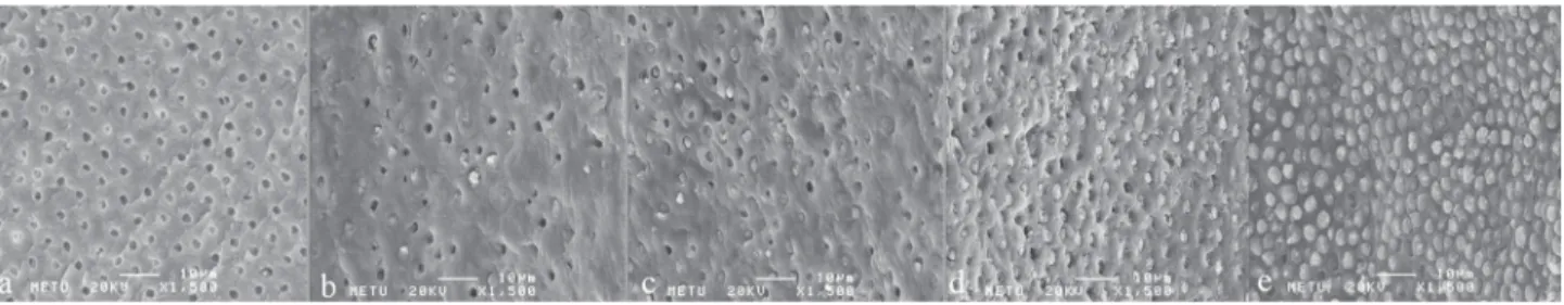
evaluation of the smear layer using SEM (Figure 1).
Root canal sealants were prepared according to the
manufacturers’ instructions and inserted inside the
canal by a size 40 lentulo spiral (Produits Dentaires
SA, Vevey, Switzerland) for evenly distributing
sealers in the whole canal together with the single
cone technique. Group 1: teeth were illed with AH
Plus Root Canal Sealer (Dentsply DeTrey, Konstanz,
Germany) and 40/06 gutta-percha Dentsply Maillefer,
Ballaiges, Switzerland); Group 2: teeth were illed with
EndoSequence BC Sealer (Brasseler USA, Savannah,
Georgia, USA; and 40/06 gutta-percha (Dentsply Maillefer, Ballaiges, Switzerland). After the illing, the
roots were stored at 37°C and 100% humidity for 5
days to allow the sealer to set entirely. The sealer
volume and application followed steps previously
described11.
Micro-CT imaging acquisition
Scanning was performed using high-resolution
micro-CT Skyscan 1172 (Brüker, Kontich, Belgium) at
100 kVp, 100 mA beam current, 0.5 mm Al/Cu ilter,
13.67 µm pixel size, 0.5 step rotation, and 30% beam
hardening. To minimize ring artefacts, air calibration of
the detector was performed prior to each scan. Each
sample was rotated 360° with an integration time of
5 min. Additional settings were also implemented,
including a beam-hardening correction as previously
described, and optimal contrast limits (0–0.06) based
on prior scanning and reconstruction of the teeth. For visualization and quantiication of 1,000 × 1,000 pixel
two-dimensional (2D) axial images, NRecon software
(ver. 1.6.7.2; Brüker, Kontich, Belgium) was used with
an algorithm described by Bouxsein, et al.5 (2010). For the reconstruction, the smoothing was initially set
to zero, followed by a setting of 40% when the ring
artefact correction (lat ield correction) was applied.
The contrast limits were set according to the Skyscan
instructions.
Micro-CT imaging analysis
From the reconstructed micro-CT images, the
roots were divided into apical (0–4 mm), middle (4–8
mm), and coronal (8–12 mm) sections. For micro porosity analyses, a ixed ring area (diameter of 2
mm) was selected as region of interest (ROI) along
the different sections of tooth using CTAn software
(version 1.12.9, Brüker, Kontich, Belgium), which
included the dentin, root canal sealant, and the
gutta-percha. All software parameters and the magniication,
contrast, and imaging enhancement tools were kept
the same to analyze the 3D microarchitectures of each
sample. Once the appropriate ROI (in 3D volume)
was selected, binary images were obtained and an
appropriate greyscale threshold was manually selected
to distinguish the gutta-percha, root canal sealant,
and dentin. Despeckling was performed to remove
white speckles in the 3D images that were fewer than
10 voxels. Following this, 3D imaging analyses were
performed to calculate the porosity of the sealant
within the ROI volume.
Figure 1- Scanning electron microscopy (SEM) images showing (a) open dentin tubules after EDTA treatment; (b) partially obturated
Scanning electron microscopy evaluation
After micro-CT scanning, three teeth from each
group were randomly selected for SEM analysis.
Roots were sectioned longitudinally in the labiolingual
direction and divided into apical (0–5 mm) and
coronal (7–12 mm) sections. Sections were vacuum
dried, coated with gold, and then examined by SEM
(Carl Zeiss NTS GmbH, Oberkochen, Germany). The
penetration of sealants into the dentinal tubules
and adaptation of each sealant to the dentin were
examined from the coronal to apical ends at 1,500×
magniication, and inally, the microphotographs were
taken.
3D micro porosity analysis
In this study, the total ROI volume (mm3), object
volume (dentin volume, mm3), volume of closed pores
(mm3), surface of closed pores (mm2), volume of open
pores (mm3), and open porosity (%) were measured
using CTAn software (version 1.12.9, Brüker, Kontich,
Belgium).
An open pore was defined as the pore that
intersects the boundary of the ROI, which means
that an open pore was connected to the outside in
2D or 3D. Therefore, open pores were a property of
the ROI, where the root canal sealant penetrated into
dentinal tubules and connected from the inside to the
outside of the ROI. In contrast, a closed pore was the
one without connecting to the outside in 2D or 3D.
Closed pores were viewed as black pixels surrounded
by a border of white pixels. Such pores were taken
as dentin tubules already illed or sealed. The open
porosity was calculated as the volume of open pores
(as deined above) as a percentage of the total ROI volume. Following those calculations, CTVol software
(version 2.2.3.0; Brüker, Kontich, Belgium) was used
to generate a 3D model of each sample and assess
the distribution of open and closed pores. From
the 3D models, the dentin volume, the root canal
volume penetrated by the sealant, and the total ROI
volume were calculated. As shown in Figure 2, each
segmentation was assigned different colors using
CTVol software.
Statistical analysis
The differences between the two groups were
assessed using the two independent-sample t-test and
the Mann-Whitney U-test while the normality was not
met. SPSS statistical software (ver. 20.0, Chicago, IL)
was used for all analyses, and p values <0.05 were
considered statistically signiicant.
Results
SEM analyses of root canals obturated with tested
root canal sealers revealed that their adaptation to
dentin was suficient along the length of the root canal.
Compared to the apical section, the coronal sections
showed superior sealing where the texture of the
sealers in the tubules was homogeneous (Figure 1).
Micro-CT testing showed a clear overview of
tooth dentin, sealant materials, and gutta-percha
at assigned grey values. The micro pores at the
interface could be observed between the root canal
dentin and the sealer illing in all tested groups. The
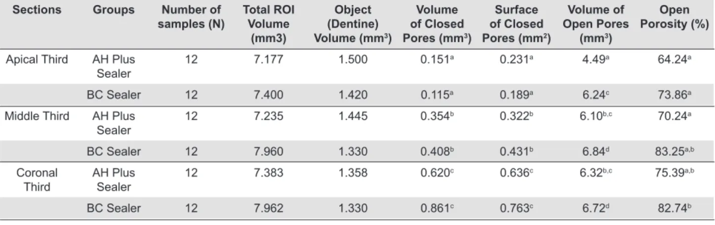
3D structural parameters of micro pores in the whole
root canal system using micro-CT were summarized
in Table 1. The volume of closed pores was larger for
the AH Plus sealant in the apical section compared
Sections Groups Number of
samples (N)
Total ROI Volume
(mm3)
Object (Dentine) Volume (mm3)
Volume of Closed Pores (mm3)
Surface of Closed Pores (mm2)
Volume of Open Pores
(mm3)
Open Porosity (%)
Apical Third AH Plus
Sealer
12 7.177 1.500 0.151a 0.231a 4.49a 64.24a
BC Sealer 12 7.400 1.420 0.115a 0.189a 6.24c 73.86a
Middle Third AH Plus
Sealer
12 7.235 1.445 0.354b 0.322b 6.10b,c 70.24a
BC Sealer 12 7.960 1.330 0.408b 0.431b 6.84d 83.25a,b
Coronal
Third AH Plus Sealer
12 7.383 1.358 0.620c 0.636c 6.32b,c 75.39a,b
BC Sealer 12 7.962 1.330 0.861c 0.763c 6.72d 82.74b
Same superscript letters indicate no statistical difference (p>0.05). Different superscript letters indicate statistical signiicance (p<0.05).
to the EndoSequence BC sealer, though without
statistical signiicance. The volume of closed pores
and the surface of closed pores showed the largest
values in the coronal sections, followed by the middle
and the apical sections for both sealants (p<0.05).
However, no signiicant difference was observed for
the volume of closed pores and the surface of closed
pores between AH Plus and BC sealers in all three
sections (p>0.05), whereas they were larger in the
apical section when the AH Plus sealant was used.
As shown in Figure 2, the BC sealer had a larger
volume of open pores than the AH Plus (p<0.05), no
matter which section of teeth. The volume of open
pores was also larger in the coronal section compared
to the apical section for both sealants (p<0.05).
Similarly, the open porosity was also larger in the
coronal section compared to the apical section for
both sealers, though signiicance only existed for the BC sealer (p<0.05); however, no signiicant difference
was observed between the coronal and middle sections
(p>0.05).
Discussion
Hermetic sealing is the primary factor associated
with the success of root canal treatment, and Ingle,
et al.4 (2008) pointed out that 58% of treatment
failures were due to incomplete obturation. Thus,
decontamination and 3D obturation are essential
following root canal treatments21. Because of this, root canal illing materials are continually improving,
and bioactive materials are becoming increasingly
popular. The EndoSequence BC sealer is one of the
ideal bioactive sealants that contains nanoparticles
(about 2 μm in diameter) facilitating the penetration
into dentinal tubules1. Previous studies suggested that
AH Plus can be considered the gold standard for root
canal sealants; therefore, we compared this sealant
to the bioactive EndoSequence BC sealant3.
Obturation quality may be influenced by the
morphology of the root canal (round or oval)24. For this reason, we only selected the oval canal that
facilitated the operation of the microscope. The inal
irrigation solution of 17% EDTA was applied for 1
min to effectively open the dentin tubules. The low
surface tension of EDTA also facilitates its access into
the dentin tubules to remove the smear layer29, which
improves access and adaptation of the sealants to the
dentin7. However, it has to be noted that because of
applying EDTA, the open porosity in the whole root
canal system - especially in the coronal section - could
be increased by removing smear layers.
Micro porosity analyses revealed that the volume
of closed pores and the surface of closed pores had
the largest values in the coronal sections and the
smallest in the apical sections for both sealants in the
entire levels of root canal. This was also conirmed by
our SEM observation. These results were consistent
to another marginal adaptation study23, which also
showed that the coronal sections had superior
adaptation compared to apical sections. On the other
side, the volume of open pores from BC sealer were
signiicantly larger than the AH Plus sealant, while for Figure 2- Micro-computed tomography (micro-CT) images showing a three-dimensional (3D) representation of AH Plus (a) and the
the open porosity, the differences between two sealers
vanished, suggesting that BC sealer may have more
penetration into the dentin tubules. This may be due
to the small particle size of the EndoSequence BC
sealer (about 2 μm in diameter) or the viscosity of the
calcium phosphate silicate ceramic-based materials
that facilitate the low of the sealant into the dentinal
tubules.
Bioceramic materials contain alumina, zirconia,
bioactive glass, glass ceramics, hydroxyapatite,
and calcium phosphates17. The alkaline nature of
bioceramic by-products has been reported to denature
collagen ibers, which facilitates the penetration of
sealers into the dentin tubules2. However, AH Plus
is naturally acidic, which may limit its bonding to
dentin. Moreover, AH Plus contains a polymer that
contracts upon polymerization, which may result in
sealant cracking and deterioration. Thus, it is likely
for these reasons that the EndoSequence BC sealer
shows superior sealing ability than AH Plus, but has yet
to be conirmed by further in-vivo follow-up studies.
In the current study, a similar volume of closed
pores was observed between the EndoSequence BC
sealer and the AH Plus, which indicated that tested
sealers adapted or penetrated equally to the dentin
in the coronal, middle, and apical sections. Similarly,
using a luid iltration method and SEM, Zhang, et
al.30 (2009) investigated the sealing ability of the
iRoot SP sealer and the AH Plus sealer to the apical
section of teeth roots. It was found that the iRoot SP
using the single-cone technique and the AH Plus using
the continuous wave condensation technique were
equivalent in luid leakage. SEM also revealed that both
sealers provided gap-free and gap-containing regions
within the canals. Consistent with those indings,
the SEM observation in this study showed that the
apical adaption of the EndoSequence BC and AH Plus
sealants were also similar. However, Al-Haddad, et
al.1 (2015) reported that the EndoSequence BC sealer was signiicantly thicker than AH Plus or MTA Fillapex
(mineral trioxide aggregates) by using the lateral
compaction technique, which improved the sealing
ability. The possible reason for this discrepancy could
be derived from the different obturation techniques
and that the results from this study were based on
the single-cone technique, which was proved to be an
effective way obturating well-tapered root canals after
adequate rotary instrumentation18.
The degree of adhesion of the sealer to the dentin
wall depends largely on the intermolecular surface
energy and cleanliness of the dentin, as well as the
surface tension and wetting ability of the sealant.
Dentin at the coronal, middle, and apical sections has
different surface energies and cleanliness. Cleanliness
is an important factor for sealer adaption, which
could be dificult to achieve in the apical region due to dificulties in removing the smear layer. The smear
layer often blocks sealer entry to the dentin tubules,
and it was suggested that differences between the
apical and coronal regions may be due to the lower
density and diameter of dentin tubules in the apical
regions10. This suggestion may also explain the
increased porosity of the coronal and middle regions
- shown as the volume of open pores - compared to
those observed in the apical region in our study.
High-resolution micro-CT is a nondestructive,
highly accurate method of imaging that is becoming
increasingly used for the noninvasive assessment
of 3D microstructures. In addition to measuring
volume, micro-CT facilitates qualitative analyses of
images and differentiates between illing materials,
voids, and tooth structures, based on the imaging
grayscale8,15. Additionally, it is found that the
root-filling materials and the voxel resolution applied
during scan can inluence the presence of artifacts and thus the observed results6. Thus, it is preferable
to apply smaller voxel size and FOV for minimizing the presence of artifacts and improving the diagnostic or
evaluation accuracy in root illed teeth. As pointed by another study22, a resolution of 34 and 68 µm would be suficient for endodontic micro-CT studies with respect of root anatomy. The resolution of micro-CT used in
the current study is as high as 13.67 µm voxel size and
the grayscale-based imaging segmentation, making it
possible to differentiate a clear porosity structure and
further assess their dimensions in the sealer region.
So far yet, none of the previous work on root
sealant has focused on the characterization of either
open or closed porosity. This study shows the ability
of micro-CT to observe open- and closed-pore size,
surface, and location in the interface between dentin
and sealants in 3D. However, while assessing
micro-CT images from endodontic cases, it is necessary
to consider beam-hardening effects. As the result
of the poly-chromaticity of the X-ray source, this
imaging artefact can cause visual distortions of the
reconstructed objects such as edge enhancement
artefacts, a beam-harding correction was performed
during imaging reconstruction, which inevitably
lead to a decrease of the image quality at certain
level. Moreover, one should be aware of the voxel
size limitation of micro-CT because of its inability to
analyze the resulting smear layer and debris retained
in the dentin tubules; SEM can be used to expatiate
the surface morphology of root canals, enabling the
conirmation of illing materials presented on the root
canal wall. Thus, the combination of SEM and micro-CT
analyses can be a powerful approach for the studies
assessing the sealing ability14.
In summary, within the limitation of the study, the
null hypothesis that no differences would be observed
between the abilities of EndoSequence and AH Plus to
seal dentin tubules is accepted while using the single
cone technique, which suggests that EndoSequence
BC sealer has a similar sealing ability in the entire
root canal as the AH Plus sealer does. A better sealing
effect could be obtained in the coronal and middle
sections than the apical part by using any of those
tested sealers.
Acknowledgement
Fellowship support from Research Foundation – Flanders (FWO) from the Belgian government.
Conlict of interest
The authors declare that they have no competing
interests.
References
1- Al-Haddad A, Abu Kasim NH, Che Ab Aziz ZA. Interfacial adaptation
and thickness of bioceramic-based root canal sealers. Dent Mater J. 2015;34(4):516-21.
2- Balguerie E, van der Sluis L, Vallaeys K, Gurgel-Georgelin M, Diemer F. Sealer penetration and adaptation in the dentinal tubules: a scanning electron microscopic study. J Endod. 2011;37(11):1576-9.
3- Barrieshi KM, Walton RE, Johnson WT, Drake DR. Coronal leakage of
mixed anaerobic bacteria after obturation and post space preparation. Oral Surg Oral Med Oral Pathol Oral Radiol Endod. 1997;84(3):310-4. 4- Benenati FW. Obturation of the radicular space. In: Ingle JI, Bakland LK, Baumgartner JC, editors. Ingle’s Endodontics. 6th ed. Hamilton: BC Decker; 2008. p.1053-87.
5- Bouxsein ML, Boyd SK, Christiansen BA, Guldberg RE, Jepsen
KJ, Müller R. Guidelines for assessment of bone microstructure
in rodents using micro-computed tomography. J Bone Miner Res. 2010;25(7):1468-86.
6- Brito-Júnior M, Santos LA, Faria-e-Silva AL, Pereira RD, Sousa-Neto MD. Ex vivo evaluation of artifacts mimicking fracture lines on
cone-beam computed tomography produced by different root canal sealers. Int Endod J. 2014;47(1):26-31.
7- Çalt S, Serper A. Time-dependent effects of EDTA on dentin structures. J Endod. 2002;28(1):17-9.
8- Celikten B, Uzuntas CF, Orhan AI, Tufenkci P, Misirli M, Demiralp KO, et al. Micro-CT assessment of the sealing ability of three root canal illing techniques. J Oral Sci. 2015;57(4):361-6.
9- Ersahan S, Aydin C. Dislocation resistance of iRoot SP, a
calcium silicate-based sealer, from radicular dentine. J Endod. 2010;36(12):2000-2.
10- Ferrari M, Mannocci F, Vichi A, Cagidiaco MC, Mjör IA. Bonding to root canal: structural characteristics of the substrate. Am J Dent. 2000;13(5):255-60.
11- Gandoli M, Parrilli A, Fini M, Prati C, Dummer PM. 3D micro CT analysis of the interface voids associated with Thermail root illings used with AH Plus or a lowable MTA sealer. Int Endod J. 2013;46(3):253-63.
12- Gomes-Filho JE, Moreira JV, Watanabe S, Lodi CS, Cintra LT, Dezan Junior E, et al. Sealability of MTA and calcium hydroxidecontaining sealers. J Appl Oral Sci. 2012;20(3):347-51.
13- Hammad M, Qualtrough A, Silikas N. Evaluation of root
canal obturation: a three-dimensional in vitro study. J Endod. 2009;35(4):541-4.
14- Hülsmann M, Bluhm V. Eficacy, cleaning ability and safety of different rotary NiTi instruments in root canal retreatment. Int Endod J. 2004;37(7):468-76.
15- Jung M, Lommel D, Klimek J. The imaging of root canal obturation using micro-CT. Int Endod J. 2005;38(9):617-26.
16- Khayat A, Lee SJ, Torabinejad M. Human saliva penetration of coronally unsealed obturated root canals. J Endod. 1993;19(9):458-61. 17- Koch K, Brave D. Bioceramics, part I: the clinician’s viewpoint. Dent Today. 2012;31(1):130-5.
18- Krug R, Krastl G, Jahreis M. Technical quality of a matching-taper single-cone illing technique following rotary instrumentation compared with lateral compaction after manual preparation: a retrospective study. Clin Oral Investig. 2017;21(2):643-52.
19- Oliver C, Abbott P. Correlation between clinical success and apical dye penetration. Int Endod J. 2001;34(8):637-44.
20- Patel D, Sherriff M, Ford T, Watson T, Mannocci F. The penetration of RealSeal primer and Tubliseal into root canal dentinal tubules: a confocal microscopic study. Int Endod J. 2007;40(1):67-71. 21- Pawar SS, Pujar MA, Makandar SD. Evaluation of the apical sealing
ability of bioceramic sealer, AH Plus & Epiphany: an in vitro study. J Conserv Dent. 2014;17(6):579-82.
22- Peters OA, Laib A, Rüegsegger P, Barbakow F. Three-dimensional analysis of root canal geometry by high-resolution computed tomography. J Dent Res. 2000;79(6):1405-9.
23- Polineni S, Bolla N, Mandava P, Vemuri S, Mallela M, Gandham
VM. Marginal adaptation of newer root canal sealers to dentin: a SEM study. J Conserv Dent. 2016;19(4):360-3.
24- Robberecht L, Colard T, Claisse-Crinquette A. Qualitative evaluation
of two endodontic obturation techniques: tapered single-cone method
versus warm vertical condensation and injection system an in vitro study. J Oral Sci. 2012;54(1):99-104.
25- Sevimay S, Kalayci A. Evaluation of apical sealing ability and
adaptation to dentine of two resin-based sealers. J Oral Rehabil. 2005;32(2):105-10.
26- Trope M, Chow E, Nissan R. In vitro endotoxin penetration of
coronally unsealed endodontically treated teeth. Endod Dent Traumatol. 1995;11(2):90-4.
27- Tsesis I, Goldberger T, Taschieri S, Seifan M, Tamse A, Rosen E.
The dynamics of periapical lesions in endodontically treated teeth
that are left without intervention: a longitudinal study. J Endod. 2013;39(12):1510-5.
29- Yilmaz Z, Basbag B, Buzoglu HD, Gümüsderelioglu M. Effect of
low-surface-tension EDTA solutions on the wettability of root canal dentin. Oral Surg Oral Med Oral Pathol Oral Radiol Endod. 2011;111(1):109-14. 30- Zhang W, Li Z, Peng B. Assessment of a new root canal sealer’s