Abstract
Submitted: February 21, 2017
Modiication: May 11, 2017 Accepted: May 28, 2017
Effect of erosive challenges on
deciduous teeth undergoing
restorative procedures with different
adhesive protocols – an
in vitro
study
Objective: To evaluate the effect of erosive challenges on the tooth-restoration interface of deciduous teeth treated with different adhesive protocols. Material and Methods: Deciduous molars were cut mesiodistally, then embedded, abraded and polished (n=80). Samples were randomly divided according to the adhesive system used into: G1 (Adper Single
Bond2®, etch-and-rinse), G2 (Universal Single Bond®, self-etching), G3
(OptibondFL®, etch-and-rinse with Fluoride) and G4 (BondForce®, self-etching
with Fluoride). After standardized cavity preparation (2 mm diameter x 2 mm depth), adhesive systems were applied and samples were restored (composite
resin Z350®). Half of the samples were exposed to erosive/abrasive cycles
(n=10, each adhesive group), and the other half (control group; n=10)
remained immersed in artiicial saliva. For microleakage analysis, samples were submersed in methylene blue and analyzed at 40x magniications.
Cross-sectional microhardness (CSMH) was carried out (50 g/5 s) at 25 µm, 50 µm, and 100 µm from the eroded surface and at 25 µm, 75 µm, and
125 µm from the enamel bond interface. Results: Regarding microleakage, 7.5% of the samples showed no dye iniltration, 30% showed dye iniltration only at the enamel interface, and 62.5% showed dye iniltration through the dentin–enamel junction, with no difference between groups (p≥0.05). No signiicant difference was observed in CSMH at different depths (two-way ANOVA, p≥0.05). Conclusions: We did not observe signiicant changes in microleakage or CSMH after erosive/abrasive challenges in deciduous teeth
treated with different adhesive protocols (etch-and-rinse and self-etching
adhesives, with and without luoride).
Keywords: Deciduous tooth. Erosive tooth wear. Adhesive. Tooth erosion. Tooth wear.
Cristiane Meira ASSUNÇÃO1 Marcelo GOULART2 Tattiana Enrich ESSVEIN2 Nicole Marchioro dos SANTOS1 Maria Carolina Guilherme ERHARDT2 Adrian LUSSI3 Jonas de Almeida RODRIGUES1
http://dx.doi.org/10.1590/1678-7757-2017-0053
1Universidade Federal do Rio Grande do Sul, Faculdade de Odontologia, Divisão de Odontopediatria, Porto Alegre, RS, Brasil.
2Universidade Federal do Rio Grande do Sul, Faculdade de Odontologia, Divisão de Dentística, Porto Alegre, RS, Brasil.
3Universität Bern, Zahnmedizinische Kliniken, Klinik für Zahnerhaltung, Präventiv- und Kinderzahnmedizin, Bern, Switzerland.
Introduction
Erosive tooth wear (ETW) is a chemical-mechanical
process that leads to the cumulative loss of hard dental tissue without the involvement of bacteria4. Enamel dissolution occurs both at the enamel/acid interface, as well as within a thin, softened, and partly demineralized layer of enamel, leading to mineral loss, and consequently to tooth substance loss27.
Tooth structure loss can cause tooth sensitivity, esthetics impairment, and loss of occlusal vertical dimension, leading to the indication of restorative treatment29. On the other hand, when exposing teeth with previous restorations to erosive and abrasive challenges, this can interfere in their durability29.
Despite ETW being an emerging theme in recent
studies, there are aspects that still need to be better explored, especially regarding the adhesive systems properties, restorative materials, and their application in deciduous teeth. The effect of erosive and abrasive challenges on enamel–restoration interfaces has not been deeply investigated up until now.
To obtain an adequate margin seal, it is necessary to apply adhesive systems under ideal conditions, thus ensuring the best restoration function
without any breakdown between the tooth and the
restoration6,19,28. Any failure at the bond interface can
lead to microleakage, characterized by the iniltration of bacteria, luids, chemical substances or ions between
the tooth and the restorative material, as well as
margin discoloration and even pulp inlammation19,28.
Erosive tooth wear lesions in restored teeth are known
by margin degradation and restorations rising above the level of the adjacent tooth surface.. This process starts at enamel and can develop until dentin exposure (rounding of cusps and grooves)4.
It is possible to assume the bonding success not only depends on adhesive proprieties, but on a combination of important aspects of the tooth substrate and the adhesive system19,28. Considering the enamel of deciduous teeth strongly reacts to acid etching, self-etching adhesive systems that have a higher pH and are less aggressive to the substrate, can be good for pediatric patients31. Besides these histological aspects, a systematic review, including in
vitro studies that evaluated enamel and dentin bond
strength, suggests that etch-and-rinse adhesives have a better performance in deciduous teeth compared to self-etch systems15.
Fluoride has been added to different dental materials to protect dental tissues. Some studies have investigated the effect of adhesive systems
with luoride on the inhibition of secondary caries,
using pH cycling models to simulate demineralization and remineralization processes13,14,22,23. These studies showed the resistance of the tooth-restoration
interface to acid increased when luoride was present
in the adhesive systems. A similar effect might be observed using erosive/abrasive cycles, but up until now, no study has tested this hypothesis in deciduous teeth.
Considering this knowledge gap, the hypothesis
of this study was that the effect of erosive challenge on the enamel–restoration interface of deciduous teeth would be different from the selected adhesive protocols (etch-and-rinse and self-etching adhesives, with and without fluoride). The purpose was to evaluate the effect of erosive challenge on the enamel– restoration interface of deciduous teeth treated with different adhesive protocols using cross-sectional
microhardness and microleakage.
Material and methods
Experimental design
The sample size measurement was based on Azevedo, et al.3 (2012), whose average difference percentage of CSMH loss was 26.5%. The statistical
power was calculated at 80%, with 95% of conidence
interval, resulting in 10 samples for each group (test and control, and 4 different adhesives). Consequently, 80 enamel samples from deciduous molars were selected for the experimental phase.
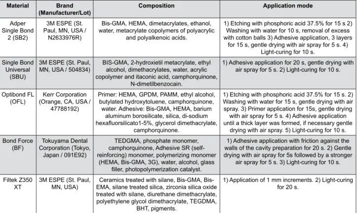
The samples were randomly and equally divided according to the adhesive system used: G1 (etch-and-rinse, Adper Single Bond2®, 3M ESPE; St.
Paul, MN, USA), G2 (self-etching, Universal Single
Bond®, 3M ESPE; St. Paul, MN, USA), G3 (etch-and-rinse with Fluoride, OptibondFL®, Kerr Corporation;
Orange, CA, USA) and G4 (self-etching with luoride,
BondForce®, Tokuyama Dental Corporation; Tokyo, Japan). Standard cavities were prepared, adhesive systems were applied and the samples were restored
with composite resin (Filtek Z350 XT®, 3M ESPE; St.
Paul, MN, USA) (Figure 1). Half of the samples were
n=10) remained immersed in artiicial saliva17 during the experimental phase. In the experimental phase, the samples were stored in relative humidity at 4°C. At the end of the experimental phase, the group samples under test were exposed to 20 erosion cycles and 5 abrasion cycles. The tested variables were mineral loss
(measured using CSMH) and marginal microleakage,
which was measured by dye penetration degree.
Sample preparation
In this study, sound deciduous molars were randomly selected from a group of extracted teeth stored at mineral solution (1.5 mmol/l CaCl2, 1.0 mmol/l KH2PO4, 50 mmol/l NaCl, pH=7.0)34. The children’s parents or legal guardians were informed on the use of the teeth for research purposes and their consent was obtained. The protocol was approved by the Research Ethics Committee of the Federal University of Rio Grande do Sul (Registration number 327.244).
The crowns were separated from the roots and cut mesiodistally, using a diamond disc at an Isomet® Low Speed Saw (Buehler; Düsseldorf, Germany), so both lingual and buccal sides were used. The teeth fragments were embedded in polystyrene resin (Paladur®, Heraeus Kulzer GmbH; Hanau, Germany)
inside PVC cylindrical molds. The samples were
abraded using silicon carbide paper (grits of 1200,
2400, and 4000; Metadi – II®, Buehler Ltda; Lake Bluff, IL, USA) under constant irrigation with distilled water and polished with a diamond abrasive cloth (1¼ µm for 1 minute) under constant cooling (APL4®, Arotec Indústria e Comércio S/A; Cotia, SP, Brazil)12. This procedure removed approximately 200 µm of the enamel surface.
To select samples with the same mineral content,
surface microhardness (KHN, 50 g/5 s, HMV-2T®, Shimadzu; Kyoto, Japan) was performed in the enamel surface, resulting in three indentations at 100 µm from each other8,21. The mean value of initial microhardness
was KHN 332.79 (SD±1.89). Samples with KHN values
different from the mean standard deviation values, scratches, fractures, exposed dentin or any other
visible law, were excluded.
Restorative procedures
Standardized cavity preparation was performed by perpendicularly introducing a cylindrical bur in the active area (diamond bur KG# 3131®, KG Sorensen Ind. e Com. Ltda; Barueri, SP, Brazil); when reaching
enamel and dentin, the cavity depth was checked with
a periodontal probe (2 mm diameter x 2 mm depth). Adhesive systems were applied and the samples were restored with composite resin, using the incremental technique (Figure 1). The light-curing was performed
using an LED device (470 mW/cm2, Ortholux LED
Material Brand
(Manufacturer/Lot)
Composition Application mode
Adper Single Bond
2 (SB2)
3M ESPE (St. Paul, MN, USA /
N2633976R)
Bis-GMA, HEMA, dimetacrylates, ethanol, water, metacrylate copolymers of polyacrylic
and polyalkenoic acids.
1) Etching with phosphoric acid 37.5% for 15 s 2) Washing with water for 10 s, removal of excess with cotton balls 3) Adhesive application, 3 layers
for 15 s, gentle drying with air spray for 5 s. 4) Light-curing for 10 s.
Single Bond Universal
(SBU)
3M ESPE (St. Paul,
MN, USA / 504834) BIS-GMA, 2-hydroxietil metacrylate, ethyl alcohol, dimethacrylates, water, acrylic copolymer and itaconic acid, camphorquinone,
N-dimetilbenzocain.
1) Adhesive application for 20 s, gentle drying with air spray for 5 s. 2) Light-curing for 10 s.
Optibond FL
(OFL) (Orange, CA, USA / Kerr Corporation 47788192)
Primer: HEMA, GPDM, PAMM, ethyl alcohol, butylated hydroxytoluene, camphorquinone, water. Adhesive: Bis-GMA, HEMA, barium
aluminum borosilicate, silica, di-sodium hexaluorsilicato1-5%, glycerol dimethacrylate,
camphorquinone.
1) Etching with phosphoric acid 37.5% for 15 s. 2) Washing with water for 15 s, gentle drying with air spray. 3) Primer application for 15s, gentle drying
with air spray for 5 s. 4) Adhesive application until a thick layer was formed, if necessary gentle
drying with air spray. 5) Light-curing for 10 s. Bond Force
(BF) Corporation (Tokyo, Tokuyama Dental Japan / 091E92)
TEDGMA, phosphate monomer, camphorquinone, Adhesive SR (self-reinforcing) monomer, polymerizing monomer
(HEMA, Bis-GMA, 3G), water, alcohol, glass iller, photopolymerization catalyst.
1) Adhesive application with friction against the walls of the cavity preparation for 20 s. 2) Gentle drying with air spray for 5s followed by a stronger
air spray for 5 s. 3) Light-curing for 10 s.
Filtek Z350
XT 3M ESPE (St. Paul, MN, USA) EMA, silane treated silica, zirconia silica oxide Ceramics treated with silane, GMA, Bis-treated with silane, diurethane dimethacrylate, polyethylene glycol dimethacrylate, TEGDMA,
BHT, pigments.
1) Application of 1 mm increments. 2) Light-curing for 20 s.
Curing Light®, 3M Unitek; Monrovia, CA, USA). Then, the samples were abraded (Sof-Lex, 3M ESPE; St.
Paul, MN, USA) and polished (with felt discs and
polish pastes, DiamondR FGM; Joinville, SC, Brazil). All samples were stored at 4°C under relative humidity until all measurements were performed5.
Erosive and abrasive challenges
In the erosive challenge, the test group samples were immersed in 50 ml of Coca-Cola® (pH 2.6, Coca-Cola Company; Curitiba, PR, Brazil) for 1 minute, at
25°C, under constant shaking, for four times a day, during ive days. Between the cycles, the samples
were washed with deionized water. The control group
samples remained immersed in artiicial saliva at room
temperature (25°C).
All the samples of test groups were brushed using an electric toothbrush after the last cycle of the day (200 g force, for 1 minute), with a paste
with luoridated toothpaste (NaF, 1450 ppm, Colgate
Total 12®, Colgate – Palmolive Comercial Ltda; São
Bernardo do Campo, SP, Brazil) and artiicial saliva
(1:1)7,16,20,32,33.
Microleakage analysis
For microleakage analysis, the area around the
restoration was protected with nail varnish to only
allow dye iniltration through the bonding margin. All
samples were immersed in methylene blue 1% (pH 6.8) for 1 hour. Then, they were washed with deionized water and cross-sectioned (Isomet 1000®, Buehler
Ltda; Lake Bluff, IL, USA). A parallel slice of each sample
was obtained, containing half of the restoration.. Two trained and blinded examiners analyzed the samples
using an optical microscope at 40x magniication. The qualitative microleakage analysis used the following
scores: 0=no dye penetration, 1=dye penetration limited to the enamel, 2=dye penetration through the dentin-enamel junction11.
Cross-sectional microhardness (CSMH)
After the microleakage evaluation, the samples
were evaluated on microhardness. Cross-sectional microhardness (CSMH) was performed with nine
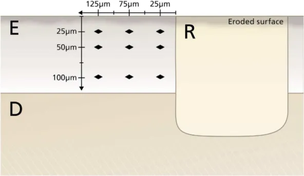
indentations (KHN, 50 g/5 s, HMV-2T®, Shimadzu; Kyoto, Japan) in enamel located at 25 µm, 50 µm and 100 µm from the eroded surface and at 25 µm, 75 µm and 125 µm from the tooth–restoration interface (Figure 2)13.
Statistical analysis
The normality of data distributions were evaluated using the Kolmogorof-Smirnov test. Considering data presented a non-normal distribution, non-parametric
tests were used. Microleakage data were analyzed using the Kruskal-Wallis test and using CSMH between
adhesive systems. The control and test groups were
analyzed using two-way ANOVA non-parametric test.
All the analyses were performed with the software
program SPSS (Statistical Package for the Social
Sciences, version 18).
Figure 2- Schematic drawing of the cross-sectional surface microhardness (CSMH) measurements. E=enamel, D=dentin, R=restoration
Results
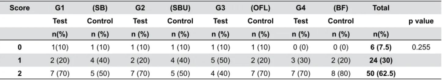
Regarding microleakage, 7.5% of the samples showed no dye iniltration, 30% showed dye iniltration
only at the enamel surface, and 62.5% showed dye
iniltration with amelo–dentin junction. We observed no signiicant difference in microleakage in groups when using the Kruskal-Wallis test (p≥0.05; Table 1).
We observed no signiicant differences in CSMH
between the control and test groups at different depths, neither between different adhesive systems
(two-way ANOVA, p≥0.05; Table 2).
Discussion
Erosive tooth wear (ETW), considered an emerging
problem in oral health, have been increasingly prevalent among adults, adolescents and children29. Despite this fact, few studies have explored this subject in deciduous teeth, especially regarding the properties of restorative materials and their resistance to the erosive challenge. This study showed no
signiicant differences in microleakage or CSMH after
erosive/abrasive challenges in deciduous teeth treated with different adhesives.
Microleakage tests that have used organic and
inorganic dyes to evaluate the tooth–restoration interface have been widely used because they are
easy and quick to perform2. However, the results of
these tests might have been inluenced by variations in
Score G1 (SB) G2 (SBU) G3 (OFL) G4 (BF) Total
Test Control Test Control Test Control Test Control p value
n(%) n (%) n (%) n (%) n (%) n (%) n (%) n (%) n(%)
0 1(10) 1 (10) 1 (10) 1 (10) 1 (10) 1 (10) 0 (0) 0 (0) 6 (7.5) 0.255
1 2 (20) 4 (40) 2 (20) 4 (40) 5 (50) 2 (20) 3 (30) 2 (20) 24 (30)
2 7 (70) 5 (50) 7 (70) 5 (50) 4 (40) 7 (70) 7 (70) 8 (80) 50 (62.5)
Table 1- Distribution frequency of microleakage scores in different adhesives systems, and in control and test groups (n=10;
Kruskal-Wallis)
Distance from bond
margin
G1 (SB2) G2 (SBU) G3 (OFL) G4 (BF)
Depth Test Control Test Control Test Control Test Control * p value †p value
25µm
25 µm 228.58
(60.27) (47.22)237.68 240.29 (76.38) (100.63)230.22 (38.62)262.81 (94.28)152.27 (46.99)263.75 210.27 (40.15) 0.607 0.103 50 µm 249.89
(57.9) (42.24)272.7 247.09 (55.78) 255.89 (97.82) (41.60)278.1 (107.77)159.91 275.49 (56.50) 252.52 (37.56) 0.960 0.634 100 µm 280.26
(44.2) (30.36)275.49 278.24 (38.46) 245.64 (80.59) (32.65)277.58 (120.38)158.35 278.72 (39.31) 280.12 (41.62) 0.419 0.112
75 µm
25 µm 259.7
(71.81) (37.97)253.02 263.54 (38.88) (65.45)247.52 (62.79)253.99 (106.11)155.47 250.06 (48.38) 253.85 (56.79) 0.947 0.797 50 µm 289.94
(52.23) (31.81)258.37 (44.59)238.7 (64.13)268.81 (43.51)286.95 (117.75)157.91 264.38 (65.01) (63.79)268.27 0.533 0.581 100 µm 264.85
(50.15) (42.86)271.92 266.68 (54.80) (72.22)281.34 (43.74)277.46 (116.16)162.60 280.47 (38.19) (46.20)271.93 0.852 0.613
125 µm
25 µm 259.75
(72.44) (39.10)263.25 271.69 (56.59) (53.73)230.55 (44.80)265.3 (108.79)155.72 240.21 (47.87) (36.56)230.06 0.321 0.166 50 µm 279.75
(64.04) 289.99 (59.96) 261.68 (44.52) (60.07)266.77 (27.01)278.75 (118.88)163.25 269.78 (74.20) (53.81)251.69 0.735 0.478 100 µm 243.22
(55.48) 268.16 (26.1) 243.21 (36.50) (61.96)269.15 (53.81)279.63 (113.65)153.77 270.27 (48.47) (44.66)263.36 0.387 0.574
Mean (DP)
* p value Comparsions among groups of adhesive protocols. †p value Comparsions between test and control groups.
Table 2- Mean values of cross-sectional surface microhardness (CSMH) measurements in different adhesive systems and control and
methodology (dye type and concentration, immersion duration, method of analysis, cavity preparation
dimension). Therefore, these variables could make result comparison more dificult, which might lead to
uncertain and incorrect conclusions2,10,25,31. Despite
the existence of such variations, microleakage tests
using dyes seem to evaluate the differences between materials in laboratory studies adequately, thus providing an improved basis for clinical trials. Choosing methylene blue 1% (pH 6.8) ensured that no other acidic exposure would interfere in the outcome of this study10.
In our study, signiicant dye penetration was found
in all groups. Some studies have tested the same adhesive systems and have also observed a high degree of dye penetration2,30. Other authors have not observed
statistically signiicant differences in microleakage
when comparing different adhesives1,2,24,30.
Some experimental models with longer immersion in dyes or a long-term evaluation of these restorations
could better compare the microleakage of the tooth–
restoration interface in different adhesive systems. In our study, we exposed the samples to 20 erosion cycles and 5 abrasion cycles, leading to initial erosive
tooth wear, which was not signiicantly different in the
tested adhesive systems.
Considering the different adhesives protocols, a recent systematic review of in vitro studies evaluated bond strength in deciduous teeth. The statistical analysis of the grouped immediate bond strength data showed that etch-and-rinse adhesives bonded better to sound enamel and dentin substrates than self-etch systems15. It described a wide range of sample sizes and adhesive protocols, so studies with less bias should be considered by professionals when deciding for one
of the many adhesives options. The luoride addition
in adhesive systems showed protective effects on the enamel-restoration interface, considering pH-cycling models13,14,22,23. Guedes, et al.9(2016) evaluated the effect of erosive pH cycling with solutions that simulate dental erosion on Martens hardness of bovine dentin
restored with luoride-releasing adhesive systems. This study concluded that luoride from self-etching
adhesive systems One Up Bond F® (Tokuyama Dental
Corporation; Tokyo, Japan) and Clearil SE Protect®
(Kuraray America, Inc.; New York, NY, USA) could
have some positive effect on erosive lesions early-stages9. Sato, et al.26(2016) evaluated the acid-base resistant zone at the adhesive/enamel interface of
self-etching adhesives with or without prior phosphoric acid etching26. They restored samples of third molars and pre molars by carrying out different self-etching adhesives protocols and pH cycling. The authors concluded that enamel beneath the bonding interface was more susceptible to acid dissolution in Scotchbond Universal® adhesive (3M ESPE; St. Paul, MN, USA)
and Clearil BOND SE ONE® (Kuraray America, Inc.;
New York, NY, USA). In the case of the self-etching
adhesives and universal adhesives, enamel etching is useful to improve the interfacial quality26. This study evaluated deciduous enamel bonding margin
after erosive/abrasive challenges, and no signiicant
difference was demonstrated in etch-and-rinse or
self-etching adhesives, with or without luoride on
composition.
With the erosive/abrasive challenge used in this study, we observed no statistically signiicant differences regarding microleakage or CSMH in
the different adhesive systems used (with and
without luoride, etch-and-rinse, and self-etching).
Microhardness evaluations, either superficial or cross-sectional, imply quantitative measures that can evaluate minimum changes on mineral content; it is a widely used method to compare different treatments in erosive/abrasive protocols. By using Coca-Cola® (pH 2.6, Coca-Cola Company; Curitiba, PR, Brazil) and following the previously described protocol (1
minute at 25°C under constant shaking), we aimed
at getting closer to in vivo conditions, simulating
the children’s acid beverage intake. A study with
bovine teeth using an erosion model showed some significant differences in microhardness values,
especially in sample restoration with luoride releasing
material, such as glass ionomer35. A pH cycling study
that simulated caries found signiicant differences between adhesive systems with and without luoride.
The microhardness values of dentin at 50 µm were similar between one self-etching adhesive system with fluoride and a conventional glass ionomer cement14. On the other hand, the same authors investigated different restorative techniques exposed to a cariogenic challenge in an in situ study, and have not found differences between adhesive systems
with or without luoride, and the group restored with
conventional glass ionomer cement showed higher CSMH values13.
Several studies comparing toothpastes with and
that luoride formulations applied on enamel had a
protective effect on teeth. It was possible to observe
lower surface loss on samples brushed with luoride toothpaste compared to samples with no luoride
toothpaste17,18. The main effect of luoride on erosion/ abrasion cycles is the increase in enamel resistance to future acid exposure, as there is no remineralization of
the softened layer. The luoride’s protective effect was
present both in test and control groups of this study
by applying a paste containing NaF luoride toothpaste
during abrasions cycles.
The fact that we have not observed signiicant
difference in CSMH is due to the removal of the
softened layer by the ive abrasion cycles and the short-term evaluation after 20 erosion cycles. We could
consider such characteristic as one of the limitations
of this study. The amount of luoride released from adhesives with luoride is not usually known and may
not be high enough to reduce demineralization in
erosive challenges. In this study, the luoride content
of the adhesive systems was not enough to have a protective effect on the enamel–restoration interface. The short term evaluation could be another limitation of the study. It could be expected that, after a long-term evaluation with more erosion/abrasion cycles and measurements of nanohardness closer than 25 µm from the enamel bond margin, some differences could be observed among the adhesive systems tested in this
study. The evaluation of surface loss with proilometry
analysis could provide additional information on the effect of erosive tooth wear on deciduous teeth restored with different adhesive systems.
The authors state that erosive tooth wear
(ETW) is a condition of growing importance even in
primary dentition, requiring preventive to restorative interventions. The selection of the most adequate adhesive system to restore deciduous teeth exposed
to ETW is an important step in ensuring the success
of restorative treatments.
Conclusion
Therefore, based on the results of this in vitro study,
the addition of luoride to adhesive systems did not interfere in the investigated outcomes (microleakage
and CSMH). The different adhesives protocols (etch-and-rinse or self-etching) did not show any difference on enamel bonding interface evaluation after erosive/
abrasive challenges.
Acknowledgments
The authors appreciate the support of FAPERGS and CAPES - Coordination of Higher Education and Graduate Training (Brazilian Ministry of Education)
for the PhD student scholarship. We would like to thank the Postgraduate Program in Dentistry of
the Federal University of Paraná for allowing us to use their laboratory to carry out the cross-sectional
microhardness analysis. We also thank Gabriel
Fischer and the Prof. J. Hüsler from the Institute of Mathematical Statistics and Actuarial Science, University of Bern, for all statistical analyses. The
authors declare no conlict of interest.
References
1- Amaral CM, Hara AT, Pimenta LA, Rodrigues AL Jr. Microleakage of hydrophilic adhesive systems in class V composite restorations. Am J Dent. 2011;14(1):31-3.
2- Amarante de Camargo DA, Sinhoreti MA, Correr-Sobrinho L, Sousa Neto MD, Consani S. Inluence of the methodology and evaluation criteria on determining microleakage in dentin-restorative interfaces. Clin Oral Investig. 2006;10(4):317-23.
3- Azevedo DT, Faraoni-Romano JJ, Derceli JR, Palma-Dibb RG. Effect of Nd:YAG laser combined with luoride on the prevention of primary tooth enamel demineralization. Braz Dent J. 2012;23(2):104-9.
4- Carvalho TS, Colon P, Ganss C, Huysmans MC, Lussi A, Schlueter N, et al. Consensus report of the European Federation of Conservative Dentistry: erosive tooth wear – diagnosis and management. Clin Oral Investig. 2015;19(7):1557-61.
5- Cheaib Z, Lussi, A. Impact of acquired enamel pellicle modiication on initial dental erosion. Caries Res. 2011;45(2):107-12.
6- Correr GM, Puppin-Rontani RM, Correr-Sobrinho L, Sinhoret MA, Consani S. Effect of sodium hypochlorite on dentin bonding in primary
teeth. J Adhes Dent. 2004;6(4):307-12.
7- Cruz JB, Lenzi TL, Tedesco TK, Guglielmi CA, Raggio DP. Eroded
dentin does not jeopardize the bond strength of adhesive restorative materials. Braz Oral Res. 2012;26(4):306-12.
8- Cury JA, Rebelo MA, Del Bel Cury AA, Derbyshire MT, Tabchoury CP. Biochemical composition and cariogenicity of dental plaque formed in the presence of sucrose or glucose and fructose. Caries Res.
2000;34(6):491-7.
9- Guedes AP, Moda MD, Suzuki TY, Godas AG, Sundfeld RH, Briso AL, et al. Effect of luoride-releasing adhesive systems on the mechanical properties of eroded dentin. Braz Dent J. 2016;27(2):153-9.
10- Heintze SD. Clinical relevance of tests on bond strength, microleakage and marginal adaptation. Dent Mater. 2013;29(1):59-84. 11- International Standardization Organization. ISO/TR 11405:1994: Dental materials - guidance on testing of adhesion to tooth structure.
Geneva: ISO; 1994.
12- Johansson AK, Sorvari R, Birkhed D, Meurman JH. Dental erosion in deciduous teeth - an in vivo and in vitro study. J Dent. 2001;29(5):333-40.
13- Kirsten GA, Rached RN, Mazur RF, Vieira S, Souza EM. Effect of open-sandwich vs. adhesive restorative techniques on enamel and
14- Kirsten GA, Takahashi MK, Rached RN, Giannini M, Souza EM. Microhardness of dentin underneath fluoride-releasing adhesive systems subjected to cariogenic challenge and luoride therapy. J Dent. 2010;38(6):460-8.
15- Lenzi TL, Gimenez T, Tedesco TK, Mendes FM, Rocha RO, Raggio
DP. Adhesive systems for restoring primary teeth: a systematic review and meta-analysis of in vitro studies. Int J Paediatr Dent.
2016;26(5):364-75.
16- Levy FM, Magalhães AC, Gomes MF, Comar LP, Rios D, Buzalaf MA. The erosion and abrasion inhibiting effect of TiF(4) and NaF varnishes and solutions on enamel in vitro. Int J Paediatr Dent. 2012;22(1):11-6. 17- Lussi A. Dental erosion - novel remineralizing agents in prevention
or repair. Adv Dent Res. 2009;21(1):13-6.
18- Magalhães AC, Rios D, Delbem AC, Buzalaf MA, Machado MA. Inluence of luoride dentifrice on brushing abrasion of eroded human enamel: an in situ/ex vivo study. Caries Res. 2007;41(1):77-9. 19- McDonough WG, Antonucci JM, He J, Shimada Y, Chiang MY, Schumacher GE, et al. A microshear test to measure bond strengths
of dentin-polymer interfaces. Biomaterials. 2002;23(17):3603-8. 20- Moretto MJ, Magalhães AC, Sassaki KT, Delbem AC, Martinhon CC. Effect of different luoride concentrations of experimental dentifrices on enamel erosion and abrasion. Caries Res. 2010;44(2):135-40. 21- Paes Leme AF, Tabchoury CP, Zero DT, Cury JA. Effect of luoridated dentifrice and acidulated phosphate luoride application on early artiicial carious lesions. Am J Dent. 2003;16(2):91-5.
22- Pedrosa VO, Flório FM, Turssi CP, Amaral FL, Basting RT, França FM. Inluence of pH cycling on the microtensile bond strength of self-etching adhesives containing MDPB and luoride to dentin and microhardness of enamel and dentin adjacent to restorations. J Adhes Dent. 2012;14(6):525-34.
23- Peris AR, Mitsui FH, Lobo MM, Bedran-Russo AK, Marchi GM. Adhesive systems and secondary caries formation: assessment of dentin bond strength, caries lesions depth and luoride release. Dent Mater. 2007;23(3):308-16.
24- Pilo R, Ben-Amar A. Comparasion of microleakage for three one-bottle and three multiple-step dentin bonding agents. J Prosthet Dent.
1999;82(2):209-13.
25- Raskin A, D'Hoore W, Gonthier S, Degrange M, Déjou J. Reliability of in vitro microleakage tests: a literature review. J Adhes Dent.
2001;3(4):295-308.
26- Sato T, Takagaki T, Matsui N, Hamba H, Sadr A, Nikaido T, et al. Morphological evaluation of the adhesive/enamel interfaces of two-step self-etching adhesives and multimode one-bottle self-etching
adhesives. J Adhes Dent. 2016;18(3):223-9.
27- Shellis RP, Barbour ME, Jesani A, Lussi A. Effects of buffering
properties and undissociated acid concentration on dissolution of dental enamel in relation to pH and acid type. Caries Res. 2013;47(6):601-11. 28- Silva Telles PD, Aparecida M, Machado M, Nör JE. SEM study of a self-etching primer adhesive system used for dentin bonding in primary
and permanent teeth. Pediatr Dent. 2001;23(4):315-20.
29- Taji S, Seow WK. A literature review of dental erosion in children. Aust Dent J. 2010;55(4):358-67.
30- Toledano M, Cabello I, Yamauti M, Giannini M, Aguilera FS, Osorio E, et al. Resistance to degradation of resin-dentin bonds produced by one-step self-etch adhesives. Microsc Microanal. 2012;18(6):1480-93. 31- Van Meerbeek B, Peumans M, Poitevin A, Mine A, Van Ende A, Neves A, et al. Relationship between bond-strength tests and clinical outcomes. Dental Materials. 2010;26(2):100-21.
32- Voronets J, Lussi A. Thickness of softened human enamel removed by toothbrush abrasion: an in vitro study. Clin Oral Investig. 2010;14(3):251-6.
33- Wang L, Casas-Apayco LC, Hipólito AC, Dreibi VM, Giacomini MC, Bim Júnior O. Effect of simulated intraoral erosion and/or
abrasion effects on etch-and-rinse bonding to enamel. Am J Dent. 2014;27(1):29-34.
34- Zero DT, Rahbek I, Fu J, Proskin HM, Featherstone JD. Comparison of the iodide permeability test, the surface microhardness test, and
mineral dissolution of bovine enamel following acid challenge. Caries Res. 1990;24(3):181-8.
35- Zhou SL, Zhou J, Watanabe S, Watanabe K, Wen LY, Xuan K. In vitro study of the effects of luoride-releasing dental materials on