ORIGIN
AL RESEAR
CH
Respiratory muscle strength, pulmonary function and
thoracoabdominal expansion in older adults and its
relation with nutritional status
Força muscular respiratória, função pulmonar e expansibilidade toracoabdominal em idosos e
sua relação com o estado nutricional
La fuerza muscular respiratoria, la función pulmonar y la expansibilidad toracoabdominal
en adultos mayores y la relación con su estado nutricional
Fernanda dos Santos Pascotini1, Elenir Fedosse2, Mônica de Castro Ramos3, Vanessa Veis Ribeiro4,
Maria Elaine Trevisan5
Mailing address: Fernanda dos Santos Pascotini – Rua Nabuco de Araújo, nº 41, ap. 401 – Santa Maria (RS), Brazil – CEP: 97045-340 – Phone: +55 30255517
E-mail: [email protected] – Financing source: Nothing to declare – Conlict of interest: Nothing to declare – Presentation: June 2016 – Accepted for publication: Sept. 2016 – Approved by Research Ethics Committee of Universidade Federal de Santa Maria: 0356.0.243.000-11.
ABSTRACT | This study aimed at describing Respiratory Muscle Strength, Pulmonary Function and Thoracoabdominal Expansion and associating its relation with nutritional status. It is an observational, cross-sectional study with 50 older adults of both sexes, aged between 60 and 84 years, with no previous diagnosis of respiratory pathologies, invited through local media. It have been evaluated maximal inspiratory (MIP) and expiratory (MEP) pressures, forced vital capacity (FVC), forced expiratory volume in one second (FEV1), thoracoabdominal cirtometry and anthropometric measurements (weight, height, body mass index – BMI –, and nutritional status). Values obtained from MIP, MEP, FVC, and FEV1 were lower than the values intended for this population (p<0.05) as well as measurements for the thoracoabdominal expansion. Regarding nutritional status, 10 older adults were classiied as malnourished, 24 as eutrophic, and 16 as obese. Values of respiratory parameters showed no association with nutritional status (p> 0.05). It was concluded that aging inluenced the respiratory parameters evaluated in this study group. Nutritional status, on the other hand, did not inluence measures of respiratory muscle strength, pulmonary function, thoracoabdominal expansion.
Keywords | Elderly; Muscle Strenght; Respiration.
416
RESUMO | Objetivou-se descrever a força muscular respiratória (FMR), função pulmonar (FP) e expansibilidade toracoabdominal (ET) e associá-las com seu estado nutricional. Trata-se de um estudo observacional transversal com 50 idosos de ambos os sexos, com idade entre 60 e 84 anos, sem diagnóstico prévio de patologias respiratórias, convidados através de mídia local. Foram avaliadas as pressões inspiratória (PImáx) e expiratória (PEmáx) máximas, capacidade vital forçada (CVF), volume expiratório forçado no primeiro segundo (VEF1), cirtometria toracoabdominal e medidas antropométricas (peso, altura, índice de massa corporal – IMC – e estado nutricional). Os valores obtidos de PImáx, PEmáx, CVF e VFE1 apresentaram-se inferiores aos valores previstos para tal população (p<0,05), bem como as medidas de expansibilidade tóraco-abdominal. Com relação ao estado nutricional, 10 idosos foram classiicados como desnutridos, 24 eutróicos e 16 obesos. Os valores dos parâmetros respiratórios não mostraram associação com o estado nutricional (p>0,05). Concluiu-se que o envelhecimento inluenciou os parâmetros respiratórios avaliados nesse grupo de estudo. O estado nutricional, por sua vez, não inluenciou as medidas de FMR, FP e ET.
Descritores | Idosos; Força Muscular; Respiração.
1PhD candidate in Human Communication Disorders, Universidade Federal de Santa Maria (UFSM) – Santa Maria (RS), Brazil. 2Professor in the Department of Speech Therapy, Universidade Federal de Santa Maria (UFSM) – Santa Maria (RS), Brazil. 3Physical therapist, Graduated from Universidade Luterana do Brasil (Ulbra) – Santa Maria (RS), Brazil.
4 PhD candidate of PPG in Speech Therapy from Faculdade de Odontologia de Bauru (FOB), Universidade de São Paulo (USP) – São
Paulo (SP), Brazil.
5 Professor in the Department of Physical Therapy and Rehabilitation, Universidade Federal de Santa Maria (UFSM) – Santa Maria (RS),
RESUMEN | El propósito de este estudio fue describir la fuerza muscular respiratoria (FMR), la función pulmonar (FP) y la expansibilidad toracoabdominal (ET) y asociarlas con el estado nutricional de adultos mayores. Se trata de un estudio observacional transversal, del cual participaron 50 adultos mayores de ambos sexos, entre 60 y 84 años de edad, sin diagnostico anterior de patologías respiratorias, y que fueron invitados por los medios locales. Se les evaluaron la presión inspiratoria (PImax) y la presión espiratoria (PEmax) máximas, la capacidad vital forzada (CVF), el volumen espiratorio forzado en el primer segundo (VEF1), la cirtometría toracoadominal y las medidas antropométricas (peso,
altura, índice de masa corporal y estado nutricional). Los valores de la PImax, la PEmax, la CVF y el VFE1 fueron inferiores a los valores previstos para los participantes (p<0,05), así como las medidas de expansibilidad toracoabdominal. Referente al estado nutricional, se clasiicaron 10 adultos mayores en desnutridos, 24 en eutróicos y 16 en obesos. Los valores de los parámetros respiratorios no se asociaron con el estado nutricional (p>0,05). Se concluye que el envejecimiento inluyó en los parámetros respiratorios evaluados en el grupo. El estado nutricional, a su vez, no inluyó en las medidas de la FMR, FP y ET.
Palabras clave | Adulto Mayor; Fuerza Muscular; Respiración.
INTRODUCTION
According to the Brazilian Institute of Geography
and Statistics (IBGE)1, the ageing tendency of the
Brazilian population remained in the latest survey and the number of older adults (individuals with more than 60 years) will quadruplicate by the year 2060, representing nearly 27% of the whole Brazilian population.
he aging process brings a range of physiological changes that result in the reduction of muscle mass, strength, and function in many systems. In the respiratory system, changes are observed in the connective tissue, causing increased stifness of the thoracic cavity and reducing the elastic component of the lungs, directly
inluencing on respiratory mechanics2, leading to the
reduction in the mobility of the thoracic cavity, lung
elasticity, and forced vital capacity (FVC)3 as well as
of the forced expiratory volume in one second (FEV1),
thoracic complacency, and increased lung complacency,
among other changes4.
Other aging-related phenomenon is the reduction of respiratory muscle strength. herefore, older adults present decreased maximal inspiratory pressure (MIP) as a result of the weakness of the inspiratory muscles and decreased maximum expiratory pressure (MEP) due to reduction in the strength of the abdominal muscles and
intercostal5,6.
Other aspects that noticeably change during aging are associated with the body composition of older adults, who tend to gain weight steadily until around 70 years, then
losing it from this age7 and to present a redistribution of
body fat from the extremities to the visceral area8, which
may interfere with the respiratory system.
Studies related to the respiratory characteristics of older adults frequently address respiratory pathologies, and a few focus on the population of older adults without complaints. Is of fundamental importance to know the physiological changes in order to act in the prevention of diseases. In addition, with the increasing older adults’ population, health professionals must be prepared to meet this demand, in such a way to promote a better quality of life for these individuals. hus, this study aimed at describing Respiratory Muscle Strength, Pulmonary Function, and horacoabdominal Expansion and associating its relation with nutritional status.
METHODOLOGY
his is an observational, cross-sectional study, approved by the Research Ethics Committee of the institution of origin under protocol no. 0356.0.243.000-11. All participants were provided with information on the research and signed an Informed Consent Form.
Sample calculation was estimated to obtain a signiicance level (alpha) of 5% (p<0.05) and a power level (beta) of 80%, based on the study by Burneiko
et al.9. For the calculation, it was considered the result
related with the MIP percentage, since it is the variable that showed the highest standard deviation (SD=15.64). he WinPepi program version 10.5 was used and, according to the calculation, a sample of 46 individuals was expected.
neurological and/or psychiatric pathologies, symptoms of cold and/or respiratory diseases on the day of assessment, those who were alcoholics, current smokers and former smokers for less than ten years, and who undergone thoracoabdominal surgery for less than ive years.
Initially, the following evaluation procedures were performed: respiratory muscle strength, pulmonary function, and thoracoabdominal mobility.
Maximal respiratory pressures were measured by Microhard MVD500 digital manovacuometer (Globalmed – Porto Alegre/RS), in sitting position, using of a nose and mouth clip irmly pressed between the lips. Two learning procedures were used and a manual gesture was combined, which would indicate
when the lungs were inlated/delated10.
To measure the maximum inspiratory pressure (MIP), it was requested expiration in Residual Volume (RV) level, followed by rapid and strong inspiration at Total Lung Capacity (TLC) level, sustained for at least three seconds, with verbal stimulus of the examiner. For maximal expiratory pressure (MEP), it was requested maximum inspiration at TLC level followed by maximal expiration at RV level, keeping it for at least three
seconds, with verbal stimulus of the examiner11. Five
maximal procedures were performed, with range of one minute for resting and, later, selected three acceptable and reproducible procedures (10% or less diference
between the eforts), being registered the higher value12
and compared with the value expected by the equation
of Neder et al.13, according to age and sex.
he Forced Vital Capacity (FVC) and Forced
Expiratory Volume in one second (FEV1) were
obtained by the spirometer Respiradyne II (Model 5-7930P Sher Wood Medical Co), from the maximal inspiration, followed by a rapid and sustained expiration in the mouth piece of the instrument, for at least six seconds. here was encouragement for the efort to be “explosive” at the beginning of the procedure, repeated until three acceptable and reproducible procedures were
obtained14. All measurements were obtained in the
sitting position, using the nose clip. he evaluators were blinded as to the outcome of the study.
To measure thoracoabdominal expansion by cirtometry were used three measuring tapes, adapted with a strap of cotton shoelaces to serve as a guide in sliding of the tapes during respiratory movements. In dorsal decubitus, the tapes were placed in three anatomical reference points – axilla skinfold, xiphoid
appendix, and umbilical line and the measures carried out at rest, after maximal inspiration (TLC) and maximal expiration (RV), under the command of the researcher. For each point, three measures were carried out in three diferent times with one-minute intervals
between them15, and the measure with greater range
coeicient was selected.
he measurement of body mass and height to calculate the body mass index (BMI) was performed. Body mass was measured in digital scale G-Life CA4000 150 kg (Magna – São Paulo/SP) with individuals barefoot, wearing light clothes. Height was measured by a portable stadiometer with a resolution of 1 mm (Sanny – Fortaleza-CE), while individuals were barefoot, in upright position, leaning on a lat vertical surface, arms alongside the body, knees in touch, heels and toes apart, forming a 60° angle, head set according to Frankfurt horizontal plane, and in deep inspiration. For the measurement, the stadiometer slider was slowly lowered until the top of the head in its middle part, without pushing it down. he slider was set and the reading of the rule was carried out to the
nearest millimeter, registering the score16. Nutritional
status was deined through the BMI, being considered
malnourished older adults with <22Kg/m2, eutrophic
between 22 and 27 Kg/m2, and obese >2717.
Finally, data were subject to nonparametric statistics by Wilcoxon Test, Mann-Whitney Test, and Kruskal-Wallis Test, adopting a signiicance level of 5%.
RESULTS
Participated in this study 50 older adults (35 women, 15 men), with an average age of 69.48±7.02 years (68.57±6.54 years for women and 71.60±7.84 years for men), body mass of 69.42±12.67 kg (65.94±11.99 kg for women and 77.53±10.60 kg for men), and height of 1.64±0.07 m (1.61±0.05 m for women and 1.71±0.05 m for men). Values expected and obtained from respiratory parameters of older adults, according to sex, are presented in Table 1.
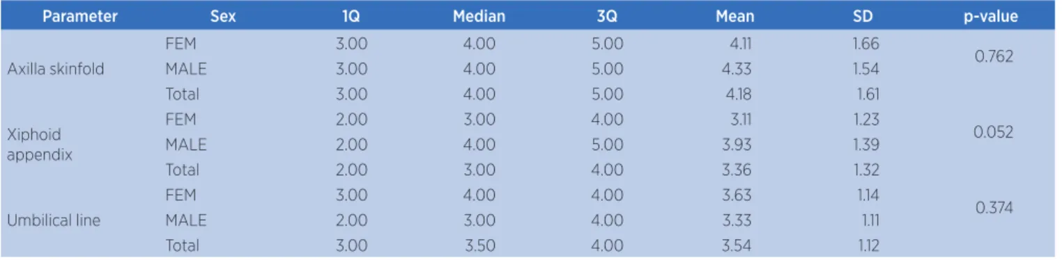
In Table 2 we observe the values of thoracoabdominal expansion for axilla skinfold, xiphoid appendix, and umbilical line, according to the sex of older adults.
Table 3 presents the characterization of older adults regarding nutritional status.
Table 1. Comparison of values expected and obtained for respiratory parameters, per sex
Parameter Sex 1Q Median 3Q Mean SD p-value
MIP
FEM Expected 74.14 77.57 79.53 76.80 3.21 <0.001*
Obtained 42.00 45.00 58.00 51.43 15.08
MALE Expected 91.30 98.10 103.30 97.53 6.21 <0.001*
Obtained 45.00 57.00 75.00 59.33 17.74
MEP
FEM Expected 70.46 74.73 77.17 73.77 4.00 <0.001*
Obtained 46.00 57.00 67.00 58.37 15.67
MALE Expected 100.50 107.79 113.46 107.62 6.47 0.001*
Obtained 63.00 67.00 80.00 73.80 23.94
FVC
FEM Expected 2.39 2.56 2.73 2.57 0.24 <0.001*
Obtained 1.75 2.16 2.60 2.17 0.57
MALE Expected 3.39 3.57 3.75 3.55 0.45 0.002*
Obtained 2.44 2.73 3.01 2.83 0.67
FEV1
FEM Expected 1.89 2.05 2.21 2.06 0.25 <0.001*
Obtained 1.44 1.67 2.09 1.77 0.47
MALE Expected 2.66 2.76 2.97 2.76 0.36 <0.001*
Obtained 1.85 2.14 2.64 2.15 0.62
*p≤0.05 – Wilcoxon test
MIP: maximal inspiratory pressure (cmH2O); MEP: maximal expiratory pressure (cmH2O); FVC: forced vital capacity (l/min); FEV1: forced expiratory volume in one second (l/s); SD: standard deviation;
1Q: irst quartile; 3Q: third quartile; FEM: female; MALE: male
Table 2. Comparison of values expected and obtained for parameters of thoracoabdominal expansion, per sex
Parameter Sex 1Q Median 3Q Mean SD p-value
Axilla skinfold
FEM 3.00 4.00 5.00 4.11 1.66
0.762
MALE 3.00 4.00 5.00 4.33 1.54
Total 3.00 4.00 5.00 4.18 1.61
Xiphoid appendix
FEM 2.00 3.00 4.00 3.11 1.23
0.052
MALE 2.00 4.00 5.00 3.93 1.39
Total 2.00 3.00 4.00 3.36 1.32
Umbilical line
FEM 3.00 4.00 4.00 3.63 1.14
0.374
MALE 2.00 3.00 4.00 3.33 1.11
Total 3.00 3.50 4.00 3.54 1.12
*p≤0.05 – Mann-Whitney Test
SD: standard deviation; 1Q: irst quartile; 3Q: third quartile; FEM: female; MALE: male
Table 3. Characterization of older adults regarding nutritional status
Nutritional status n % Age (mean±SD) years
MAL 10 20 69.6±7.42
EUT 24 48 68.6±6.76
OBE 16 32 70.62±7.43
Descriptive analysis
MAL: malnourished; EUT: Eutrophic; OBE: obese; n: number of subjects; %: percentage of subjects; SD: standard deviation
Table 4. Comparison of variables obtained regarding nutritional status
Variable Nutritional status Mean SD Q1 Median Q3 p-value
Obtained MIP
MAL 51.70 13.91 42.00 51.50 57.00
0.966
EUT 53.71 15.50 44.00 50.50 63.00
OBE 55.08 19.06 43.00 49.00 89.00
Obtained MEP
MAL 57.10 13.99 46.00 64.00 65.00
0.670
EUT 63.17 21.68 49.50 61.50 70.00
OBE 65.77 19.47 42.00 52.00 73.00
Table 4. Continuation
Variable Nutritional status Mean SD Q1 Median Q3 p-value
Obtained FVC
MAL 2.60 0.54 2.19 2.37 2.83
0.441
EUT 2.41 0.73 1.85 2.45 2.89
OBE 11.16 24.73 1.50 2.43 2.76
Obtained FEV1
MAL 1.99 0.47 1.72 1.93 2.08
0.511
EUT 1.95 0.53 1.50 1.85 2.44
OBE 12.12 28.63 1.14 2.00 2.55
AXILLA
MAL 4.60 1.78 3.00 4.00 6.00
0.693
EUT 4.17 1.63 3.00 4.00 5.00
OBE 3.88 1.50 2.00 3.00 5.00
XIPHOID
MAL 3.30 1.16 3.00 3.50 4.00
0.841
EUT 3.29 1.40 2.00 3.00 4.00
OBE 3.63 1.36 3.00 4.00 6.00
UMB
MAL 3.10 0.74 3.00 3.00 4.00
0.209
EUT 3.83 1.24 3.00 4.00 5.00
OBE 3.29 1.07 1.75 3.00 4.00
*p ≤ 0.05 Kruskal-Wallis Test
MIP: maximal inspiratory pressure (cmH2O); MEP: maximal expiratory pressure (cmH2O); FVC: forced vital capacity (l/min); FEV1: forced expiratory volume in one second (l/s); malnourished; EUT: Eutrophic; OBE: obese; SD: standard deviation; 1Q: irst quartile; 3Q: third quartile
DISCUSSION
Maximal respiratory pressures were below the values
expected for age and sex13. Previous studies4,18,19,21,22
also found that respiratory muscle strength showed reduced compared with expected values for older adults,
according to the equation regarding age and sex13. With
advancing age, the reduction in respiratory muscle strength is similar to the reduction in the strength of
the skeletal muscles, especially after 60 years of age21.
It is worth noting that even though MIP decreased, no older adult presented symptoms related to decreased inspiratory force. On the other hand, MEP, although indirectly related to ventilatory activities, should be evaluated, since it closely participates in non-ventilatory activities, such as sneeze and cough, that, when not
efective, can afect the pulmonary health21.
Nabil23 noted in his study on 325 older adults an
inverse relationship of the maximal respiratory pressures with age, that is, to the extent that age increases, the respiratory pressure decreases. he reduction in respiratory muscle strength was also observed in a
study24 on 100 individuals aged between 40 and 89
years, of both sexes, concluding that with advancing age there is a reduction in inspiratory and expiratory muscle
strength. Simões et al.20 highlight that the reduction
in blood pressure values are evidence of the reduction in respiratory muscle strength that occurs in older adults who are in an age group considered older, being
directly related to the aging phenomenon. his fact was conirmed by the strong negative correlations between age and respiratory pressures found in their study with 60 older adults subdivided in three age groups: From 60 to 69; 70 to 79; and 80 to 90 years.
hese changes may be related to aging, which, in turn, includes physiological changes that show decreased muscle mass (sarcopenia) and muscle ibers, mainly the
type II one22. hus, with the reduction in muscle mass,
respiratory muscles lose their eiciency in generating force, relecting in lower maximal respiratory pressure values2,20.
Aging comprises changes in postural structure, joints start getting more rigid, there is a calciication, and degeneration of the cartilage, causing bones to be merged, thereby changing the posture of the individual, leading to decreased mobility of the rib cage, thus causing a reduction in maximal respiratory pressures. In addition, the diaphragm muscle has primary function in the respiratory system, being responsible for about 60% of the respiratory function; however, its satisfactory performance depends on the structures that surrounds it. If there is a postural imbalance, it will inluence on the action of gravity on such a structure, and consequently, deicits in its performance will occur, hindering the
process of inspiration and expiration18,22,25.
he degree of thoracic mobility observed in this study is below normal parameters for a healthy young
researched literature values of speciic reference to the older adult population, only to healthy young adults. his value reduces in older people due to changes in
the thoracic structure26. Authors point out that the
reduction in thoracic compliance in healthy individuals is most evident from 80 years, although it was already
present since 50 years old27.
he maximal pulmonary functional performance is reached, on average, at 20 years old in woman and 25 years old in men. Subsequently, a slow and gradual reduction in lung capacity begins, which remains, however, in a condition to provide adequate gas exchange even in extreme ages in healthy individuals. he FVC shows a 25% drop between 20 and 70 years old, and at 70 years old its value is 75% of the 20 years
old value28.
In our study, FVC and FEV1 decreased in older
adults, corroborating other studies29,30. he decreased
thoracic expansion found can explain, in part, the reduction in FVC, since this is the index of the ability
of the dilation of thoracic and pulmonary system30, in
addition to the reduction in respiratory muscle strength. Nutritional status did not afect respiratory parameters. A correlational study veriied the relationship between body fat and lung function of 17 female older adults aging 60 to 80 years, selected by convenience, of a group of this population, and found no association between body composition and BMI
variables with the pulmonary function31. We found
no other studies relating BMI/nutritional status and respiratory parameters of the population studied in our article. One hypothesis is that this result may have been determined by the use of BMI as a determinant of obesity, rather than the use of other more reliable methods. It is suggested that the location of fat could afect the respiratory system, since the excess of abdominal fat could compress the diaphragm and compromise breathing.
As a limitation of the study, we highlight the absence of a control group (composed of adults) for comparison purposes.
CONCLUSION
Aging inluenced the respiratory parameters evaluated in this study group, regardless of sex. here was also a reduction in maximal respiratory pressures, FVC,
FEV1, and thoracoabdominal expansion. Nutritional
status, on the other hand, did not inluence measures of respiratory muscle strength, pulmonary volumes, and thoracoabdominal expansion. We highlight the importance of this knowledge to health professionals, in order that, in clinical practice, actions are directed to the prevention and promotion of the health of older adults.
REFERENCES
1. Instituto Brasileiro de Geograia e Estatística. Síntese de indicadores sociais: uma análise das condições de vida da população brasileira – 2012. Rio de Janeiro: IBGE; 2012. 2. Pegorari MS, Ruas G, Patrizzi LJ. Relationship between frailty
and respiratory function in the community-dwelling elderly. Braz J Phys Ther. 2013;17(1):9-16.
3. Francisco PMSB, Donalisio MR, Barros MBA, César CLG, Carandina L, Goldbaum M. Fatores associados à doença pulmonar em idosos. Rev Saúde Pública. 2006;40(3):428-35. doi: dx.doi.org/10.1590/S0034-89102006000300010. 4. Freitas FS, Ibiapina CC, Alvim CG, Britto RR, Parreira VF.
Relação entre força de tosse e nível funcional em um grupo de idosos. Rev Bras Fisioter. 2010;14(6):470-6. doi: dx.doi. org/10.1590/S1413-35552010000600004.
5. Borges JBC, Santos DF, Munhoz F, Carvalho SMR. Pressões e volumes pulmonares em idosos institucionalizados. Rev Bras Med. 2009;15(72):27-32.
6. Gusmão MFS, Duarte SFP, Lago LS, Nascimento CP, Almeira RFF, Reis LA. Mensuração das pressões respiratórias máximas em idosos participantes de grupos de convivência. InterScientia. 2015;3(2):133-41.
7. Matsudo SM, Matsudo VKR, Barro Neto TL. Impacto do envelhecimento nas variáveis antropométricas, neuromotoras e metabólicas da aptidão física. R Bras Ciênc Mov. 2000;8(4):21-32.
8. World Health Organization. Physical status: the use and interpretation of anthropometry [Internet]. 1995 [citado em 10 ago 2016]. Disponível em: http://whqlibdoc.who.int/trs/ WHO_TRS_854.pdf
9. Burneiko RCVM, Melatto T, Padulla SAT, Matta MV, Giacomassi IWS, Sato KT. Efeitos da inspiração fracionada ou incentivador a volume no pós-operatório revascularização do miocárdio. Rev Eletrônica Fisioter FCT/UNESP. 2009;1(1):124-38. 10. Pascotini FS, Ramos MC, Silva AMV, Trevisan ME. Espirometria
de incentivo a volume versus a luxo sobre parâmetros respiratórios em idosos. Fisioter Pesq. 2013;20(4):355-60. doi: dx.doi.org/10.1590/S1809-29502013000400009. 11. Rocha CBJ, Araújo S. Avaliação das pressões respiratórias
máximas em pacientes renais crônicos nos momentos pré e pós-hemodiálise. J Bras Nefrol. 2010;32(1):107-13. doi: dx.doi. org/10.1590/S0101-28002010000100017.
12. Souza RB. Pressões respiratórias estáticas máximas. J Pneumol. 2002;28(Supl 3):155-65.
14. Pereira CAC, Neder JA. Diretrizes para testes de função pulmonar. J Pneumol. 2002;28(Supl 3):S1-S41.
15. Caldeira VS, Starling CCD, Britto RR, Martins JA, Sampaio RF, Parreira VF. Precisão e acurácia da cirtometria em adultos saudáveis. J Bras Pneumol. 2007;33(5):519-26. doi: dx.doi. org/10.1590/S1806-37132007000500006.
16. Cielo CA, Pascotini FS, Haefner LSB, Ribeiro VV, Christmann MK. Tempo máximo fonatório de /e/ e /ė/ não-vozeado e sua relação com índice de massa corporal e sexo em crianças. Rev CEFAC. 2016;18(2):491-7. doi: dx.doi. org/10.1590/1982-021620161825915.
17. Lipschitz DA. Screening for nutritional status in the elderly. Prim Care. 1994;21(1):55-67.
18. Simões RP, Castello V, Auad MA, Dionísio J, Mazzonetto M. Prevalência de redução da força muscular respiratória em idosas institucionalizadas. São Paulo Med J. 2009;127(2):78-83. doi: dx.doi.org/10.1590/S1516-31802009000200005. 19. Oyarzún M. Función respiratoria en la senectud. Rev
Méd Chile. 2009;137(3):411-8. doi: dx.doi.org/10.4067/ S0034-98872009000300014.
20. Fonseca MA, Cader SA, Dantas EHM, Bacelar SC, Silva EB, Leal SMO. Programas de treinamento muscular respiratório: impacto na autonomia funcional de idosos. AMB Rev Assoc Med Bras. 2010;56(6):642-8. doi: dx.doi.org/10.1590/ S0104-42302010000600010.
21. Almeida RFF, Nascimento CP, Lago LS, Gusmão MFS, Duarte SFP, Reis LA. Relação entre força muscular respiratória e faixa etária em idosos participantes de grupos de convivência. Rev Enferm Contempor. 2015;4(1):33-8.
22. El Hajjar, N. Avaliação da força muscular respiratória em idosos. Pleiade. 2007;1(1):95-112.
23. Simões RP, Auad MA, Dionísio J, Mazzonetto M. Inluência da idade e do sexo na força muscular respiratória. Fisioter Pesqui. 2007;14(1):36-41.
24. Simões RP, Castello V, Auad MA, Dionísio J, Mazzonetto M. Força muscular respiratória e sua relação com a idade em idosos de sessenta a noventa anos. RBCEH. 2010;7(1):52-61. 25. Pettenon R, Milano D, Bittencourt DC, Schneider RH.
Adaptação funcional do aparelho respiratório e da postura no idoso. RBCEH. 2008;5(2):64-77.
26. Guimarães ACA, Pedrini A, Matte DL, Monte FG, Parcias SR. Ansiedade e parâmetros funcionais respiratórios de idosos praticantes de dança. Fisioter Mov. 2011;24(4):683-8. doi: dx.doi.org/10.1590/S0103-51502011000400012.
27. Parreira VF, Bueno CJ, França DC, Vieira DSR, Pereira DR, Britto RR. Padrão respiratório e movimento toracoabdominal em indivíduos saudáveis: inluência da idade e do sexo. Rev Bras Fisioter. 2010;14(5):411-6. doi: dx.doi.org/10.1590/ S1413-35552010000500010.
28. Barreto SSM. Volumes pulmonares. J Pneumol. 2002; 28(Supl 3):83-94.
29. Ide MR. Estudo comparativo dos efeitos de um protocolo de cinesioterapia respiratória desenvolvido em dois diferentes meios, aquático e terrestre, na função respiratória de idosos [dissertação]. São Paulo (SP): Faculdade de Medicina da Universidade de São Paulo; 2004.
30. Ruivo S, Viana P, Martins C, Baeta C. Efeito do envelhecimento cronológico na função pulmonar: comparação da função respiratória entre adultos e idosos saudáveis. Rev Port Pneumol. 2009;15(4):629-53.