ARTICLE
Monitoring respiratory muscle strength
assists in early diagnosis of respiratory
dysfunction as opposed to the isolated use of
pulmonary function evaluation in amyotrophic
lateral sclerosis
Monitoramento da força muscular respiratória em detrimento do uso isolado da avaliação
da função pulmonar para o diagnóstico precoce de disfunção respiratória em pacientes
com esclerose lateral amiotrófica
Guilherme Fregonezi1, Palomma Russelly Saldanha Araújo1, Tathiana Lindemberg Ferreira Macêdo1, Mario
Emilio Dourado Junior2, Vanessa Regiane Resqueti3, Armele de Fátima Dornelas de Andrade3
1PneumoCardioVascular and Respiratory Muscle Performance, Department of Physical Therapy, Federal University of Rio Grande do Norte (UFRN), Natal RN, Brazil; 2Onofre Lopes University Hospital, UFRN, Natal RN, Brazil;
3Department of Physiotherapy, Laboratory of Cardiopulmonary Physiotherapy, Federal University of Pernambuco (UFPE), Pernambuco PE, Brazil.
Correspondence: Guilherme Fregonezi; Departamento de Fisioterapia, Universidade Federal do Rio Grande do Norte, Campus Universitário Lagoa Nova Caixa Postal 1524; 59072-970 Natal RN - Brasil; E-mail: fregonezi@ufrnet.br
Support: This work was supported by the CNPq Brazil (number: 301661/2009-0) and Programa Nacional de Cooperação Acadêmica - Ação Novas Fronteiras (Procad-NF) number 764/2010-UFRN-UFMG-UFPE.
Conflict of interest: There is no conlict of interest to declare.
Received 04 May 2012; Received in inal form 08 August 2012; Received in inal form 15 August 2012.
ABSTRACT
Objective: It was study the relationship between respiratory muscle strength and forced vital capacity (FVC) in patients with amyotrophic lateral sclerosis (ALS) versus healthy subjects. Methods: Pulmonary function and respiratory muscle strength [maximal inspiratory (PImax), maximal expiratory (PEmax) and sniff nasal inspiratory pressure (SNIP)] were assessed in patients with ALS and healthy subjects, matched using cutoffs established in the literature for impaired pulmonary function and respiratory muscle weakness. Results: Twenty-eight ALS pa-tients and 28 healthy subjects were studied. We found sensitivity and speciicity for PImax, PEmax and SNIP of 75/58%, 81/67% and 75/67%. The Receiver Operating Characteristic curve (ROC curve) indicated that the variables PImax, PEmax and SNIP can identify differences in respiratory muscle strength between ALS and healthy individuals at 0.89, 0.9 and 0.82, respectively. A positive correlation was recorded between FVC (%) versus SNIP, PImax and PEmax. Conclusion: In ALS, monitoring respiratory muscle strength assists in early diagnosis of respiratory dysfunction as opposed to the isolated use of FVC.
Key words: muscle weakness, neuromuscular diseases, pulmonary function test, respiratory muscles.
RESUMO
Objetivo: Estudar a relação entre a força dos músculos respiratórios e a capacidade vital forçada (CVF) em pacientes com esclerose lateral amiotróica (ELA) e sujeitos saudáveis. Métodos: Avaliamos a função pulmonar e a força dos músculos respiratórios [pressão inspiratória (PImax), pressão expiratória (PEmax) e pressão inspiratória nasal de sniff (SNIP)] utilizando pontos de corte estabelecidos na literatura para diagnóstico de fraqueza muscular respiratória. Resultados: Foram estudados 28 pacientes com ELA e 28 sujeitos saudáveis. Encontramos sensibilidade e especiicidade para PImax, PEmax e SNIP de 75/58%, 81/67% e 75/67%. A curva ROC (Receiver Operating Characteristic) in-dicou que as variáveis PImax, PEmax e SNIP podem identiicar diferenças na força dos músculos respiratórios em pacientes com ELA versus sujeitos saudáveis em 0,89, 0,9 e 0,82 respectivamente. Foi encontrada uma correlação positiva entre CVF (%) e SNIP, PImax e PEmax. Con-clusão: Em pacientes com ELA, o monitoramento da força muscular respiratória auxilia no diagnóstico precoce da disfunção em detrimento do uso da CVF isolada.
Amyotrophic lateral sclerosis (ALS) is a neurodegenera-tive disease that causes loss or death of the upper and lower motor neurons1. Symptoms typically appear in the 50th de-cade of life, and the disease follows a speciic clinical course. In general, survival does not exceed three years in 76% of cas-es and ive to ten years in 8 to 16% of cascas-es2. Clinically, ALS is characterized by loss of function in the skeletal muscles; with regard to prognosis of the disease, recent data in the lit-erature have identiied the primary clinical cause of death as respiratory failure in 70% of cases3-5. Respiratory failure is characterized by hypoxemia and/or hypercapnia, or a com-bination of both, and is directly associated to weakness in re-spiratory muscles6-9.
Evaluation of forced vital capacity (FVC) through spirom-etry has been widely used as an important tool for the moni-toring of pulmonary function in ALS patients. However, the assessment of respiratory muscle strength through maximal inspiratory and experitory pressures (PImax, PEmax) and snif nasal inspiratory pressure (SNIP) has become increas-ingly relevant in the early diagnosis of respiratory muscle weakness, as demonstrated in other neurological disease10,11. Despite the complementarity of respiratory muscle strength tests, recent studies have shown that these tests and pulmo-nary function assessments are not commonly used to mon-itor the respiratory health of patients with ALS. A recent investigation12 demonstrated that only 38% of clinical neu-rologists conduct some type of respiratory evaluation in their irst contact with patients, and these tests are almost never repeated at reassessment. Among clinicians who requested some form of written information, evaluation of respiratory muscle strength was rare. PImax/PEmax was requested by only 6% of physicians and SNIP in 17% of cases, either at each visit or only when symptoms were present. he present study aimed to analyze the relationship between forced vital capac-ity and measurements of respiratory muscle strength in pa-tients with ALS and matched healthy subjects.
METHOD
Subjects
his is a cross-sectional study of patients diagnosed with ALS by a neurologist, treated at the Multidisciplinary Clinic for Neuromuscular Diseases, and healthy individuals recruit-ed from the university community. Patients were includrecruit-ed by sample of convenience and assessed from January 2009 to July 2011, and healthy subjects were recruited and evaluated over the same period, paired by gender, age, height and body weight. All patients monitored by the multidisciplinary team and healthy participants had no respiratory or cardiac diseases. Healthy individuals and those sufering from ALS were includ-ed in the study after being informinclud-ed about its nature and pur-pose and giving written informed consent. he investigation
was conducted in accordance with Resolution 196/96 of the National Health Council and approved by the Onofre Lopes Hospital Research Ethics Committee (Protocol no 239/08).
Study design
All selected participants were submitted to anthropo-metric evaluation, forced vital capacity and respiratory mus-cle strength testing. Examinations of patients and healthy subjects took place on one day and were performed by the same examiner.
Anthropometric assessment
Weight was determined on a WELMY electronic bal-ance (WELMY, Santa Bárbara do Oeste, Paraná, Brazil). Both height and weight were assessed with individuals barefoot, standing upright and wearing lightweight clothing. his was followed by the calculation of Body Mass Index (BMI) using the formula: BMI: weight (kg)/height2 (m).
Forced spirometry
Technical procedures, acceptability and reproducibil-ity criteria, interpretive values, standardization and equip-ment were in accordance with American horacic Society/ European Respiratory Society recommendations13, and all patients were valued in seated position. Forced expiratory volume in the irst second (FEV1), FVC and the FEV1/FVC relationship in their absolute and relative values were consid-ered, with the latter obtained by comparison with the normal curve for all spirometric variables in relation to the Brazilian population14. A DATOSPIR® 70 (Siblemed, Barcelona, Spain) device was used, calibrated daily. With respect to cutofs for impaired lung function according to FVC, we used FVC <75% of the predicted value, as described in the literature15.
Respiratory muscle strength and sniff nasal inspiratory pressure
in accordance with reference values for maximal respiratory pressures and snif nasal inspiratory pressure published in pre-vious studies21,22. Respective values for male and female were: PImax: 56.1 cmH2O and 53.4 cmH2O; PEmax: 70.6 cmH2O and 57.2 cmH2O; and SNIP 61.1 cmH2O and 57.2 cmH2O.
Statistical analysis
Results are expressed as mean and standard deviation. he unpaired t-test was applied to compare anthropometric, lung function and respiratory muscle strength values between ALS patients and healthy subjects. Pearson’s correlation was used to verify the association between the variables pulmo-nary function and respiratory muscle strength, and simple linear regression was employed to analyze the relationship between PImax and PEmax in ALS patients and healthy sub-jects. A study of speciicity and sensitivity was performed for each variable of respiratory muscle strength (PImax, PEmax and SNIP) in relation to FVC, in addition to an analysis of the Receiver Operating Characteristic curve (ROC curve) for respiratory muscle strength values of ALS patients in rela-tion to those of healthy subjects. GraphPad Prism 4 software (GraphPad Software Inc., San Diego Califórnia USA) was used for all analyses, with a signiicance level of p<0.05 and bilat-eral approximation.
RESULTS
We evaluated 31 patients (19 men) with ALS and 28 healthy subjects (16 men). hree ALS patients were excluded for being unable to perform the maneuver required for respi-ratory muscle assessment. he inal sample consisted of 28 individuals sufering from ALS (16 men) and 28 healthy sub-jects (16 men).
he average time since diagnosis was 44.8±5.9 months. According to El Escorial criteria, 15 patients (7 males and 8 females) were diagnosed with deinite ALS and 13 (9 men and 4 women) with probable ALS. With regard to type of classiication, 5 individuals were classiied as sufering from bulbar/irst order motor neuron ALS and 23 (12 males and 11 females) with spinal/second order motor neuron ALS. Eight patients used orthosis (wheelchairs) for locomotion and 20 were able to walk unaided or with partial assis-tance provided by an orthosis brace. Regardless of the use
of orthoses, all patients were capable of standing for weight and height analysis.
Anthropometric characteristics
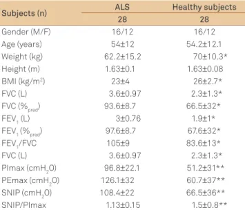
Signiicant diferences were recorded between the group of ALS suferers and healthy volunteers with respect to weight (p=0.029) and BMI (p=0.004). Among females, signii-cant diferences were also found between weight (p=0.029) and BMI (p=0.001) for ALS patients in relation to healthy sub-jects. Anthropometric characteristics of the sample are de-scribed in Table 1.
Spirometry and inspiratory and expiratory muscle strength
In regard to pulmonary function, signiicant diferences were observed for both male and female patients in rela-tion to healthy participants for the variables FVC and FEV1, in absolute values and percentages of the predicted FEV1/ FVC ratio (Table 2). ALS patients were classiied as sufer-ing from mild restrictive ventilatory dysfunction. Regardsufer-ing respiratory muscle strength, considering the cutof points
Table 2. Cross-Tab between forced vital capacity and cutoff points limits for diagnosis of respiratory muscle weakness.
PImax PEmax SNIP
Limits Normal Limits Normal Limits Normal
FVC≤75%pred
16 patients 12 (75%) 4 (25%) 13 (81.25%) 3 (18.75%) 12 (75%) 4 (25%)
FVC >75%pred
12 patients 5 (41.6%) 7 (58.3%) 4 (38.3%) 8 (66.6%) 4 (38.3%) 8 (66.6%)
FVC: forced vital capacity; SNIP: sniff nasal inspiratory pressure; PImax: maximal inspiratory pressure; PEmax: maximal expiratory pressure. Cutoff points limits: PImax ♂=56.1 cmH2O e ♀=53.4 cmH2O; PEmax ♂=70.6 cmH2O and ♀=57.2 cmH2O; SNIP ♂=61.1 cmH2O and ♀=57.2 cmH2O.
Table 1. Anthropometric characteristics and pulmonary
function in both groups.
Subjects (n) ALS Healthy subjects
28 28
Gender (M/F) 16/12 16/12
Age (years) 54±12 54.2±12.1
Weight (kg) 62.2±15.2 70±10.3*
Height (m) 1.63±0.1 1.63±0.08
BMI (kg/m2) 23±4 26±2.7*
FVC (L) 3.6±0.97 2.3±1.3*
FVC (%pred) 93.6±8.7 66.5±32*
FEV1 (L) 3±0.76 1.9±1*
FEV1 (%pred) 97.6±8.7 67.6±32*
FEV1/FVC 105±9 83.6±13*
FVC (L) 3.6±0.97 2.3±1.3*
PImax (cmH2O) 96.8±22.1 51.2±31**
PEmax (cmH2O) 126.1±32 60.7±37**
SNIP (cmH2O) 108.4±22 66.5±36**
SNIP/PImax 1.13±0.15 1.5±0.8**
Data are expressed as mean±standard deviation. *non-paired t-test p≤0.05 and **non-paired t-test p≤0.01. M: male; F: female; BMI: body mass index; FVC: forced vital capacity; FEV1: forced expiratory volume in the irst second;
FEV1/FVC: ratio between forced expiratory volume in the irst second and
applied, all healthy volunteers exhibited respiratory muscle strength within the normal range. In the ALS group, 9 men (32%) and 8 women (28%) were classiied with inspiratory muscle weakness, considering only PImax. When apply-ing cutof points for inspiratory muscle weakness tested by SNIP, we detected 9 men (32%) and 7 women, while expira-tory muscle weakness categorized by PEmax analysis iden-tiied 9 male (32%) and 8 female patients (28%). Results are shown in Fig 1.
After subdividing patients, just for descriptive analy-sis, into bulbar (5 subjects) and spinal ALS (23 individuals), FVC was found to be reduced in both groups (68.7±32.2 ver-sus 66.2±32.3%pred, respectively). However, PImax, PEmax
and SNIP values indicative of muscular weakness were low-er in the bulbar ALS group when compared with the spinal ALS group (PImax: 45.6±35.7 cmH2O versus 52.5±30 cmH2O; SNIP: 60.8±38.9 cmH2O versus 67.7±37.6 cmH2O; PEmax: 49.6±21.7 cmH2O vs 63.1±40 cmH2O) respectively.
Analysis of cutof points suggested to diagnose inspira-tory and expirainspira-tory muscle weakness versus reduced FVC <75%pred identiied sensitivity and speciicity for PImax, PEmax and SNIP of 75%/58%, 81%/67% and 75%/67%, respectively (Table 2). he ROC curve demonstrated high ac-curacy for PImax, PEmax and SNIP tests in detecting weak-ness in the respiratory muscles of ALS patients. he likeli-hood of randomly chosen patients obtaining PEmax, PImax and SNIP results lower than those recorded for a randomly selected healthy individuals is 0.90 (90%), 0.89 (89%) and 0.82 (82%) respectively (Fig 2).
Relationship between respiratory muscle strength and pulmonary function
Considering the relationship between the strength of diferent respiratory muscles, Fig 1 shows an alteration in strength equilibrium between inspiratory and expiratory muscles in ALS patients. In healthy subjects, a relationship was observed in which PEmax = 16.46+1.13*PImax, whereas in ALS patients PEmax = 28.9+0.73*PImax.
PImax was positively correlated to SNIP in healthy indi-viduals and ALS patients, with r=0.802 and r=0.872, respec-tively, and p<0.001 for both groups. A positive correlation was recorded between FVC (%)/SNIP, FVC %/PImax and FVC (%)/PEmax only among ALS patients, with r=0.748, r=0.724 and r=0.826, respectively, and p<0.001 (Fig 3).
DISCUSSION
he present study aimed to investigate the relationship between measurements of respiratory muscle strength and FVC in patients with amyotrophic lateral sclerosis versus healthy subjects. Furthermore, we sought to establish the importance of the correlation between respiratory muscle
strength and spirometric variables, particularly forced vital capacity, in identifying early-onset respiratory muscle weak-ness in ALS patients. Despite the slight change in pulmonary function, the strength of respiratory muscles was already moderately to severely impaired.
In light of the need for early diagnosis of reduced lung function in patients with neuromuscular diseases, several measures to achieve this purpose have been described in the literature. Determining FVC by spirometry has been reported and applied as a simple and efficient means23 of monitoring declining lung function. However, it is not sen-sitive to detecting early respiratory damage since respira-tory symptoms are often not yet evident, despite the pres-ence of alterations in respiratory muscle strength. Patients assessed in the present study exhibited restrictive venti-latory pattern, with 57% of patients showing diminished FVC, 75% displaying reduced SNIP and 81% demonstrat-ing a decrease in PEmax. Likewise, accorddemonstrat-ing to the cutoff points applied in this investigation to diagnose respiratory muscle weakness, approximately 40% of patients exhibit-ed loss of inspiratory and/or expiratory muscle strength, even when pulmonary function was preserved. Results found in the present study reinforce this fact. In a previous study, Tsara et al.24 evaluated lung function through FVC in 28 individuals suffering from ALS and observed that 78.5% of patients showed reduced FVC and 89.5% of these demonstrated a decline in maximal respiratory pressure. Analysis of maximal respiratory pressures depicts the re-spiratory muscles as a whole, which is complemented by sniff nasal inspiratory pressure as an excellent assessment of muscular contraction in the diaphragm.
functional and respiratory assessment. In patients with spinal ALS, the authors found correlations between PImax and PEmax of r=-0.76; PEmax and pulse oximetry of r=0.58; ALSFRS-R score and percentage weight loss (%WL) r=0.59 and between PImax and ALSFRS-R score r=0.65. In the bulbar group, a correlation was observed between PEmax and BMI (r=0.97). Moreover, the bulbar group also exhibited severely compromised respiratory muscles and pulmonary function, with mean PImax of 24.1 cmH2O and PEmax of 35 cmH2O and FVC of 54%pred. In the spinal group, respiratory muscle strength and lung function showed a moderate decrease, with PImax of 61.1 cmH2O, PEmax at 62.3 cmH2O and FVC of 84%pred. As in our study, this investigation demonstrates that, al-though loss of strength was already present in respirato-ry muscles, pulmonarespirato-ry function in the spinal ALS group showed a slower decline, in contrast with the bulbar ALS group, which was evident even in a smaller sample of patients.
Fig 2. Receiver Operating Characteristic curve (ROC curve)
for respiratory muscle strength values of amyotrophic lateral sclerosis patients in relation to those of healthy subjects.
0.0 0.2 0.4 0.6 0.8 1.0
0.0 0.2 0.4 0.6 0.8 1.0
PEmax PImax SNIP
1 - Specificity
S
e
n
s
iti
vit
y
Healhty subjects
0 25 50 75 100 125 150 175 200
0 25 50 75 100 125 150 175 200
P
E
max
(c
m
H2
O)
ALS
25 50 75 100 125 150 175 200
0 25 50 75 100 125 150 175 200
0 25 50 75 100 125 150 175
0 25 50 75 100 125 150 175
0 25 50 75 100 125 150 175
0 25 50 75 100 125 150 175
P
E
max
(c
m
H2
O)
S
N
IP
(c
m
H2
O)
S
N
IP
(c
m
H2
O)
Plmax (cmH2O) Plmax (cmH2O)
Plmax (cmH2O) Plmax (cmH2O)
Fig 1. Relationship between inspiratory maximal pressure (PImax) versus expiratory maximal pressure (PEmax) and sniff nasal
inspiratory pressure (SNIP) in healthy subjects and amyotrophic lateral sclerosis patients.
Male: close circle; Female: open circle. Cut of points for female are represented with continues line and for male dotted line.
In a similar study, Almeida et al.27 evaluated 16 patients di-agnosed with probable or deinite ALS over 8 months, using spi-rometry, maximal respiratory pressures, arterial gasometry, pulse oximetry, BMI and percentage weight loss. he authors found that PCO2 was a signiicant parameter to monitor the evolution of the disease during the study period (p=0.051). Signiicant cor-relations were also observed between PImax and PEmax (r=0.83); BMI and PImax (r=0.70); BMI and PEmax (r=0.72); pulse oxim-etry and forced vital capacity (r=0.57). At the same time, this study shows moderate alterations in respiratory muscle strength with PImax of 65.6 cmH2O and PEmax at 74.2 cmH2O, with few changes in FVC% (82%pred). Although both studies identify im-portant information with respect to respiratory elements, they present little new evidence regarding respiratory muscle strength and spirometry. By contrast, the present study assesses the rela-tionship between respiratory muscle strength and spirometry, establishing parameters that can be used as a reference and de-termining cutof points for respiratory muscle strength, clearly demonstrating the diference between men and women in the functional loss of strength in these muscles.
Several other studies have been published with the purpose of establishing a relationship between the degree of respiratory muscle weakness and distribution of weakness (diaphragmat-ic versus overall muscle weakness) and lung volumes. Qureshi et al.28 investigated risk factors and predictors of disease progres-sion in 106 healthy subjects over 12 months. hey observed nor-mal total lung capacity (TLC), elevated residual volume, reduced
Fig 3. Relationship between forced vital capacity percentage (FVC%) of the predicted value and maximal expiratory pressure
(PEmax), maximal inspiratory pressure (PImax) and sniff nasal inspiratory pressure (SNIP) in amyotrophic lateral sclerosis patients and healthy subjects.
Healhty subjects
0 25 50 75 100 125 150 175 200 225
0 25 50 75 100 125 150
0 25 50 75 100 125 150 175 200 0
25 50 75 100 125 150
Healthy ects
0 25 50 75 100 125 150 175 200
0 25 50 75 100 125 150
0 25 50 75 100 125 150 175 200 0
25 50 75 100 125 150
0 25 50 75 100 125 150 175 0
25 50 75 100 125 150
Healthy ects
0 25 50 75 100 125 150 175 200
200 0
25 50 75 100 125 150
H
2O)
Plmax (cmH2O)
PEmax (cmH2O)
PEmax (cmH2O) Plmax (cmH2O)
CVF
%
SNIP (cmH2O)
vital capacity and diminished respiratory muscle strength (in-spiratory and expiratory), with a greater decline in PEmax than maximal PImax. he study showed that a marked reduction in PEmax determines increased residual volume (RV), which clini-cally translates into lower expiratory eiciency. Other authors have conirmed the relationship between muscular alteration already present and pulmonary function. In 213 patients, Fallat et al.29 reported preserved FVC and TLC associated with an ac-centuated change in RV and maximal voluntary ventilation. he same authors established a relationship between symptoms and lung function, observing that, despite existing muscular altera-tion, respiratory symptoms were only reported by patients after a signiicant decrease in pulmonary function28.
A potential limitation of our study is the small sample size due to the limited time and few resources available dur-ing the study period well deined. Furthermore, the heteroge-neity of sample size that represents the ALS cases in health care in our outpatients clinics. However the possible weak point of the study, the results add new perspective in terms of respiratory assessment in ALS patients.
1. Lima NMFV, Nucci A. Clinical attention and assistance proile of patients with amyotrophic lateral sclerosis. Arq Neuropsiquiatr 2011;69:170-175.
2. Hirtz D, Thurman DJ, Gwinn-Hardy K, Mohamed M, Chaudhuri AR, Zalutsky R. How common are the common neurological disorders? Neurology 2007:68;326-337.
3. Spataro R, LoRe M, Piccoli T, Piccoli F, La Bella V. Causes and place of death in Italian patients with amyotrophic lateral sclerosis. Acta Neurol Scand 2010;122:217-223.
4. Kurian KM, Forbes RB, Colville S, Swingler RJ. Cause of death and clinical grading criteria in a cohort of amyotrophic lateral sclerosis cases undergoing autopsy from the Scottish Motor Neurone Disease Register. J Neurol Neurosurg Psychiatry 2009;80:84-87.
5. Corcia P, Pradat PF, Salachas F, et al. Causes of death in a post-mortem series of ALS patients. Amyotroph Lateral Scler 2008;9:59-62.
6. Resqueti VR, Araújo PRS, Dourado Junior ME, Fregonezi GAF. Esclerose lateral amiotróica (ELA) e músculos respiratórios. Ter Man 2011;9:503-508.
7. Uldry C, Janssens JP, de Muralt B, Fitting JW. Sniff nasal inspiratory pressure in patients with chronic obstructive pulmonary disease. Eur Respir J 1997;10:1292-1296.
8. Nava S, Ambrosino N, Crotti P, Fracchia C, Rampulla C. Recruitment of some respiratory muscles during three maximal inspiratory maneuvers. Thorax 1993;48:702-707.
9. Katagiri M, Abe T, Yokoba M, Dobashi Y, Tomita T, Easton PA. Neck and abdominal muscle activity during a sniff. Respir Med 2003;97:1027-1035.
10. Hart N, Polkey MI, Sharshar T, et al. Limitations of sniff nasal pressure in patients with severe neuromuscular weakness Neurol Neurosurg Psychiatry 2003;74:1685-1687.
11. Azevedo IG, Severino FG, Araújo TL, Resqueti VR, Bruno S, Fregonezi GAF. Relação entre pressão inspiratória nasal e pressão inspiratória máxima em pacientes com distroia miotônica. Ter Man 2010;8:224-230.
12. O’Neill CL, Williams TL, Peel ET, et al. Non-invasive ventilation in motor neuron disease: an update of current UK practice. J Neurol Neurosurg Psychiatry 2012;83:371-376.
13. Miller MR, Hankinson J, Brusasco V, et al. ATS/ERS Task Force. Standardisation of spirometry. Eur Respir 2005;26:319-338.
14. Pereira CAC, Sato T, Rodrigues SC. New reference values for forced spirometry in white adults in Brazil. J Bras Pneumol 2007;33:397-406.
15. Lechtzin N, Wiener CM, Shade DM, Clawson L, Diette GB. Spirometry in the supine position improves the detection of diaphragmatic weakness in* patients with amyotrophic lateral sclerosis. Chest 2002;121;436-442.
16. Black LF, Hyatt RE. Maximal respiratory pressures: normal values and relationship to age and sex. Am Rev Respir Dis 1969;99:696-702.
17. Neder JA, Andreoni S, Lerario MC, Nery LE. References values for lung function tests. Maximal respiratory pressures and voluntary ventilation. Braz J Med Biol Res 1999;32:719-727.
18. Severino FG, Resqueti VR, Bruno SS, Azevedo IG, Vieira RHG, Fregonezi GAF. Comparação entre o manovacuômetro nacional e o importado para medida da pressão inspiratória nasal. Rev Bras Fisioter 2010;14:426-431.
19. Lofaso F, Nicot F, Lejaille M, et al. Sniff nasal inspiratory pressure: what is the optimal number of sniffs? Eur Respir J 2006;27:980-982.
20. Steier J, Kaul S, Seymour J, et al. The value of multiple tests of respiratory muscle strength. Thorax 2007;62:975-980.
21. Araujo PRS, Resqueti VR, Nascimento Jr JF, et al. Maximal sniff nasal inspiratory pressure in Brazilian healthy subjects: a multicenter study. Maximal respiratory pressure in Brazilian health population: a multicenter study (in press).
22. Fregonezi GAF, Araujo PRS, Dornelas de Andrade AF, et al. Maximal sniff nasal inspiratory pressure in Brazilian healthy subjects: a multicenter study. In: XXI European Annual Congress, 2011, Amsterdam. Eur Resp J 2011;239.
23. Lechtzin N, Wiener CM, Shade DM, et al. Spirometry in the supine position improves the detection of diaphragmatic weakness in patients with amyotrophic lateral sclerosis. Chest 2002;121:436-442.
24. Tsara V, Serasli E, Steiropoulos P, Tsorova A, Antoniadou M, Zisi P. Respiratory function in amyotrophic lateral sclerosis patients. The role of sleep studies. Hippokrattia 2010;14:33-36.
25. Singh D, Verma R, Garg RK, et al. Assessment of respiratory functions by spirometry and phrenic nerve studies in patients of amyotrophic lateral sclerosis. J Neurol Sci 2011;306:76-81.
26. Silva LBC, Mourão LF, Silva AA, et al. Amyotrophic lateral sclerosis: combined nutritional, respiratory and functional assessment. Arq Neuropsiquiatr 2008;66:354-359.
27. Almeida SRM, Silva LBC, Guerreiro CAM, Nucci A. Amyotrophic lateral sclerosis: prospective study on respiratory parameters. Arq Neuropsiquiatr 2010;68:258-262
28. Qureshi MM, Hayden D, Urbinelli L, et al. Analysis of factors that modify susceptibility and rate of progression in amyotrophic lateral sclerosis (ALS). Amyotrophic Lateral Sclerosis 2006;7:173-182.
29. Fallat RJ, Jewitt B, Bass M, Kamm B, Norris FH Jr. Spirometry in amyotrophic lateral sclerosis. Arch Neurol 1979;36:74-80.