MODELS, BIOLOGICAL
Glutamine and ornithine alpha-ketoglutarate supplementation on malate dehydrogenases
expression in hepatectomized rats
1Artur Guimarães FilhoI, Rodrigo Maranguape Silva da CunhaII, Paulo Roberto Leitão de VasconcelosIII, Sergio Botelho
GuimarãesIV
DOI: http://dx.doi.org/10.1590/S0102-86502014000600003
IFellow PhD degree, Postgraduate Program in Surgery, Department of Surgery, Federal University of Ceara (UFC), Fortaleza-CE, Brazil. Conception
and design of the study, technical procedures, acquisition and interpretation of data.
IIPhD, Associate Professor, Vale do Acarau University (UVA). Coordinator, Sobral Biotechnology Nucleus. Fortaleza-CE, Brazil. Acquisition and
interpretation of data.
IIIPhD, Full Professor, Coordinator, Postgraduate Program in Surgery, Department of Surgery, UFC, Fortaleza-CE, Brazil. Interpretation of data, critical
revision.
IVPhD, Associate Professor, Department of Surgery. Head, Experimental Surgical Research Laboratory (LABCEX), UFC, Fortaleza-CE, Brazil.
Conception, design, intellectual and scientiic content of the study, manuscript writing, critical revision.
ABSTRACT
PURPOSE: To evaluate the relative gene expression (RGE) of cytosolic (MDH1) and mitochondrial (MDH2) malate dehydrogenases enzymes in partially hepatectomized rats after glutamine (GLN) or ornithine alpha-ketoglutarate (OKG) suplementation.
METHODS: One-hundred and eight male Wistar rats were randomly distributed into six groups (n=18): CCaL, GLNL and OKGL and fed calcium caseinate (CCa), GLN and OKG, 0.5g/Kg by gavage, 30 minutes before laparotomy. CCaH, GLNH and OKGH groups were likewise fed 30 minutes before 70% partial hepatectomy. Blood and liver samples were collected three, seven and 14
days after laparotomy/hepatectomy for quantiication of MDH1/MDH2 enzymes using the real-time polymerase chain reaction (PCR)
methodology. Relative enzymes expression was calculated by the 2-ΔΔCT method using the threshold cycle (CT) value for normalization.
RESULTS: MDH1/MDH2 RGE was not different in hepatectomized rats treated with OKG compared to rats treated with CCa. However, MDH1/MDH2 RGE was greater on days 3 (321:1/26.48:1) and 7 (2.12:1/2.48:1) while MDH2 RGE was greater on day 14 (7.79:1) in hepatectomized rats treated with GLN compared to control animals.
CONCLUSION: Glutamine has beneicial effects in liver regeneration in rats by promoting an up-regulation of the MDH1 and MDH2
relative gene expression.
Introduction
Potential mechanisms and pathways of liver regeneration have been extensively investigated in recent reviews1,2. Holecek3
studied the effects of carbohydrates, lipids, and amino acids on partial hepatectomy and concluded that administration of a standard amino acid mixture without energy substrate has an inhibitory effect on liver regeneration. Birkhahn et al.4 studies demonstrated
that the inhibitory effect of glucose on liver regeneration can be diminished by the simultaneous administration of aminoacids and other substances.
Experimental studies have shown that ornithine alpha-ketoglutarate (OKG) supplementation of enteral feeding
signiicantly shortens wound healing time in severe burn patients5.
It has been reported that OKG stimulates the production on insulin and growth hormone promoting intracellular aminoacid transport and protein synthesis6. Glutamine (GLN) is the most
abundant amino acid in the circulation and the main source of nitrogen for various metabolic processes including purine and pyrimidine synthesis necessary for DNA replication and cellular proliferation7,8. Yoshida et al.8 treated partially hepatectomized rats
with a parenteral solution containing GLN, resulting in increased uptake of the aminoacid by the hepatocytes and enterocytes and an increase in DNA and protein synthesis.
Two essential malate dehydrogenases, MDH1 (cytosolic malate dehydrogenase) and MDH2 (mitochondrial malate dehydrogenase), play important roles in the Krebs cycle for energy production through aerobic respiration9. MDH1 transports NADH
equivalents across the mitochondrial membrane, controlling tricarboxylic acid (TCA) cycle pool size. MDH1 mRNA is expressed primarily in cardiac and skeletal muscle and in the brain, at intermediate levels in the spleen, kidney, intestine, liver, and testes and at low levels in lung and bone marrow10. MDH2 is the
inal enzyme in the mitochondrial tricarboxylic acid (TCA) cycle.
It catalyzes the inter-conversion of L-malate and oxaloacetate using nicotinamide adenine dinucleotide (NAD) as a cofactor to generate reducing equivalents11.
Considering the importance of MDH1 and MHD2 in
the metabolic processes and the absence of speciic studies of the
role of these enzymes in hepatic regeneration, this study aims to evaluate the expression of MDH1 and MHD2 in an experimental partial hepatectomy model after administration of OKG and GLN.
Methods
Approval for experimental use of laboratory animals was obtained from the local Ethics Committee on Animal Use (CEUA, former CEPA) (protocol 54/08, 30 Sept 2008) and is in compliance with the Federal Law No. 11794 of October 8, 2008, and the Decree nº 6,689, July 15, 2009 that regulated the law in 11,794, available at: http: www.planalto.gov.brccivil03Ato2007-20102008LeiL11794.htm. The study was designed to minimize the number of animals required for the experiments.
In this controlled experimental study, after one week of acclimatization, 36 adult male Wistar rats weighting 170-325g, provided by the Sobral Zoonosis Center Vivarium were used. Rats were kept in a climate controlled room under 12-hour light/ dark cycles with free access to food and tap water throughout the studies. Before the experiments the animals were randomly distributed into six groups (n =18) and treated (Table 1) as follows: groups CCaL, GLNL and OKGL were fed calcium caseinate (CCa), GLN and OKG, 0.5g/Kg by gavage, 30 minutes before laparotomy; the 3 remaining groups (CCaH, GLNH and OKGH) were likewise fed CCa, GLN and OKG, 0.5g/Kg by gavage, 30 minutes before 70% partial hepatectomy. Next, each group was divided into three subgroups (n=6) according to sample collection time (3, 7 and 14 days after laparotomy/hepatectomy) (Table 1).
Groups Subgroups (n) Drugs and Procedures
CCaL CCaL3D (6) CCa L 7D (6) CCaL14D (6) CCa + laparotomy GLNL GLN L3D (6) GLN L 7D (6) GLN L14D (6) GLN + laparotomy OKGL OKG L3D (6) OKG L7D (6) OKG L14D (6) OKG + laparotomy CCaH CCaH3D (6) CCaH7D (6) CCaH14D (6) CCa + hepatectomy (70%) GLNH GLN H3D (6) GLN H7D (6) GLN H14D (6) GLN + hepatectomy (70%) OKGH OKG H3D (6) OKG H7D (6) OKG H14D (6) OKG + hepatectomy (70%)
TABLE 1 - Experimental groups and subgroups classiied according to drugs used and surgical procedures.
Drugs
Drugs used in the study were purchased from Nutrimed Industrial Ltda, Fortaleza, Brazil (Nutri Protein Nutrimed and L-glutamine), Chiesi S.A, Courbevoie, France (Cétornan - OKG), Promega Corporation, Promega Biotecnologia do Brasil, Ltda, Sao Paulo, Brazil (M-MLV Reverse Transcriptase), Life Technologies, New York, USA (Invitrogen™),Sigma-Aldrich Corp.(Sao Paulo, Brazil) SyBr Green™.
Surgical procedure
Anesthesia was induced with an intramuscular injection of ketamine hydrochloride (50 mg/kg) and xylazine (10 mg/kg). The anesthetic regimen was the same throughout the experiment. At the end of three, seven or 14 days as scheduled, the animals were anesthetized for sample collection (1.0 ml of arterial blood and 1.0 g of liver tissue). Another small fragment of liver (150 mg) was individualized to be processed
with speciic reagents for analysis and quantiication of malate
dehydrogenase enzymes using the real-time PCR (polymerase chain reaction) methodology.
Laboratory procedures
Real-time PCR was used for quantiication of malate
dehydrogenase enzymes. In brief, 150 mg of liver tissue was homogenized in 1 ml of Trizol (Invitrogen™), compound containing phenol and guanidine thiocyanate. Next, 200 µl of chloroform was added to 1 ml of the homogenate. After centrifugation and separation of the phases, the RNA retained in the aqueous phase was transferred to another tube containing 500 µl of isopropyl phenol alcohol. After spinning, the liquid was discarded and the pellet (dehydrated RNA) was rehydrated with 1 ml of 75% ethanol and 50 µl of UltraPure™ DEPC-treated Water to 0.01 and stored at -80º C 12. The production of cDNA was performed in 02
subsequent reactions. Reaction: 1 Oligo dT (Invitrogen™) (0.5 µg µl) -1 µL; Total RNA -1 µL; dNTP Mix (Invitrogen™) (1 mm) -10 µl; sterile H2O - 1 µL. Reaction 2: First-Stand Buffer 5 x-4 µl; Dithiothreitol (DTT) 0.1 M-2 µl; M-MLV Reverse Transcriptase 1 µL. The resulting solutions were stored at -20°C. As soon as the
CDNA was defrosted, glyceraldehyde 3-phosphate dehydrogenase (GAPDH), MDH1 e MDH2 (3.6µL) forward and reverse primers were added as well as other components (Sigma-Aldrich SyBr Green™ 10 µL, cDNA 10 ng e sterile H2O 1 µL) before taking the mixture to the thermal cycler for initiation of PCR reaction13,14. Each
test was conducted in triplicate and the inal result was obtained by calculating the average of these results. The inal values were
obtained considering the calculations of delta threshold cycle (CT) value (ΔCT) and delta/delta C
T (ΔΔC
T
) values15,16. Relative enzymes
expression was calculated by 2-ΔΔCT method using the threshold cycle (CT) value for normalization15,16. The extracted RNA
quantiication was carried out spectrophotometrically GeneQuant
Pro™ - Amersham Biosciences Corp NJ, USA) measuring the absorbance index at 260 nm and 280 nm wavelengths. Values greater than 1.6 were considered valid17.
Statistical analyses
To ensure the appropriateness of parametric testing, all data were examined for normality, using Kolmorogov-Smirnov test (with Dalal-Wilkins Lilliefor P Value). Data (expressed as mean ± standard deviation) were examined for signiicance using ANOVA followed by Tukey multiple comparisons test. GraphPad Prism 4.0 (GraphPad Software, San Diego, California, USA, www.graphpad.com) was used for statistical analysis and graphics design. Values of p<0.05 were accepted
as statistically signiicant.
Results
MDH1 and MDH2 relative gene expression (RGE) was calculated by 2-ΔΔCt method using the Ct value for normalization15.
No differences were found in MDH1 RGE in rats submitted to laparotomy only when comparing the animals treated with CCa,
GLN or OKG. Also, no signiicant differences were found in
Discussion
Analysis of data from real-time quantitative PCR experiments may be carried out using different methods. The two
most commonly used methods are absolute quantiication and relative quantiication. The irst method (absolute quantiication)
determines the input copy number, usually by relating the PCR signal
to a standard curve. The second method (relative quantiication)
relates the PCR signal of the target transcript in a treatment group to that of another sample such as an untreated control15. A reliable
method to analyze the relative changes in gene expression from real-time quantitative PCR experiments is known as the 2-ΔΔCt. This method may be used to calculate relative changes in gene expression determined from real-time quantitative PCR experiments15.
According to Livak and Schimittgen15, the endpoint of
real-time PCR analysis is the threshold cycle of Ct. Considering that Ct is determined from a log–linear plot of the PCR signal versus the cycle number, the value obtained is an exponential and not a linear term. Therefore, any statistical presentation using the raw CT values should be avoided. Based on the assumptions, the 2
-Ct calculation was used here for results analyses.
Liver regeneration mechanism has been extensively studied. Hepatic cells are normally quiescent. However, after partial hepatectomy approximately 95% of hepatic cells rapidly re-enter the cell cycle. In the rat liver, the rate of DNA synthesis in hepatocytes begins to increase after about 12 hours and peaks around 24 hours. Non-parenchymal tissues have a later start for DNA synthesis.
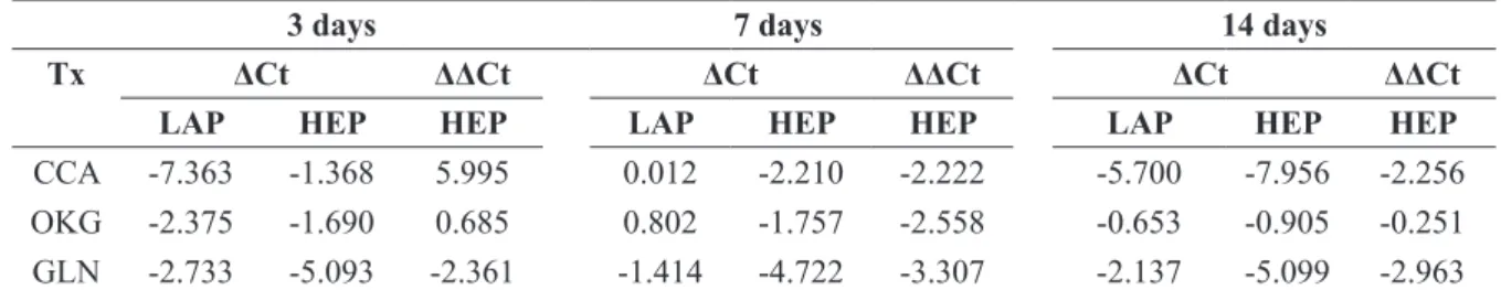
3 days 7 days 14 days
Tx ΔCt ΔΔCt ΔCt ΔΔCt ΔCt ΔΔCt
LAP HEP HEP LAP HEP HEP LAP HEP HEP
CCA -7.475 -6.707 0.768 -3.885 -5.845 -1.960 -6.763 -6.617 0.146 OKG -5.045 -2.322 2.723 -3.100 -1.882 1.218 -1.658 -3.201 -1.543 GLN -4.735 -8.693 -3.958 -4.219 -7.492 -3.272 -3.325 -6.141 -2.816
TABLE 3 - Liver enzyme MDH2 calculated values (ΔCT, ΔΔCT) on the 3rd, 7th and 14th day of the experiment.
Tx = Treatment; CCA = Calcium caseinate; OKG = ornithine alpha-ketoglutarate; GLN = Glutamine; LAP= Laparotomy; HEP= Hepatectomy. The ΔCts were obtained by normalization with GAPDH gene. The ΔΔCts were obtained using data from laparotomy animals as calibrator.
FIGURE 1 - Liver enzymes (MDH1, MDH2) relative gene expression (2-ΔΔCT) on the 3rd, 7th and 14th day of the experiment. Results
were obtained using the 2-ΔΔCT calculation (Livak and Schimittgen, 2001). MDH1 Relative Gene Expression (RGE) was greater on days 3 and 7, compared with CCA. MDH2 RGE was greater in all timepoints, compared with CCA in GLN-treated rats.
Tx = Treatment; CCA = Calcium caseinate; OKG = ornithine alpha-ketoglutarate; GLN = Glutamine; LAP= Laparotomy; HEP= Hepatecto-my. The ΔCts were obtained by normalization with GAPDH gene. The ΔΔCts were obtained using data from laparotomy animals as calibrator
3 days 7 days 14 days
Tx ΔCt ΔΔCt ΔCt ΔΔCt ΔCt ΔΔCt
LAP HEP HEP LAP HEP HEP LAP HEP HEP
CCA -7.363 -1.368 5.995 0.012 -2.210 -2.222 -5.700 -7.956 -2.256 OKG -2.375 -1.690 0.685 0.802 -1.757 -2.558 -0.653 -0.905 -0.251 GLN -2.733 -5.093 -2.361 -1.414 -4.722 -3.307 -2.137 -5.099 -2.963
Subsequent levels of DNA synthesis in hepatocytes are lower, as complete restoration of liver mass requires an average of ~1.6 cycles of replication in all cells18. Most of the increase in
liver mass has occurred by three days after partial hepatectomy and mass restoration is complete in 5–7 days19.
In order to maintain the metabolic homeostasis during regeneration the liver undergoes an adaptive response to regulate the differentiated functions of the hepatocyte. This is accomplished by the interplay between different sets of transcription factors, induced by the regenerative response20. Liver regeneration requires large
amounts of energy to take place. One of the mechanisms involved in supplying energy to the hepatocyte is the autophagy seen in acute liver injury. However, hyperactivation of autophagy induces cell death, and the necrosis rate is actually a predictor of liver failure21.
The malate/aspartate shuttle is the major pathway by which cytosolic reducing equivalents from NADH can enter the mitochondria and be oxidized22. MDH1 and MDH2 enzymes play
important roles in the Krebs cycle for energy production through aerobic respiration9. The malate-aspartate shuttle translocates
electrons produced during glycolysis across the semipermeable inner membrane of the mitochondrion to the electron transport chain via NADH to generate ATP. The shuttle system is required since the mitochondrial inner membrane is impermeable to NADH. To circumvent this, malate carries the reducing equivalents across the membrane23.
This study has demonstrated that the use of GLN in partially hepatectomized rats promotes an over expression of MDH1 and MDH2 enzymes during liver regeneration. The
explanation for the these indings is the same already duly
proven in previous study23 increased GLN availability enhances
the malate-aspartate shuttle requiring an up-regulation of gene expression of both MDH1 and MDH2 enzymes.
The results presented here show that GLN supplementation
in this model of partial hepatectomy has beneicial effects in liver
regeneration considering that there was an up-regulation of the MDH1 and MDH2 gene expression in rats treated with GLN. On the other hand, the use of OKG did not lead to an overexpression of MDH1/MDH2.
Conclusions
Glutamine has beneicial effects in experimental liver
regeneration by promoting an up-regulation of MDH1 and MDH2 gene expression. The use of ornithine alpha-ketoglutarate does not change MDH1 and MDH2 gene expression in experimental liver regeneration in rats.
References
1. Fujiyoshi M, Ozaki M. Molecular mechanisms of liver regeneration and protection for treatment of liver dysfunction and diseases. J Hepatobiliary Pancreat Sci. 2011 Jan;18(1):13-22. doi: 10.1007/ s00534-010-0304-2.
2. Riehle KJ, Dan YY, Campbell JS, Fausto N. New concepts in liver regeneration. J Gastroenterol Hepatol. 2011 Jan;26 Suppl 1:203-12. doi: 10.1111/j.1440-1746.2010.06539.x.
3. Holecek M. Nutritional modulation of liver regeneration by carbohydrates, lipids, and amino acids: a review. Nutrition. 1999 Oct;15(10):784-8. PubMed PMID: 10501293.
4. Birkhahn RH, Awad S, Thomford NR. Parenteral monoacetoacetin and liver regeneration interaction after partial hepatectomy in the rat. JPEN J Parenter Enteral Nutr. 1994 May-Jun;18(3):219-24. PubMed PMID: 80649956.
5. Coudray-Lucas C, Le Bever H, Cynober L, De Bandt JP, Carsin H. Ornithine alpha-ketoglutarate improves wound healing in severe burn patients: a prospective randomized double-blind trial versus isonitrogenous controls. Crit Care Med. 2000 Jun;28(6):1772-6. PubMed PMID: 10890617.
6. Jeevanandam M, Holaday NJ, Petersen SR.
Ornithine-alpha-ketoglutarate (OKG) supplementation is more effective than its component salts in traumatized rats. J Nutr. 1996 Sep;126(9):2141-50.PubMed PMID: 8814202.
7. Nurjhan N, Bucci A, Perriello G, Stumvoll M, Dailey G, Bier DM, Toft I, Jenssen TG, Gerich JE. Glutamine: a major gluconeogenic precursor and vehicle for interorgan carbon transport in man. J Clin Invest. 1995 Jan;95(1):272-7. PubMed PMID: 7814625.
8. Yoshida S, Yunoki T, Aoyagi K, Ohta J, Ishibashi N, Noake T, Kakegawa T. Effect of glutamine supplement and hepatectomy on DNA and protein synthesis in the remnant liver. J Surg Res. 1995 Oct;59(4):475-81. PubMed PMID: 7564320.
9. Bourneuf E, Hérault F, Chicault C, Carré W, Assaf S, Monnier A, Mottier S, Lagarrigue S, Douaire M, Mosser J, Diot C. Microarray analysis of differential gene expression in the liver of lean and fat chickens. Gene. 2006 May 10;372:162-70. PubMed PMID: 14965116. 10. Lo AS, Liew CT, Ngai SM, Tsui SK, Fung KP, Lee CY, Waye
MM. Developmental regulation and cellular distribution of human cytosolic malate dehydrogenase (MDH1). J Cell Biochem. 2005 Mar 1;94(4):763-73. PubMed PMID: 11225055.
11. Dupourque D, Kun E. Malate dehydrogenases of ox kidney. 2. Two substrate kinetic and inhibition analyses. Eur J Biochem. 1969 Jan;7(2):247-52. PubMed PMID:4303911.
12. Chomczynski P, Sacchi N. Single-step method of RNA isolation by acid guanidinium thiocyanate-phenol-chloroform extraction. Anal Biochem. 1987 Apr;162(1):156-9. PubMed PMID: 2440339. 13. Gibson UE, Heid CA, Williams PM. A novel method for real time
quantitative RT-PCR. Genome Res. 1996 Oct;6(10):995-1001. PubMed PMID: 8908519.
14. Heid CA, Stevens J, Livak KJ, Williams PM. Real time quantitative PCR. Genome Res. 1996 Oct;6(10):986-94. PubMed PMID: 8908418.
15. Livak KJ, Schmittgen TD. Analysis of relative gene expression data using real-time quantitative PCR and the 2(-Delta Delta C(T)) Method. Methods. 2001 Dec;25(4):402-8. PubMed PMID: 11846609.
16. Kubista M, Andrade JM, Bengtsson M, Forootan A, Jonák J, Lind K, Sindelka R, Sjöback R, Sjögreen B, Strömbom L, Ståhlberg A, Zoric N. The real-time polymerase chain reaction. Mol Aspects Med. 2006 Apr-Jun;27(2-3):95-125. PubMed PMID: 16460794. 17. Sambrook J, Russel W. Molecular cloning - A laboratory manual.
18. Sigal SH, Rajvanshi P, Gorla GR, Sokhi RP, Saxena R, Gebhard DR Jr, Reid LM, Gupta S. Partial hepatectomy-induced polyploidy attenuates hepatocyte replication and activates cell aging events. Am J Physiol. 1999 May;276(5 Pt 1):G1260-72. PubMed PMID: 10330018.
19. Grisham JW. A morphologic study of deoxyribonucleic acid synthesis and cell proliferation in regenerating rat liver; autoradiography with thymidine-H3. Cancer Res. 1962 Aug;22:842-9. PubMed PMID: 13902009.
20. Costa RH, Kalinichenko VV, Holterman AX, Wang X. Transcription factors in liver development, differentiation, and regeneration. Hepatology. 2003 Dec;38(6):1331-47. PubMed PMID: 14647040. 21. Rautou PE, Mansouri A, Lebrec D, Durand F, Valla D, Moreau R.
Autophagy in liver diseases. J Hepatol. 2010 Dec;53(6):1123-34. doi: 10.1016/j.jhep.2010.07.006.
22. McKenna MC, Waagepetersen HS, Schousboe A, Sonnewald U. Neuronal and astrocytic shuttle mechanisms for cytosolic-mitochondrial transfer of reducing equivalents: current evidence and pharmacological tools. Biochem Pharmacol. 2006 Feb 14;71(4):399-407. PubMed PMID: 16368075.
23. Shi Q, Gibson GE. Up-regulation of the mitochondrial malate
dehydrogenase by oxidative stress is mediated by miR-743a. J Neurochem. 2011 Aug;118(3):440-8. doi: 10.1111/j.1471-4159.2011.07333.x
Correspondence:
Prof. Sergio Botelho Guimarães
Rua Professor Costa Mendes, 1608/3º andar 60430-140 Fortaleza – CE Brasil
Tel.: (55 85)3366-8083 Fax: (55 85)3366-8064 [email protected]
Received: Jan 14, 2014 Review: March 13, 2014 Accepted: April 17, 2014
Conlict of interest: none
Financial source: none
1Research performed at Experimental Surgical Research Laboratory