Histological and immunohistochemical findings of the action of
botulinum toxin in salivary gland: systematic review
J. B. Oliveira
a,b, J. Evêncio-Neto
b,cand L. Baratella-Evêncio
d*
aDepartment of Anatomy, Biological Sciences Center – CCB, Universidade Federal de Pernambuco – UFPE,
Av. Prof. Moraes Rego, 1235, Cidade Universitária, CEP 50670-901, Recife, PE, Brazil
bPost-graduate Program in Bioscience Animal – PPGBA, Universidade Federal Rural de Pernambuco – UFRPE,
Rua Dom Manoel de Medeiros, s/n, Dois Irmãos, CEP 52171-900, Recife, PE, Brazil
cDepartment of Animal Morphology and Physiology, Universidade Federal Rural de Pernambuco – UFRPE,
Rua Dom Manoel de Medeiros, s/n, Dois Irmãos, CEP 52171-900, Recife, PE, Brazil
dDepartment of Histology and Embryology, Biological Sciences Center – CCB, Universidade Federal de Pernambuco –
UFPE, Av. Prof. Moraes Rego, 1235, Cidade Universitária, CEP 50670-901, Recife, PE, Brazil
*e-mail: Liriane@uol.com.br
Received: July 24, 2015 – Accepted: February 15, 2016 – Distributed: May 31, 2017
Abstract
The treatment of sialorrhea is necessary for the constant risks posed by hypersalivation. A new therapeutic option comes up with the application of botulinum toxin in salivary glands. However, little is known about its mechanism of action in glandular tissue. Based on the above, this work had the objective to systematically review the literature about the action of botulinum toxin on submandibular and parotid salivary glands tissues. Electronic search was performed in databases of great relevance for this study (PubMed, SciELO, HighWire, Crossref, Scopus, Science Direct, MEDLINE,
OLDMEDLINE, Serials Database, NLM Catalog, LILACS and IBECS). Inclusion and exclusion criteria for articles were established, and a total number of 14 articles were selected and used. There are few publications that clarify how the salivary gland acini behave with application of botulinum toxin. Although, the immunohistochemical findings were consistent among authors, showing weak immunoreactivity in glands treated with botulinum toxin. Histometric data are divergent, requiring more detailed studies to answer the questions about the efficacy and safety of botulinum toxin in salivary glands.
Keywords: botulinum toxins, salivary glands, sialorrhea, botulinum toxin type A.
Achados histológicos e imunohistoquímicos da ação da toxina botulínica
em glândula salivar: revisão sistemática
Resumo
O tratamento da sialorreia se faz necessário pelos constantes riscos trazidos por este estado de hipersalivação. Uma nova opção terapêutica surge com a aplicação da toxina botulínica em glândulas salivares. Entretanto, pouco se sabe sobre o seu mecanismo de ação no tecido glandular. Com base no exposto, este trabalho teve o objetivo de revisar sistematicamente na literatura a ação da toxina botulínica sobre o tecido das glândulas salivares submandibular e parótida. Foi realizada uma busca eletrônica em bases de dados de grande relevância para este estudo (PubMed,
SciELO, HighWire, Crossref, Scopus, Science Direct, MEDLINE, OLDMEDLINE, Serials Database, NLM Catalog, LILACS e IBECS). Foram estabelecidos critérios de inclusão e exclusão para os artigos, e um “n” de 14 trabalhos foram selecionados e utilizados. São poucas as publicações que esclarecem como os ácinos das glândulas salivares se comportam mediante aplicação da toxina botulínica. Apesar de os achados imunohistoquímicos entre os autores serem concordantes, com imunorreatividade mais fracas nas glândulas tratadas com a toxina botulínica, os dados histométricos são divergentes e há questionamentos metodológicos, necessitando de mais estudos pormenorizados para responder as questões sobre a eficácia e segurança da toxina botulínica nas glândulas salivares.
1. Introduction
Sialorrhea or hypersalivation is a common phenomenon in children during the development of oral neuromuscular control, ranging from 18 to 24 months of life. However, after 4 years of age this condition is considered abnormal and hence, pathological (Augusto and Perez, 2006).
Hypersalivation is the result of hypersecretion of salivary glands, but it is commonly associated with the loss of neuromuscular control with impaired oral motor activity and increased saliva flow (Yang et al., 2006). It can also occur as a side effect of drugs that act by increasing the activity of specific receptors in the secretomor pathway, resulting in hypersecretion. However, most patients suffering from sialorrhea show poor oral neuromuscular control (Jongerius et al., 2001).
The consequences of hypersalivation include facial and perioral dermatitis (Yang et al., 2006; Bloem et al., 2009); increased perioral and oral infections, halitosis, hygiene difficulties, social isolation, aspiration risk and loss of fluids and electrolytes (Yang et al., 2006; Jankovic, 2009); difficulties to speak and pneumonia (Alter, 2010); and risk of lung infections (Ellies et al., 2002), which generate great impact on the patient’s life. On the other hand, the decrease in salivary flow and xerostomia, the person may develop severe weakness in oral health, difficulty with speech, chewing, swallowing, changes in the mucous membrane, tooth loss (Sanioto et al., 2013), among others as microbial infections such as candidiasis caused by pathogenic species of Candida (Rodrigues et al., 2004).
As this is a multifactorial disease, there are several therapeutic approaches. Anticholinergic drug substances such as Atropine, Benztropine, Glycopyrrolate, and Benzhexol Hydrochloride are some options, which reduce the volume of saliva in the oral cavity by blocking the action of parasympathetic autonomic nervous system on acetylcholine receptors on the salivary glands (Ellies et al., 2006a; Coskun et al., 2007; Bavikatte et al., 2012). Other ways include antihistaminic drugs (Alter, 2010); surgery such as ablation of salivary glands, tympanic neurectomy, transposition or retropositioning of excretory ducts or ligation of excretory ducts (Ellies et al., 2002; Savarese et al., 2004; Manrique et al., 2007); radiotherapy (Bavikatte et al., 2012; Kasarskis et al., 2011; Corso et al., 2011); speech therapy (Crysdale, 1980); techniques of body position, and the “biofeedback” (Tscheng, 2002; Savarese et al., 2004; Bloem et al., 2009; Valencia and Mendoza, 2011).
Currently, botulinum toxin has been used in the treatment of sialorrhea (Savarese et al., 2004; Lagalla et al., 2009; Intiso, 2012) because it is able to depress the secretory activity of salivary glands (Ellies et al., 2004). These are neurotoxins produced by Clostridium botulinum (Prevot, 1953), an anaerobic bacterium (Sposito, 2009). The bacterium Clostridium botulinum produces many serological types of toxins (A, B, C1, D, E, F and G) (Tsui, 1996; Sposito, 2004, 2009; Poulain et al., 2008; WHO, 2013) as a complex mixture of neurotoxic polypeptides and nontoxic protein components, and type A and B are
commercialized and available for medical use (Sposito, 2004, 2009; Poulain et al., 2008). There are still some authors mentioning an eighth serotype of botulinum toxin, the C2 (Bhayani and Suskind, 2008).
The application of botulinum toxin type A as a treatment for sialorrhea was first proposed in 1997 through intraglandular injection (Bushara, 1997). The intraglandular application of botulinum toxin type A or B has been used to treat hypersalivation (Lagalla et al., 2009; Intiso, 2012) because it is able to depress secretory activity of the salivary glands (Ellies et al., 2004) and saliva production can be effectively reduced by botulinum toxin (Turk-Gonzales and Odderson, 2005). Its action is based on the inhibition of acetylcholine (ACh) release at the presynaptic level, by acting on the cholinergic nerve terminals (parasympathetic nerve terminals), causing local chemical blocking and the loss of neuronal activity in the target organ (Bushara, 1997). All serotypes of botulinum neurotoxin interfere with acetylcholine exocytosis, but act in different intracellular targets. Botulinum toxin type A has targeted the SNAP-25 protein, while the type B cleaves VAMP/synaptobrevin, protein comprising a complex called SNARE, responsible for the fusion of synaptic acetylcholine vesicles at the synaptic membrane (Schiavo et al., 2000; Aoki and Guyer, 2001).
Salivary glands produce two distinct types of saliva, a more serous (secreted by acinar serous, it is a fluid secretion rich in electrolytes and enzymes, but containing low glycoprotein content as the saliva produced by the parotid glands) and a more mucosal (more viscous and has, beyond the components already described above, highly glycosylated proteins called mucin, as that produced by sublingual glands) (Nakamura et al., 2004; Proctor, 2006; Sanioto et al., 2013). The fluid secretion rich in electrolytes (glandular fluid and ion secretion) is secreted mainly by stimulation of M3 muscarinic receptors for acetylcholine (Nakamura et al., 2004; Proctor, 2006).
Even though there are many clinical trials in the literature showing the efficacy of the drug, experimental studies are scarce. There is the need for detailed studies on its safety and effect on glandular tissue. Thus, this study aims to review the existing literature of experimental studies in animals, verifying data on the action of botulinum toxin in the major salivary glands tissues and the efficiency and safety of this drug for sialorrhea treatment.
2. Material and Methods
An electronic search was performed in the Portal de
Periódicos da Coordenação de Aperfeiçoamento de Pessoal de Nível Superior (CAPES) which brings together highly relevant databases for this study (SciELO, HighWire,
search in reference lists was performed in the period from February 2013 to November 2015.
To perform this research, the descriptors “Botulinum
Toxins” AND “Salivary Glands” AND “Drooling OR
Sialorrhea OR Hypersalivation” were used, all included in the Medical Subject Headings (MeSH) and Descritores em
Ciências da Saúde (DeCS). The research was conducted by two reviewers, checking the intersection of these descriptors and their correspondents in English. This research did not count on language restriction and a filter of time was not used. The additional papers were selected for inclusion and exclusion criteria.
Literature review articles, case reports, conference abstracts, non-experimental studies and experiments not performed in laboratory animals were excluded from this search. The selection had as inclusion criteria the application of intraglandular botulinum toxin type A or type B, or type A and type B associated, in salivary glands (parotid or submandibular) of animals, and which have had subsequent histopathological, immunohistochemical and/or ultrastructural analysis.
3. Results
A total of 216 peer-reviewed journals were found during the search in CAPES; in PubMed the search resulted in 277 works, using a filter which limits the species in “other animals” excluding “humans”; and in BVS/BIREME a number of 106 articles were found. In the manual search, based on the list of references in the articles, a number of 184 articles were obtained, totalizing 783 scientific articles. In the first analysis, 82 articles were discarded by being repeated and 406 by the title. The remaining 295 articles have gone through a critical analysis of the evaluators, and after application of exclusion and inclusion criteria, 14 articles were selected and used in this work.
All articles were evaluated independently by three reviewers, resulting in the elaboration of Table 1. Selected articles were organized by author and the year of publication; type and dose of applied botulinum toxin (A or B); animal and gland used in the study; type of processing methods used to evaluate drug action; and main results.
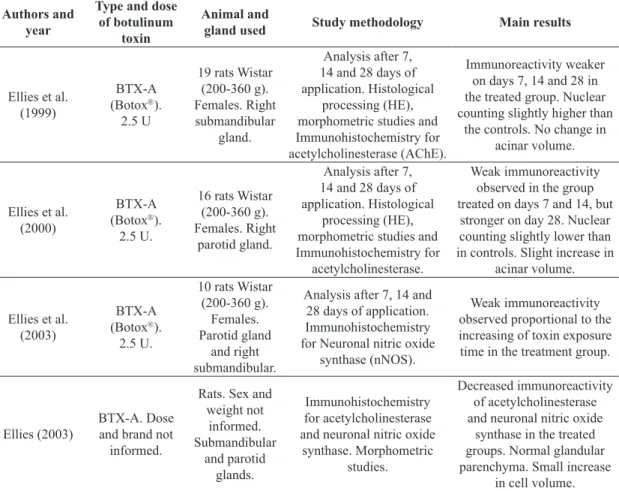
Table 1. Studies on the action of botulinum toxin in the salivary gland of rats, identified by author, year, type and dose of toxin, salivary gland and study methodology used and main results.
Authors and year
Type and dose
of botulinum
toxin
Animal and
gland used Study methodology Main results
Ellies et al. (1999)
BTX-A (Botox). 2.5 U
19 rats Wistar (200-360 g). Females. Right submandibular
gland.
Analysis after 7, 14 and 28 days of application. Histological
processing (HE), morphometric studies and Immunohistochemistry for acetylcholinesterase (AChE).
Immunoreactivity weaker on days 7, 14 and 28 in the treated group. Nuclear counting slightly higher than
the controls. No change in acinar volume.
Ellies et al. (2000)
BTX-A (Botox). 2.5 U.
16 rats Wistar (200-360 g). Females. Right
parotid gland.
Analysis after 7, 14 and 28 days of application. Histological
processing (HE), morphometric studies and Immunohistochemistry for
acetylcholinesterase.
Weak immunoreactivity observed in the group treated on days 7 and 14, but
stronger on day 28. Nuclear counting slightly lower than in controls. Slight increase in
acinar volume.
Ellies et al. (2003)
BTX-A (Botox). 2.5 U.
10 rats Wistar (200-360 g).
Females. Parotid gland
and right submandibular.
Analysis after 7, 14 and 28 days of application. Immunohistochemistry for Neuronal nitric oxide
synthase (nNOS).
Weak immunoreactivity observed proportional to the increasing of toxin exposure time in the treatment group.
Ellies (2003)
BTX-A. Dose and brand not
informed.
Rats. Sex and weight not
informed. Submandibular
and parotid glands.
Immunohistochemistry for acetylcholinesterase and neuronal nitric oxide
synthase. Morphometric studies.
Decreased immunoreactivity of acetylcholinesterase and neuronal nitric oxide
synthase in the treated groups. Normal glandular parenchyma. Small increase
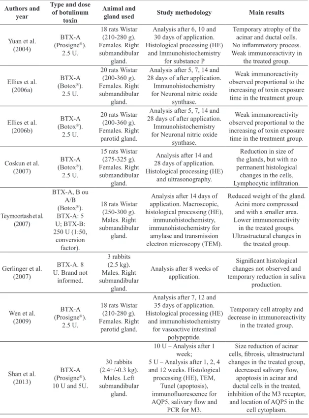
Table 1. Continued...
Authors and year
Type and dose
of botulinum
toxin
Animal and
gland used Study methodology Main results
Yuan et al. (2004)
BTX-A (Prosigne).
2.5 U.
18 rats Wistar (210-280 g). Females. Right submandibular
gland.
Analysis after 6, 10 and 30 days of application. Histological processing (HE)
and Immunohistochemistry for substance P
Temporary atrophy of the acinar and ductal cells. No inflammatory process. Weak immunoreactivity in
the treated group.
Ellies et al. (2006a)
BTX-A (Botox). 2.5 U.
20 rats Wistar (200-360 g). Females. Right submandibular
gland.
Analysis after 5, 7, 14 and 28 days of after application.
Immunohistochemistry for Neuronal nitric oxide
synthase.
Weak immunoreactivity observed proportional to the increasing of toxin exposure time in the treatment group.
Ellies et al. (2006b)
BTX-A (Botox). 2.5 U.
20 rats Wistar (200-360 g). Females. Right
parotid gland.
Analysis after 5, 7, 14 and 28 days of after application.
Immunohistochemistry for Neuronal nitric oxide
synthase.
Weak immunoreactivity observed proportional to the increasing of toxin exposure time in the treatment group.
Coskun et al. (2007)
BTX-A (Botox). 2.5 U.
15 rats Wistar (275-325 g). Females. Right submandibular
gland.
Analysis after 14 and 28 days of application. Histological processing (HE)
and ultrasonography.
Reduction in size of the glands, but with no permanent histological changes in the cells. Lymphocytic infiltration.
Teymoortash et al. (2007)
BTX-A, B ou A/B (Botox). BTX-A: 5 U; BTX-B: 250 U (1:50,
conversion factor).
18 rats Wistar (250-300 g). Males. Right submandibular
gland.
Analysis after 14 days of application. Macroscopic, histological processing (HE),
immunohistochemistry, immunohistochemistry for
amylase and transmission electron microscopy (TEM).
Reduced weight of the gland. Acini more compressed and with a smaller area. Lower immunoreactivity
in the treated groups. Ultrastructural changes in
the treated group.
Gerlinger et al. (2007)
BTX-A. 8 U. Brand not
informed. 3 rabbits (2.5 kg). Males. Right submandibular gland.
Analysis after 8 weeks of application.
Significant histological changes not observed and temporary reduction in saliva
production.
Wen et al. (2009)
BTX-A (Prosigne).
2.5 U.
18 rats Wistar (210-280 g). Females. Right
parotid gland.
Analysis after 7, 12 and 35 days of application. Histological processing (HE)
and immunohistochemistry for vasoactive intestinal
polypeptide.
Temporary cell atrophy and decrease in immunoreactivity
in the treated group.
Shan et al. (2013)
BTX-A (Prosigne). 10 U and 5U.
30 rabbits (2.4+/-0.3 kg).
Males. Left submandibular
gland.
10 U – Analysis after 1 week;
5 U – Analysis after 1, 2, 4 and 12 weeks. Histological processing (HE), TEM,
Tunel (apoptosis), immunofluorescence for AQP5, salivary flow and
PCR for M3.
Size reduction of acinar cells, fibrosis, ultrastructural changes in the treated group, decreased salivary flow,
apoptosis in acinar and ductal cells in the treated, inhibition of the M3 receptor,
4. Discussion
There are few studies in literature reporting the action of botulinum toxin in parotid or submandibular salivary glands tissues. However, Emmelin (1961) investigated the effect of botulinum toxin in parotid and submandibular glands of cats, in which noticed a decrease in acetylcholine release and an increase in sensitivity of the glandular tissue by other stimulating agents of salivary secretion.
The use of the toxin in sialorrhea treatment was based on a publication in 1923, when Dickson and Shevsky observed that the tympanic nerve, which induces salivation, was blocked in cats infected by Clostridium botulinum (Savarese et al., 2004; Ellies et al., 1999). As a therapeutic resource, the toxin was released to patient administration just in 1989, when the FDA (Food and Drugs Administration) classified it as safe and effective drug for the treatment of movement disorders. In 1990, the consent of the National Institutes of Health included botulinum toxin type A in the list of safe and efficient medications (Sposito, 2004).
There are few studies involving histological processing with histometric and histopathological data. Currently, experimental studies of the action of botulinum toxin in salivary glands follow a pattern, in rats, and the majority refers to immunohistochemical studies. Immunohistochemical investigations were made mainly for the enzyme acetylcholinesterase (AChE) and neuronal nitric oxide synthase (nNOS) in submandibular and/or parotid glands of female rats, such as publications of Ellies et al. (1999, 2000, 2003, 2006a, b) and Ellies (2003).
The choice of acetylcholinesterase is justified by the fact that intraglandular treatment with botulinum toxin is a pharmacological “denervation” of salivary glands, which causes inhibition of acetylcholine release (ACh) in the neuroglandular junction (chemical parasympathectomy) and produces a reduction in saliva flow, since botulinum toxin selectively inhibit cholinergic componentes (Intiso, 2012; Teymoortash et al., 2007). The AChE activity depends on the concentration and availability of its substrate, acetylcholine, and they both vary according to the degree of cholinergic innervation of exocrine glands (Ellies et al., 1999).
Nitric oxide (NO) is a small simple molecule, intercellular messenger in higher mammals, an important neurotransmitter, with potentiating capacity, endocrine, autocrine and paracrine actions (Flora-Filho and Zilberstein, 2000). Nitric oxide acts as a possible vascular neuromodulator in the regulation of specific secretory processes in the upper aerodigestive tract (Ellies et al., 2006a). The enzyme neuronal nitric oxide synthase is an important marker of nerve terminals in salivary glands (Chiba and Tanaka, 1998; Takai et al., 1999). Thus, besides acetylcholine transmitters, other neurotransmitters, such as NO neuromodulator may also be involved in regulating the function of the salivary gland (Ellies et al., 2003, 2006a, b).
Ellies et al. (1999, 2000, 2003, 2006a, b) and Ellies (2003), tested the immunoreactivity for acetylcholinesterase and neuronal nitric oxide synthase in parotid and submandibular glands of female rats and demonstrated weak immunoreactivity for these enzymes in the groups Table 1. Continued...
Authors and year
Type and dose
of botulinum
toxin
Animal and
gland used Study methodology Main results
Younis et al. (2013)
BTX-A (Botox).
2 U.
15 rats Wistar (150-200 g). Males. Right parotid gland.
Analysis after 20 days of application. Histological processing (HE) and TEM.
Reduction in the size of the acini, with less secretory granules and with extensive
and coarse vacuoles. Larger interlobular spaces.
Ultrastructural changes in the treated group.
Xu et al. (2015)
BTX-A (Prosigne). 1, 2, 3 and 10 U.
30 rats Sprague-Dawley
(230-250 g). Males. Left submandibular
gland.
Analysis 1, 2, 4, 12 and 24 weeks after application.
Measurement of Saliva Secretion; Western Blotting;
Immunohistochemistry for SNAP-25 and immunofluorescence for aquaporin 5 (AQP5); Cell Culture and Transfection; Cell Surface Biotinylation and Western Blotting; Cell Surface Biotinylation and Immunocytofluorescence; Preparation of Cytoplasm and Membrane Fractions.
Reduced salivary flow in a dose-dependent; SNAP-25
immunostaining was decreased; proteolysis of
SNAP-25 in the treated groups; decreased AQP5 immunofluorescence; and
redistribution of AQP5 (diffuse cytoplasmic distribution) in the treated
treated with botulinum toxin, becoming proportional with the increase of the time exposure to the toxin.
According to histological studies in submandibular glands of female rats made by Ellies et al. (1999), there is no clear difference of serous acini of glands injected with the toxin and saline in their histometric measurements, although both had a slightly higher nuclear counting than in the acini control group. They did not find apoptosis and morphometric measurements revealed no acinar volume.
Parotid glands of rats on the 7th day after drug application
showed nuclear number a little lower than in the control group. Histometric measurements found a greater volume in the acinar cells, especially on day seven, resulting in a smaller number of nuclei per cross‑sectional area. This finding has been described as a result of parasympathectomy and can refer to the retention of the cell body waste products. Important side effects, such as apoptosis due to a toxic reaction, was not observed (Ellies et al., 2000). Ellies (2003), says that after injecting the toxin into the parotid glands of rats, the integrity of glandular parenchyma remained unchanged, with just a slight increase in cell volume explained by the temporary retention of excretory material.
Yuan et al. (2004), in addition to conventional histological processing, the authors also performed immunohistochemistry for substance P, a bioactive peptide that modulates the secretion on salivary glands. They concluded in their work that there was no cellular infiltration, no inflammatory process and nor necrosis around acinar cells and ducts of submandibular glands of rats, although it temporarily induces atrophy of acinar and ductal cells. They also observed decreased immunoreactivity to substance P.
Teymoortash et al. (2007), treated submandibular glands of rats with botulinum toxin type A, B and A/B, performed conventional histological studies, immunohistochemical and ultrastructural analysis, and tested the immunoreactivity for amylase. They showed a reduction in weight of the submandibular gland. Structural changes were observed in all treated groups. More significant changes were observed in glands removed from animals that received simultaneous administration of botulinum toxin type A and B, showing the most densely compressed acini, with slightly elongated shape and the basophilic basal area containing the most pronounced core, in which the morphometric analysis indicated that the area of acinar cells were lower in rats treated with botulinum toxin than in controls with an increase in intersticial tissue, and ultrastructural changes were also observed, such as fewer secretory material, increased rough endoplasmic reticulum, change in size of the secretory vacuoles with occurrence of coalescenses. They also showed that the preparations for immunoreactivity of salivary amylase were strongly stained in the control group, decreasing in the treated groups.
Thus, it is clear that the results found by Teymoortash et al. (2007), are different from those found by Ellies et al. (1999), since these last authors claim to be no clear difference between the serous acini of treated and untreated groups. Teymoortash et al. (2007), affirm that in the first study performed by Ellies et al. (1999), morphometric evaluations
were limited to the description of serous acinar cell nuclei and did not use a standard method of fixation to minimize differences in shrinkage of the tissue samples.
Coskun et al. (2007), in addition to histological processing, used radiological studies before and after application of the toxin in submandibular gland of rats, observed reduction in size of the glands, but with no permanent histological changes in the cells. There was no change in the vascularization. It is believed that changes in the size of the submandibular gland may have occurred as a result of functional effect due to a decrease in secretion. Furthermore, the experiments showed lymphocytic infiltration and apoptosis was not observed. Gerlinger et al. (2007), found no remarkable histological changes in submandibular glands of rabbits, however, the authors only analyzed the gland after two months of drug injection, suggesting the completion of the same action on the glandular tissue.
Wen et al. (2009), also performed immunohistochemistry for vasoactive intestinal polypeptide (VIP) in parotid gland of rats and showed a decrease in immunoreactivity to VIP, which would act as a possible modulator of secretion in parotid glands.
Thus, these authors Yuan, Hou and Wen (2004) and Coskun et al. (2007), agree in saying that botulinum toxin type A temporarily induces atrophy of acinar cells. However, disagree on the point regarding on whether or not there is leukocyte infiltration. Wen et al. (2009), using botulinum toxin type A in parotid glands of rats, reached the same conclusions, claiming to exist a temporary cell atrophy, disagreeing with the findings of Ellies et al. (2000), who also used the parotid gland and observed an increase in acinar volume.
Shan et al. (2013), injected 10 U and 5 U of botulinum toxin type A in submandibular glands of rabbits and showed a reduction in size of acinar cells, fibrosis, ultrastructural changes in the treated group with coalescence of secretory granules, decreased salivary flow, apoptosis in acinar and ductal cells in the groups treated with the toxin. The morphology of the gland returned to normality as the botulinum toxin was losing its effect. Younis et al. (2013), which applied 2 U of botulinum toxin type A in the parotid gland of rats and observed a reduction in the size of the acini and wide variations in secretory granules with extensive and coarse intracellular vacuole, increased endoplasmic reticulum and increased number of degenerate mitochondria and interlobular wider spaces.
the gland. However, no author suggests this mechanism and further studies should occur to clarify such events.
Moreover, Shan et al. (2013), applying botulinum toxin type A in submandibular glands of rabbits and performing immunofluorescence to aquaporin 5 (AQP5) and PCR for M3, suggested that the molecular mechanism of action of botulinum toxin type A is achieved by inhibiting the muscarinic receptor (M3), blocking the expression of AQP5 protein (an aquoporine) involved in secretion of water. Xu et al. (2015), performed immunofluorescence for AQP5 in the submandibular glands of mice and showed decreased AQP5 immunofluorescence in the apical portion of the acinar cells of the groups treated with the toxin, but being reversed over time. These authors also conducted culture of acinar cells of submandibular glands. In these cultured and treated with the toxin cells, AQP5 appeared present in the cytoplasm rather than being located on its apical plasma membrane, indicating a postsynaptic effect of the drug.
The increase in cytosolic intracellular free calcium caused by acetylcholine stimulates the transport of aquaporin (AQP) to the apical plasma membrane and the rapid movement of water. In salivary glands of rats was identified aquaporin 5 (Cheidde and Schor, 1999; Melvin et al., 2005; Catalan et al., 2009).
Xu et al. (2015) also verified clinically the salivary flow of animals and concluded that salivary flow is reduced depending on the dose applied, agreeing with Shan et al. (2013). Regarding the immunohistochemistry for SNAP-25, Xu et al. (2015) observed reduced immunoreactivity and proteolysis of SNAP-25 protein in the treated groups and that it is recovered over time. These results support what is described in the literature about the mechanism of action of botulinum toxin type A, which has as something with SNAP-25 anchoring protein, and that after a certain period, the toxin begins to lose its effect and there is the regeneration of SNAP-25 proteins. According Pérez-Legaspi et al. (2015), SNAP-25 protein plays a crucial role in neuroexocytosis and transmitting of nerve impulses.
5. Conclusion
Based on the findings in literature, it is possible to conclude that there are few publications of experimental studies to determine the mechanism of action of botulinum toxin inside the salivary gland tissue. Despite the immunohistochemical findings among authors are similar, with weaker immunoreactivity in the glands treated with botulinum toxin, histometric data are different and there are methodological questions, requiring more detailed studies to answer possible questions about the effectiveness and safety of botulinum toxin in salivary gland and to explain the mechanisms behind the observed changes in transmission electron microscopy.
A better understanding of morphological, histological and immunohistochemical mechanisms of salivary glands under the influence of botulinum toxin is directly related to the understanding of professionals who could use it
as a resource in the treatment of sialorrhea. Therefore, it is necessary to encourage new experimental and clinical studies about the effect of botulinum toxin, in order to the make use of this substance more frequent and accessible, decreasing the use of more invasive techniques.
Acknowledgements
The authors thank CAPES/CNPq for financial support.
References
ALTER, K.E., 2010. High-frequency ultrasound guidance for neurotoxin injections. Physical Medicine and Rehabilitation Clinics of North America, vol. 21, no. 3, pp. 607-630. http://dx.doi. org/10.1016/j.pmr.2010.05.001. PMid:20797552.
AOKI, K.R. and GUYER, B., 2001. Botulinum toxin type A and other botulinum toxin serotypes: a comparative review of biochemical and pharmacological actions. European Journal of Neurology, vol. 8, suppl. 5, pp. 21-29. http://dx.doi.org/10.1046/ j.1468-1331.2001.00035.x. PMid:11851731.
AUGUSTO, A.G. and PEREZ, A.C., 2006. Drooling. Investigation over the best therapeutic approach. Acta ORL/Técnicas em Otorrinolaringologia, vol. 24, no. 1, p. 1-6.
BAVIKATTE, G., SIT, P.L. and HASSOON, A., 2012. Management of drooling of saliva. British Journal of Medical Practioners, vol. 5, no. 1, pp. 502-507.
BHAYANI, M.K. and SUSKIND, D.L., 2008. The use of botulinum toxin in patients with sialorrhea. Operative Techniques in Otolaryngology, vol. 19, no. 4, pp. 243-247. http://dx.doi. org/10.1016/j.otot.2008.10.008.
BLOEM, B.R., KALF, J.G., VAN DE KERKHOF, P.C. and ZWARTS, M.J., 2009. Debilitating consequences of drooling. Journal of Neurology, vol. 256, no. 8, pp. 1382-1383. http://dx.doi. org/10.1007/s00415-009-5144-0. PMid:19412723.
BUSHARA, K.O., 1997. Sialorrhea in amyotrophic lateral sclerosis: a hypothesis of a new treatment – botulinum toxin A injections of the parotid glands. Medical Hypotheses, vol. 48, no. 4, pp. 337-339. http://dx.doi.org/10.1016/S0306-9877(97)90103-1. PMid:9160288.
CATALÁN, M.A., NAKAMOTO, T. and MELVIN, J.E., 2009. The salivary gland fluid secretion mechanism. The Journal of Medical Investigation, vol. 56, suppl., pp. 192-196. http://dx.doi. org/10.2152/jmi.56.192. PMid:20224180.
CHEIDDE, L. and SCHOR, N., 1999. Revisão: transportadores de água. Revista da Associação Médica Brasileira, vol. 4, no. 1, pp. 71-78. http://dx.doi.org/10.1590/S0104-42301999000100013. PMid:10436597.
CHIBA, T. and TANAKA, K., 1998. A target specific pathway from nitric oxide synthase immunoreactive preganglionic sympathetic to superior cervical ganglion neurons innervating the submandibular salivary gland. Journal of the Autonomic Nervous System, vol. 71, no. 2-3, pp. 139-147. http://dx.doi.org/10.1016/ S0165-1838(98)00068-X. PMid:9760050.
COSKUN, B.U., SAVK, H., CICEK, E.D., BASAK, T., BASAK, M. and DADAS, B., 2007. Histopathological and radiological investigations of the influence of botulinum toxin on the submandibular gland of the rat. European Archives of Oto-Rhino-Laryngology, vol. 264, no. 7, pp. 783-787. http://dx.doi. org/10.1007/s00405-007-0254-8. PMid:17285331.
CRYSDALE, W.S., 1980. The drooling patient: evaluation and current surgical options. The Laryngoscope, vol. 90, no. 5 Pt 1, pp. 775-783. http://dx.doi.org/10.1288/00005537-198005000-00006. PMid:7374307.
ELLIES, M., 2003. Tierexperimentelle und klinische untersuchungen zur sekretionshemmung der kopfspeicheldrüsen durch botulinum Toxin A. Laryngo- Rhino- Otologie, vol. 82, no. 10, pp. 713-714. http://dx.doi.org/10.1055/s-2003-43237. PMid:14593570.
ELLIES, M., GOTTSTEIN, U., ROHRBACH-VOLLAND, S., ARGLEBE, C. and LASKAWI, R., 2004. Reduction of salivary flow with botulinum toxin: extended report on 33 patients with drooling, salivary fistulas, and sialadenitis. The Laryngoscope, vol. 114, no. 10, pp. 1856-1860. http://dx.doi.org/10.1097/00005537-200410000-00033. PMid:15454785.
ELLIES, M., LASKAWI, R., GÖTZ, W., ARGLEBE, C. and TORMÄHLEN, G., 1999. Immunohitochemical and morphometric investigations of the influence of botulinum toxin on the submandibular gland of the rat. European Archives of Oto-Rhino-Laryngology, vol. 256, no. 3, pp. 148-152. http://dx.doi. org/10.1007/s004050050129. PMid:10234485.
ELLIES, M., LASKAWI, R., SCHÜTZ, S. and QUONDAMATTEO, F., 2003. Immunohistochemical evidence of nNOS and changes after intraglandular application of botulinum toxin A in cephalic salivary glands of adult rats. Journal for Otorhinolaryngology. Head & Neck Surgery, vol. 65, no. 3, pp. 140-143. http://dx.doi. org/10.1159/000072251. PMid:12925814.
ELLIES, M., LASKAWI, R., TORMÄHLEN, G. and GÖTZ, W., 2000. The effect of local injection of botulinum toxin A on the parotid gland of the rat: an immunohistochemical and morphometric study. Journal of Oral and Maxillofacial Surgery, vol. 58, no. 11, pp. 1251-1256. http://dx.doi.org/10.1053/joms.2000.16625. PMid:11078136.
ELLIES, M., ROHRBACH-VOLLAND, S., ARGLEBE, C., WILKEN, B., LASKAWI, R. and HANEFELD, F., 2002. Successful managenment of drooling with botulinum toxin A in neurologically disbled children. Neuropediatrics, vol. 33, no. 6, pp. 327-330. http://dx.doi.org/10.1055/s-2002-37084. PMid:12571790.
ELLIES, M., SCHÜTZ, S., QUONDAMATTEO, F. and LASKAWI, R., 2006a. The effect of local injection of botulinum toxin A on the immunoreactivity of nNOS in the rat submandibular gland: An immunohistochemical study. International Journal of Pediatric Otorhinolaryngology, vol. 70, no. 1, pp. 59-63. http://dx.doi. org/10.1016/j.ijporl.2005.05.015. PMid:16002154.
ELLIES, M., SCHÜTZ, S., QUONDAMATTEO, F. and LASKAWI, R., 2006b. Immunohistochemical Investigations of the Influence of Botulinum Toxin A on the Immunoreactivity of nNOS in the Parotid Gland of the Rat. Journal of Oral and Maxillofacial Surgery, vol. 64, no. 3, pp. 397-401. http://dx.doi.org/10.1016/j. joms.2005.11.029. PMid:16487800.
EMMELIN, N., 1961. Supersensitivity of salivary gland caused by botulinum toxin. The Journal of Physiology, vol. 156, no. 1, pp. 121-127. http://dx.doi.org/10.1113/jphysiol.1961.sp006662. PMid:13726643.
FLORA-FILHO, R. and ZILBERSTEIN, B., 2000. Óxido nítrico: o simples mensageiro percorrendo a complexidade. Metabolismo, síntese e funções. Revista da Associação Médica Brasileira, vol. 46, no. 3, pp. 265-271. http://dx.doi.org/10.1590/S0104-42302000000300012. PMid:11070518.
GERLINGER, I., SZALAI, G., HOLLÓDY, K. and NÉMETH, A., 2007. Ultrasound-guided, intraglandular injection of botulinum toxin A in children suffering from excessive salivation. The Journal of Laryngology and Otology, vol. 121, no. 10, pp. 947-951. http:// dx.doi.org/10.1017/S0022215107006949. PMid:17391573.
INTISO, D., 2012. Therapeutic Use of Botulinum Toxin in Neurorehabilitation. Journal of Toxicology, vol. 2012, pp. 1-12. http://dx.doi.org/10.1155/2012/802893. PMid:21941544.
JANKOVIC, J., 2009. Disease-oriented approach to botulinum toxin use. Toxicon, vol. 54, no. 5, pp. 614-623. http://dx.doi. org/10.1016/j.toxicon.2008.11.013. PMid:19073203.
JONGERIUS, P.H., ROTTEVEEL, J.J., VAN DEN HOOGEN, F., JOOSTEN, F., VAN HULST, K. and GABREËLS, F.J.M., 2001. Botulinum toxin A: a new option for treatment of drooling in children with cerebral palsy. Presentation of a case serie. European Journal of Pediatrics, vol. 160, no. 8, pp. 509-512. http://dx.doi. org/10.1007/s004310100784. PMid:11548191.
KASARSKIS, E.J., HODSKINS, J. and CLAIR, W.H.S., 2011. Unilateral parotid electron beam radiotherapy as palliative treatment for sialorrhea in amyotrophic lateral sclerosis. Journal of the Neurological Sciences, vol. 308, no. 1-2, pp. 155-157. http://dx.doi.org/10.1016/j.jns.2011.06.016. PMid:21726879.
LAGALLA, G., MILLEVOLTE, M., CAPECCI, M., PROVINCIALI, L. and CERAVOLO, M.G., 2009. Long-lasting benefits of botulinum toxin type B in Parkinson’s disea.se-related drooling. Journal of Neurology, vol. 256, no. 4, pp. 563-567. http://dx.doi. org/10.1007/s00415-009-0085-1. PMid:19401804.
MANRIQUE, D., DO BRASIL, O.O. and RAMOS, H., 2007. Drooling: analysis and evaluation of 31 children who underwent bilateral submandibular gland excision and parotid duct ligation. Revista Brasileira de Otorrinolaringologia, vol. 73, no. 1, pp. 41-45. http://dx.doi.org/10.1590/S0034-72992007000100007. PMid:17505597.
MELVIN, J.E., YULE, D., SHUTTLEWORTH, T. and BEGENISICH, T., 2005. Regulation of fluid and electrolyte secretion in salivary gland acinar cells. Annual Review of Physiology, vol. 67, no. 1, pp. 445-469. http://dx.doi.org/10.1146/annurev. physiol.67.041703.084745. PMid:15709965.
NAKAMURA, T., MASTSUI, M., UCHIDAK, K., FUTATSUGI, A., KUSAKAWA, S., MATSUMOTO, N., NAKAMURA, K., MANABE, T., TAKETO, M.M. and MIKOSHIBA, K., 2004. M3 muscarinic acetylcholine receptor plays a critical role in parasympathetic control of salivation in mice. The Journal of Physiology, vol. 558, no. 2, pp. 561-575. http://dx.doi.org/10.1113/ jphysiol.2004.064626. PMid:15146045.
PÉREZ-LEGASPI, L.A., RICO-MARTÍNEZ, R. and QUINTANAR, J.L., 2015. Reduced expression of exocytotic proteins caused by anti-cholinesterase pesticides in Brachionus calyciflorus (Rotifera: Monogononta). Brazilian Journal of Biology = Revista Brasileira de Biologia, vol. 75, no. 3, pp. 759-765. http://dx.doi. org/10.1590/1519-6984.01614. PMid:26465735.
Journal, vol. 1, no. 1, pp. 14-87. http://dx.doi.org/10.1504/ TBJ.2008.018951.
PROCTOR, G.B., 2006. Muscarinic receptors and salivary secretion. Journal of Applied Physiology, vol. 100, no. 4, pp. 1103-1104. http://dx.doi.org/10.1152/japplphysiol.01546.2005. PMid:16540706.
RODRIGUES, J.A.O., HÖFLING, J.F., TAVARES, F.C.A., DUARTE, K.M.R., GONÇALVES, R.B. and AZEVEDO, R.A., 2004. Evaluation of biochemical and serological methods to identify and clustering yeast cells of oral Candida species by CHROMagar test, SDS-PAGE and ELISA. Brazilian Journal of Biology = Revista Brasileira de Biologia, vol. 64, no. 2, pp. 317-326. http://dx.doi.org/10.1590/S1519-69842004000200018. PMid:15462306.
SANIOTO, S.M., AMORIM, J.B.O., MANCINI, M.N.G. and BALDO, M.V.C., 2013. Regulação neurovegetativa do aparelho estomatognático: fisiologia da secreção salivar. In: M.V.C. BALDO and M.C. REGATÃO. Fundamentos de odontologia: fisiologia
oral. São Paulo: Santos. 192 p.
SAVARESE, R., DIAMOND, M., ELOVIC, E. and MILLIS, R.S., 2004. Intraparotid injection of botulinum toxin A as a treatment to control sialorrhea in children with cerebral palsy. American Journal of Physical Medicine & Rehabilitation, vol. 83, no. 4, pp. 304-311, quiz 312-314, 336. http://dx.doi.org/10.1097/01. PHM.0000104680.28335.B9. PMid:15024333.
SCHIAVO, G., MATTEOLI, M. and MONTECUCCO, C., 2000. Neurotoxins affecting neuroexocytosis. Physiological Reviews, vol. 80, no. 2, pp. 717-766. http://dx.doi.org/10.1.1.326.2327. PMid:10747206.
SHAN, X.-F., XU, H., CAI, Z.-G., WU, L.-L. and YU, G.-Y., 2013. Botulinum toxin A inhibits salivary secretion of rabbit submandibular gland. International Journal of Oral Science, vol. 5, no. 4, pp. 217-223. http://dx.doi.org/10.1038/ijos.2013.82. PMid:24158141.
SPOSITO, M.M.M., 2004. Toxina botulínica tipo A – propriedades farmacológicas e uso clínico. Acta Fisiátrica, suppl. 01, pp. S7-S44. SPOSITO, M.M.M., 2009. Botulinic toxin type A: action mechanism. Acta Fisiátrica, vol. 16, no. 1, pp. 25-37.
TAKAI, N., UCHIHASHI, K., HIGUCHI, K., YOSHIDA, Y. and YAMAGUCHI, M.M., 1999. Localization of neuronal-constitutive nitric oxide synthase and secretory regulation by nitric oxide in the rat submandibular and sublingual glands. Archives of Oral Biology, vol. 44, no. 9, pp. 745-750. http://dx.doi.org/10.1016/ S0003-9969(99)00064-3. PMid:10471158.
TEYMOORTASH, A., SOMMER, F., MANDIC, R., SCHULZ, S., BETTE, M., AUMÜLLER, G. and WERNER, J.A., 2007. Intraglandular application of botulinum toxin leads to structural
and functional changes in rat acinar cells. British Journal of Pharmacology, vol. 152, no. 1, pp. 161-167. http://dx.doi. org/10.1038/sj.bjp.0707375. PMid:17618309.
TSCHENG, D.Z., 2002. Sialorrhea – therapeutic drug options. The Annals of Pharmacotherapy, vol. 36, no. 11, pp. 1785-1790. http://dx.doi.org/10.1345/aph.1C019. PMid:12398577.
TSUI, J.K.C., 1996. Botulinum Toxin as a Therapeutic Agent. Pharmacology & Therapeutics, vol. 72, no. 1, pp. 13-24. http:// dx.doi.org/10.1016/S0163-7258(96)00091-5. PMid:8981568.
TURK-GONZALES, M. and ODDERSON, I.R., 2005. Quantitative reduction of saliva production with botulinum toxin type B injection into the salivary glands. Neurorehabilitation and Neural Repair, vol. 19, no. 1, pp. 58-61. http://dx.doi.org/10.1177/1545968304273201. PMid:15673844.
VALENCIA, D.V. and MENDOZA, A., 2011. Toxina botulínica tipo A, una nueva opción en el tratamiento de la sialorrea en niños con parálisis cerebral. Revista Colombiana de Medicina Física y Rehabilitación, vol. 21, no. 1, pp. 23-31.
WEN, W.-D., YUAN, F. and HOU, Y.P., 2009. The mechanism of inhibitory effect on parotid gland secretion with local injection of botulinum toxin type A in the rat. Zhonghua Kou Qiang Yi Xue Za Zhi, vol. 44, no. 1, pp. 38-40. http://dx.doi.org/10.3760/cama .j.issn.1002-0098.2009.01.011. PMid:19489258.
WORLD HEALTH ORGANIZATION – WHO, 2013 [viewed 21 June 2014]. Governments to agree increased focus on people with disabilities in development strategies [online]. Geneva: WHO. Available from: http://www.who.int/mediacentre/news/ notes/2013/disability_and_development_20130920/en/.
XU, H., SHAN, X.F., CONG, X., YANG, N.Y., WU, L.L., YU, G.Y., ZHANG, Y. and CAI, Z.G., 2015. Pre- and post-synaptic effects of botulinum toxin A on submandibular glands. Journal of Dental Research, vol. 94, no. 10, pp. 1454-1462. http://dx.doi. org/10.1177/0022034515590087.
YANG, P.-Y., HAN, T.-I., CHOU, L.-W., JOU, H.-J., CHOU, Y.-C. and MENG, N.-H., 2006. Botulinum toxin A in the treatment of sialorrhea in children with cerebral palsy. Mid-Taiwan Journal of Medicine, vol. 11, pp. 261-266.
YOUNIS, R.E., ABOU ELKHIER, M.T., MOURAD, M.I. and ELNAHAS, W., 2013. The ultrastructural changes of the parotid gland of rats after intraglandular injection of botulinum toxin A. Annals of Oral & Maxillofacial Surgery, vol. 1, no. 4, pp. 38. http://dx.doi.org/10.13172/2052-7837-1-4-1095.